- murmur

Содержание
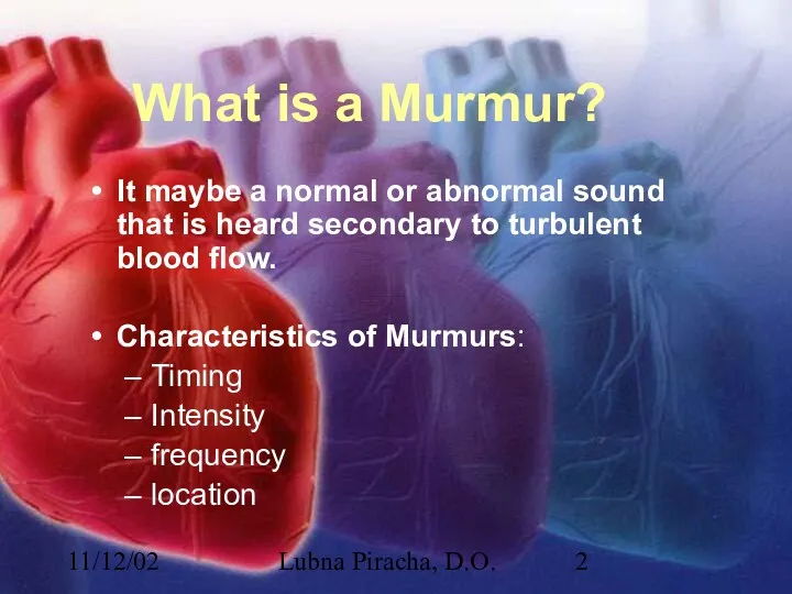
- 2. 11/12/02 Lubna Piracha, D.O. What is a Murmur? It maybe a normal or abnormal sound that
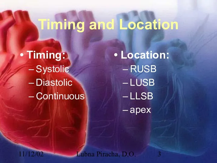
- 3. 11/12/02 Lubna Piracha, D.O. Timing and Location Timing: Systolic Diastolic Continuous Location: RUSB LUSB LLSB apex
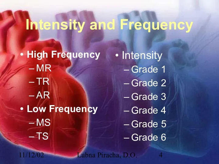
- 4. 11/12/02 Lubna Piracha, D.O. Intensity and Frequency High Frequency MR TR AR Low Frequency MS TS
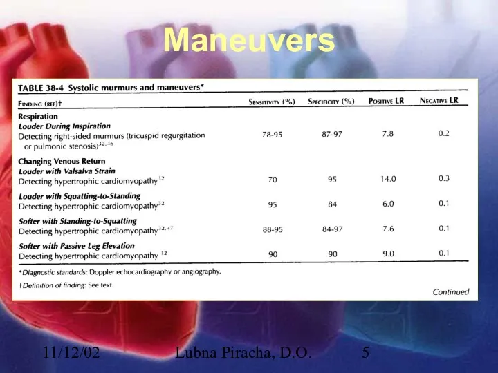
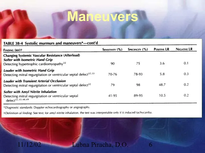
- 5. 11/12/02 Lubna Piracha, D.O. Maneuvers
- 6. 11/12/02 Lubna Piracha, D.O. Maneuvers
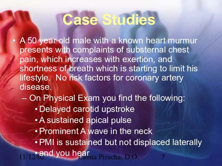
- 7. 11/12/02 Lubna Piracha, D.O. Case Studies A 50 year old male with a known heart murmur
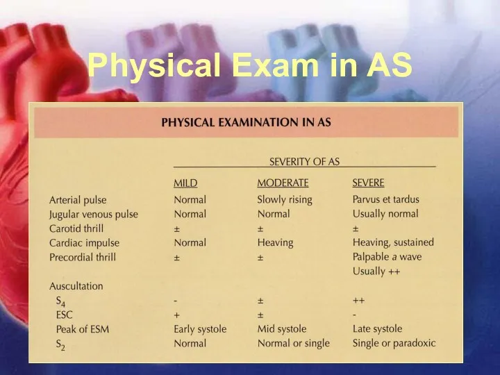
- 8. 11/12/02 Lubna Piracha, D.O. Physical Exam in AS
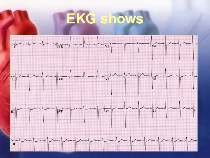
- 9. 11/12/02 Lubna Piracha, D.O. EKG shows
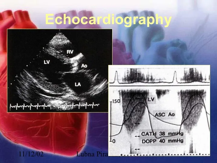
- 10. 11/12/02 Lubna Piracha, D.O. Echocardiography
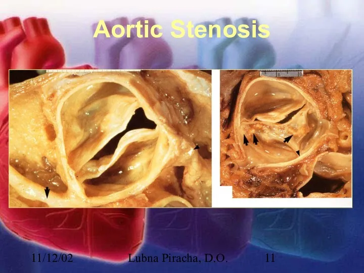
- 11. 11/12/02 Lubna Piracha, D.O. Aortic Stenosis
- 12. 11/12/02 Lubna Piracha, D.O. Aortic Stenosis There is little hemodynamic disturbance that occurs as the valve
- 13. 11/12/02 Lubna Piracha, D.O. Aortic Stenosis continued: Concentric hypertrophy develops in response to this overload. The
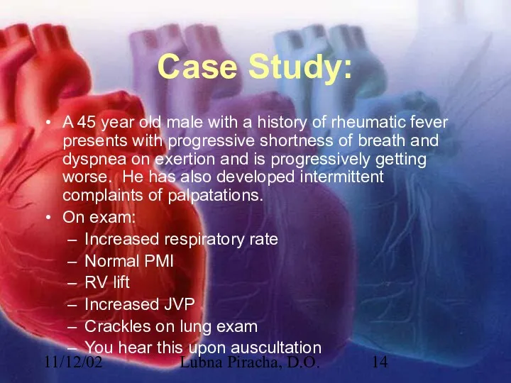
- 14. 11/12/02 Lubna Piracha, D.O. Case Study: A 45 year old male with a history of rheumatic
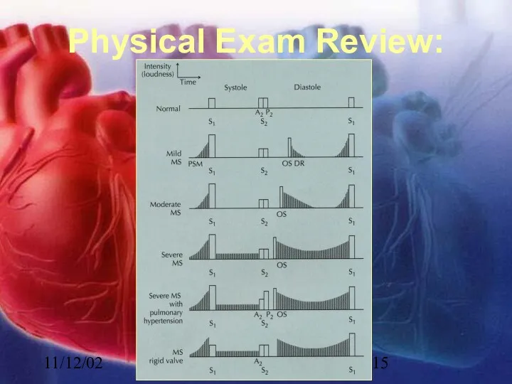
- 15. 11/12/02 Lubna Piracha, D.O. Physical Exam Review:
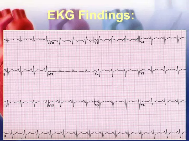
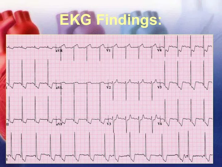
- 16. 11/12/02 Lubna Piracha, D.O. EKG Findings:
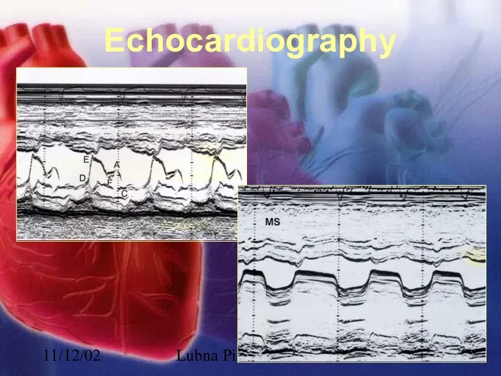
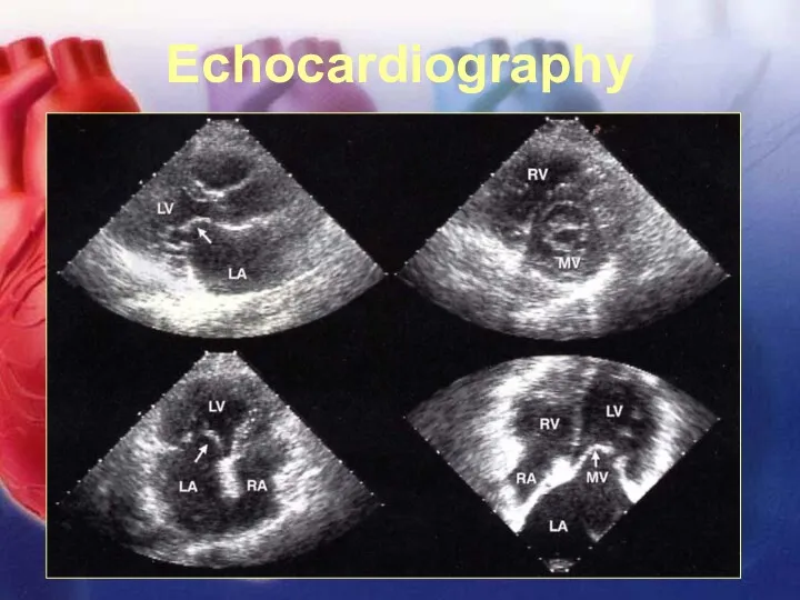
- 17. 11/12/02 Lubna Piracha, D.O. Echocardiography
- 18. 11/12/02 Lubna Piracha, D.O. Echocardiography
- 19. 11/12/02 Lubna Piracha, D.O. Echocardiography
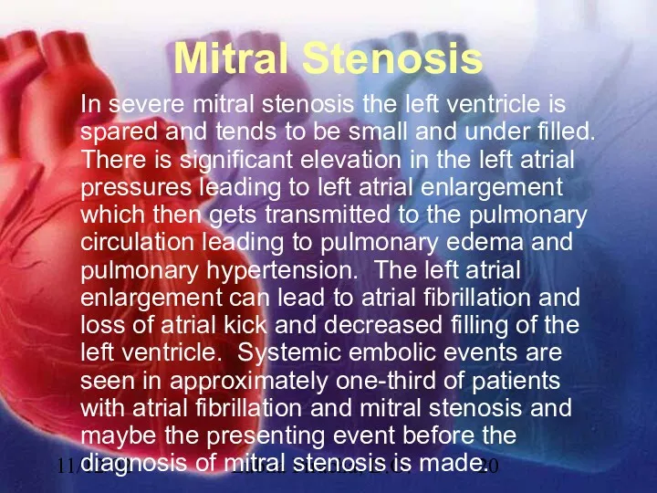
- 20. 11/12/02 Lubna Piracha, D.O. Mitral Stenosis In severe mitral stenosis the left ventricle is spared and
- 21. 11/12/02 Lubna Piracha, D.O. Case Studies: A 52 year old female presents with complaints of slowly
- 22. 11/12/02 Lubna Piracha, D.O. Physical Exam Review Early diastolic murmur of regurgitation blowing, and high frequency,
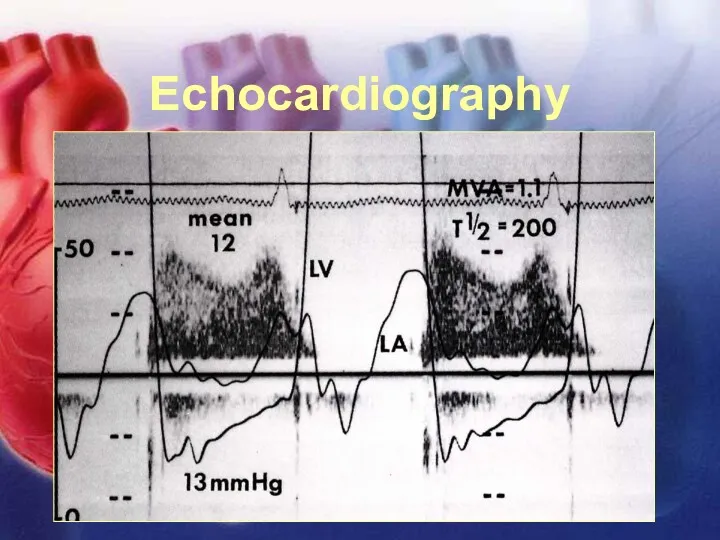
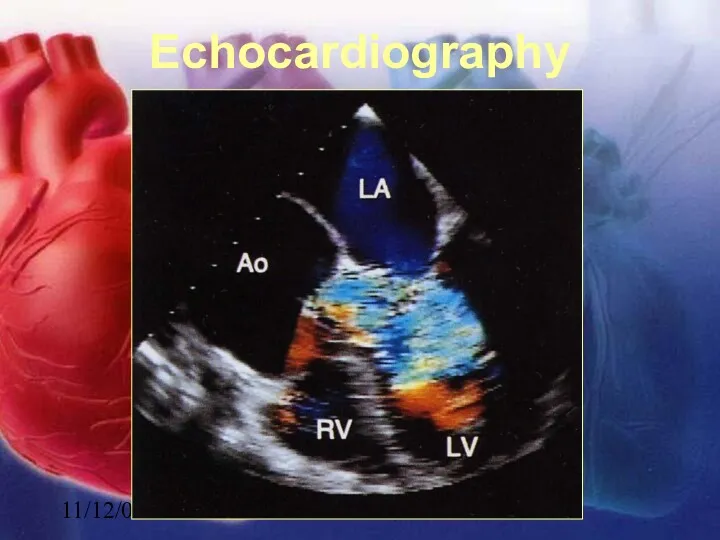
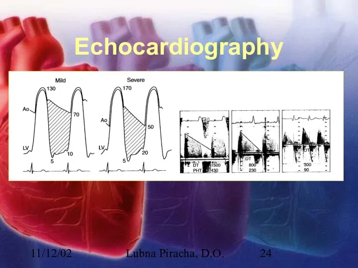
- 23. 11/12/02 Lubna Piracha, D.O. Echocardiography
- 24. 11/12/02 Lubna Piracha, D.O. Echocardiography
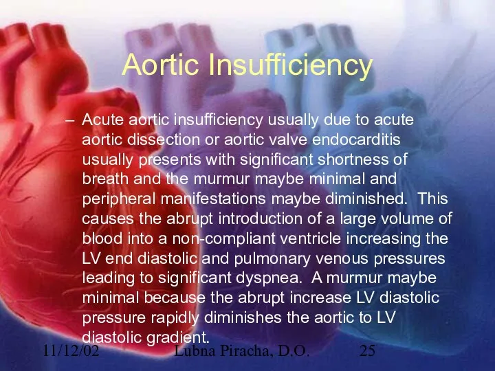
- 25. 11/12/02 Lubna Piracha, D.O. Aortic Insufficiency Acute aortic insufficiency usually due to acute aortic dissection or
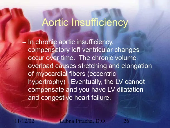
- 26. 11/12/02 Lubna Piracha, D.O. Aortic Insufficiency In chronic aortic insufficiency, compensatory left ventricular changes occur over
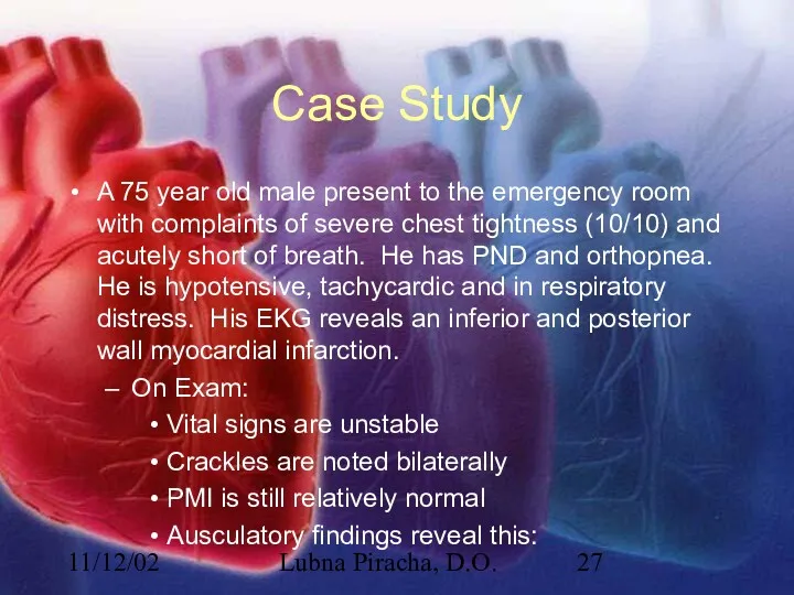
- 27. 11/12/02 Lubna Piracha, D.O. Case Study A 75 year old male present to the emergency room
- 28. 11/12/02 Lubna Piracha, D.O. Physical Exam Review In acute MR, there is tachycardia, the murmur maybe
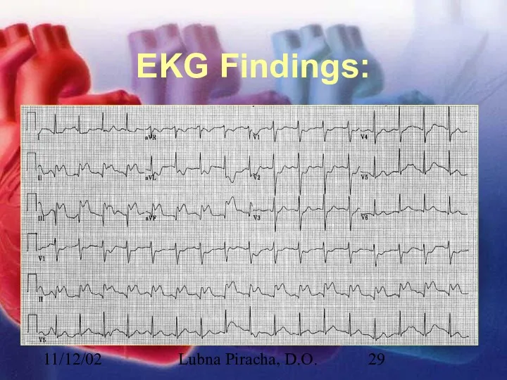
- 29. 11/12/02 Lubna Piracha, D.O. EKG Findings:
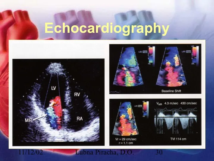
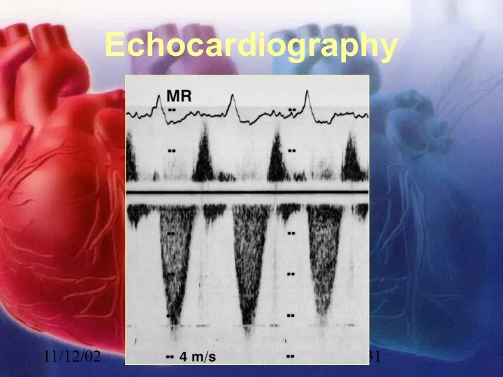
- 30. 11/12/02 Lubna Piracha, D.O. Echocardiography
- 31. 11/12/02 Lubna Piracha, D.O. Echocardiography
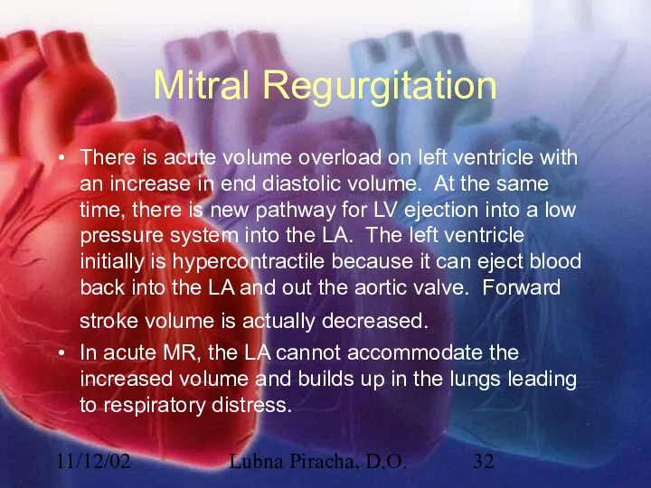
- 32. 11/12/02 Lubna Piracha, D.O. Mitral Regurgitation There is acute volume overload on left ventricle with an
- 33. 11/12/02 Lubna Piracha, D.O. Mitral Regurgitation In chronic MR, the LA will slowly dilate, the LV
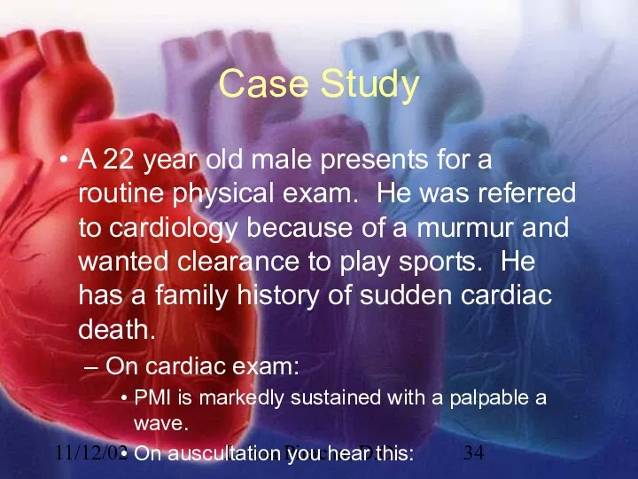
- 34. 11/12/02 Lubna Piracha, D.O. Case Study A 22 year old male presents for a routine physical
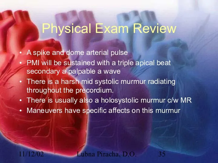
- 35. 11/12/02 Lubna Piracha, D.O. Physical Exam Review A spike and dome arterial pulse PMI will be
- 36. 11/12/02 Lubna Piracha, D.O. EKG Findings:
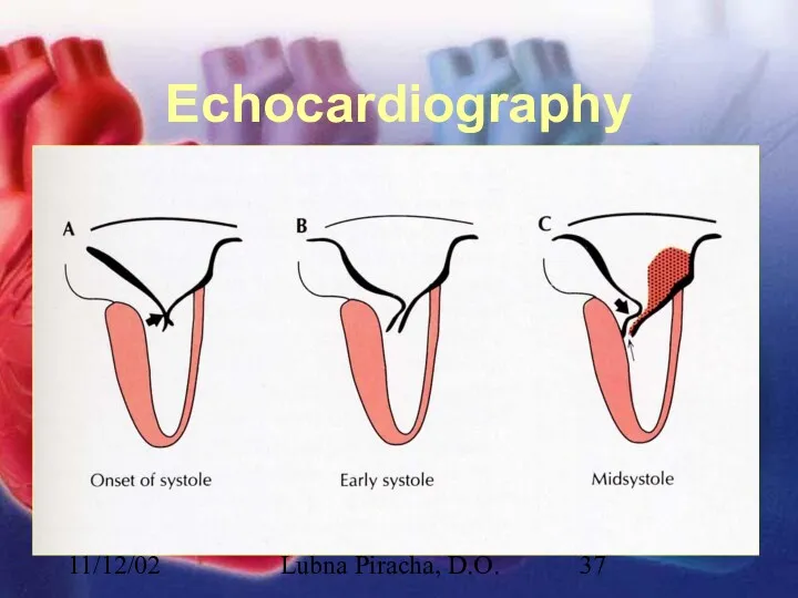
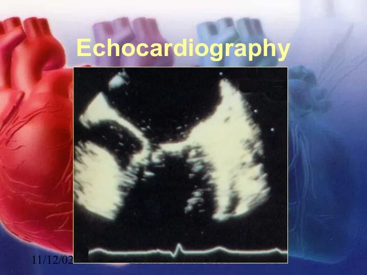
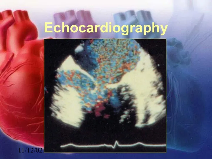
- 37. 11/12/02 Lubna Piracha, D.O. Echocardiography
- 38. 11/12/02 Lubna Piracha, D.O. Echocardiography
- 39. 11/12/02 Lubna Piracha, D.O. Echocardiography
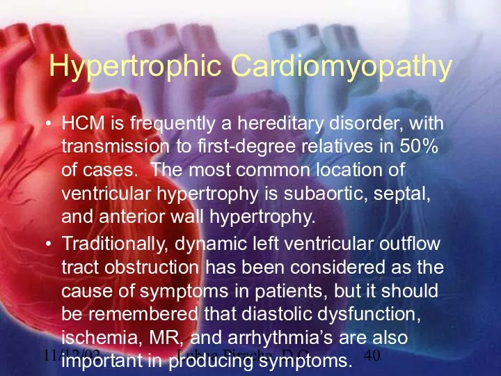
- 40. 11/12/02 Lubna Piracha, D.O. Hypertrophic Cardiomyopathy HCM is frequently a hereditary disorder, with transmission to first-degree
- 42. Скачать презентацию
11/12/02
Lubna Piracha, D.O.
What is a Murmur?
It maybe a normal or abnormal
11/12/02
Lubna Piracha, D.O.
What is a Murmur?
It maybe a normal or abnormal
11/12/02
Lubna Piracha, D.O.
Timing and Location
Timing:
Systolic
Diastolic
Continuous
Location:
RUSB
LUSB
LLSB
apex
11/12/02
Lubna Piracha, D.O.
Timing and Location
Timing:
Systolic
Diastolic
Continuous
Location:
RUSB
LUSB
LLSB
apex
11/12/02
Lubna Piracha, D.O.
Intensity and Frequency
High Frequency
MR
TR
AR
Low Frequency
MS
TS
Intensity
Grade 1
Grade 2
Grade 3
Grade
11/12/02
Lubna Piracha, D.O.
Intensity and Frequency
High Frequency
MR
TR
AR
Low Frequency
MS
TS
Intensity
Grade 1
Grade 2
Grade 3
Grade
11/12/02
Lubna Piracha, D.O.
Maneuvers
11/12/02
Lubna Piracha, D.O.
Maneuvers
11/12/02
Lubna Piracha, D.O.
Maneuvers
11/12/02
Lubna Piracha, D.O.
Maneuvers
11/12/02
Lubna Piracha, D.O.
Case Studies
A 50 year old male with a known
11/12/02
Lubna Piracha, D.O.
Case Studies
A 50 year old male with a known
11/12/02
Lubna Piracha, D.O.
Physical Exam in AS
11/12/02
Lubna Piracha, D.O.
Physical Exam in AS
11/12/02
Lubna Piracha, D.O.
EKG shows
11/12/02
Lubna Piracha, D.O.
EKG shows
11/12/02
Lubna Piracha, D.O.
Echocardiography
11/12/02
Lubna Piracha, D.O.
Echocardiography
11/12/02
Lubna Piracha, D.O.
Aortic Stenosis
11/12/02
Lubna Piracha, D.O.
Aortic Stenosis
11/12/02
Lubna Piracha, D.O.
Aortic Stenosis
There is little hemodynamic disturbance that occurs as
11/12/02
Lubna Piracha, D.O.
Aortic Stenosis
There is little hemodynamic disturbance that occurs as
11/12/02
Lubna Piracha, D.O.
Aortic Stenosis continued:
Concentric hypertrophy develops in response to this
11/12/02
Lubna Piracha, D.O.
Aortic Stenosis continued:
Concentric hypertrophy develops in response to this
11/12/02
Lubna Piracha, D.O.
Case Study:
A 45 year old male with a history
11/12/02
Lubna Piracha, D.O.
Case Study:
A 45 year old male with a history
11/12/02
Lubna Piracha, D.O.
Physical Exam Review:
11/12/02
Lubna Piracha, D.O.
Physical Exam Review:
11/12/02
Lubna Piracha, D.O.
EKG Findings:
11/12/02
Lubna Piracha, D.O.
EKG Findings:
11/12/02
Lubna Piracha, D.O.
Echocardiography
11/12/02
Lubna Piracha, D.O.
Echocardiography
11/12/02
Lubna Piracha, D.O.
Echocardiography
11/12/02
Lubna Piracha, D.O.
Echocardiography
11/12/02
Lubna Piracha, D.O.
Echocardiography
11/12/02
Lubna Piracha, D.O.
Echocardiography
11/12/02
Lubna Piracha, D.O.
Mitral Stenosis
In severe mitral stenosis the left ventricle
11/12/02
Lubna Piracha, D.O.
Mitral Stenosis
In severe mitral stenosis the left ventricle

11/12/02
Lubna Piracha, D.O.
Case Studies:
A 52 year old female presents with complaints
11/12/02
Lubna Piracha, D.O.
Case Studies:
A 52 year old female presents with complaints
11/12/02
Lubna Piracha, D.O.
Physical Exam Review
Early diastolic murmur of regurgitation
blowing, and high
11/12/02
Lubna Piracha, D.O.
Physical Exam Review
Early diastolic murmur of regurgitation
blowing, and high
11/12/02
Lubna Piracha, D.O.
Echocardiography
11/12/02
Lubna Piracha, D.O.
Echocardiography
11/12/02
Lubna Piracha, D.O.
Echocardiography
11/12/02
Lubna Piracha, D.O.
Echocardiography
11/12/02
Lubna Piracha, D.O.
Aortic Insufficiency
Acute aortic insufficiency usually due to acute aortic
11/12/02
Lubna Piracha, D.O.
Aortic Insufficiency
Acute aortic insufficiency usually due to acute aortic
11/12/02
Lubna Piracha, D.O.
Aortic Insufficiency
In chronic aortic insufficiency, compensatory left ventricular changes
11/12/02
Lubna Piracha, D.O.
Aortic Insufficiency
In chronic aortic insufficiency, compensatory left ventricular changes
11/12/02
Lubna Piracha, D.O.
Case Study
A 75 year old male present to the
11/12/02
Lubna Piracha, D.O.
Case Study
A 75 year old male present to the
11/12/02
Lubna Piracha, D.O.
Physical Exam Review
In acute MR, there is tachycardia, the
11/12/02
Lubna Piracha, D.O.
Physical Exam Review
In acute MR, there is tachycardia, the
11/12/02
Lubna Piracha, D.O.
EKG Findings:
11/12/02
Lubna Piracha, D.O.
EKG Findings:
11/12/02
Lubna Piracha, D.O.
Echocardiography
11/12/02
Lubna Piracha, D.O.
Echocardiography
11/12/02
Lubna Piracha, D.O.
Echocardiography
11/12/02
Lubna Piracha, D.O.
Echocardiography
11/12/02
Lubna Piracha, D.O.
Mitral Regurgitation
There is acute volume overload on left ventricle
11/12/02
Lubna Piracha, D.O.
Mitral Regurgitation
There is acute volume overload on left ventricle

11/12/02
Lubna Piracha, D.O.
Mitral Regurgitation
In chronic MR, the LA will slowly dilate,
11/12/02
Lubna Piracha, D.O.
Mitral Regurgitation
In chronic MR, the LA will slowly dilate,
11/12/02
Lubna Piracha, D.O.
Case Study
A 22 year old male presents for a
11/12/02
Lubna Piracha, D.O.
Case Study
A 22 year old male presents for a
11/12/02
Lubna Piracha, D.O.
Physical Exam Review
A spike and dome arterial pulse
PMI will
11/12/02
Lubna Piracha, D.O.
Physical Exam Review
A spike and dome arterial pulse
PMI will
11/12/02
Lubna Piracha, D.O.
EKG Findings:
11/12/02
Lubna Piracha, D.O.
EKG Findings:
11/12/02
Lubna Piracha, D.O.
Echocardiography
11/12/02
Lubna Piracha, D.O.
Echocardiography
11/12/02
Lubna Piracha, D.O.
Echocardiography
11/12/02
Lubna Piracha, D.O.
Echocardiography
11/12/02
Lubna Piracha, D.O.
Echocardiography
11/12/02
Lubna Piracha, D.O.
Echocardiography
11/12/02
Lubna Piracha, D.O.
Hypertrophic Cardiomyopathy
HCM is frequently a hereditary disorder, with transmission
11/12/02
Lubna Piracha, D.O.
Hypertrophic Cardiomyopathy
HCM is frequently a hereditary disorder, with transmission
 Пульмонологиядагы микробиологиялық зерттеу әдістері
 Средства наружной терапии в дерматологии
 Методика проведения массажа и обертывания с водорослями в боди - коррекция в области бедер и ягодиц
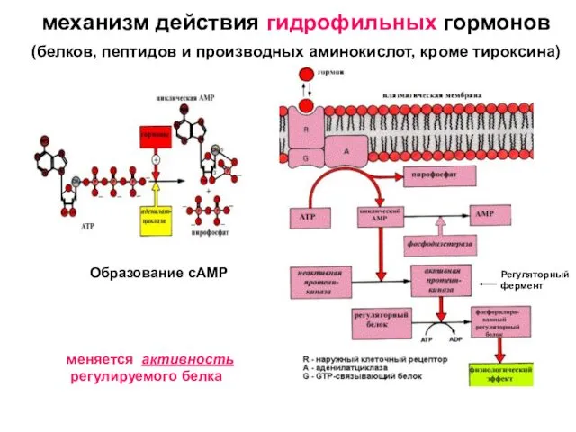 Эндокринная система. Механизм действия гидрофильных гормонов
 История развития сестринского дела в России
 Воспаление. Факторы, вызывающие воспаление
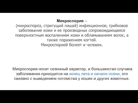 Микроспория (микроспороз, стригущий лишай)
 Здоровый образ жизни
 Материалы для обтурации корневых каналов
 Ведение в токсикологию. Основные закономерности взаимодействия организма и химических веществ
 Ешерихії, сальмонели та їх класифікація
 Медико–педагогическая реабилитация детей с врожденными расщелинами верхней губы и неба
 Сестринское дело в системе первичной медико-санитарной помощи населению
 Ламбдацизм и методы его исправления
 Лечение кишечных инфекций. Сальмонеллёз
 Генерализованный листериоз у новорожденного. Случай из практики
 День донора
 Интеграция в психотерапии. Использование уникальных условий океанской среды для лечения расстройств
 Пиодермии
 Бронхиальная астма – глобальная проблема
 Стрептомицин. Фармакотерапиялық тобы
 Заболевания прямой кишки
 Методы иммунодиагностики и иммунопрофилактики инфекционных болезней
 Cовременные синтетические материалы в хирургии и травматологии
 Перинаталді медицинаның қазіргі мәселелері
 ЭКГ қалыпты жағдайдағы
 Жедел іш синдромы кезіндегі диагностика және жедел көмек көрсету алгоритмі
 Мозжечок, синдромы поражения. Экстрапирамидная система, синдромы поражения