- Omphalocele and gastroschisis

Содержание
- 2. Description of lesion Preoperative stabilization Preanesthetic evaluation Anesthetic management Postoperative considerations OVERVIEW
- 3. GUT DEVELOPMENT Primitive gut - Divided into 3 regions Foregut- Pharynx, esophagus and stomach Midgut- Small
- 4. GUT DEVELOPMENT Week five Week ten Week eleven
- 5. OMPHALOCELE Greek- omphalos-navel, cele- hernia Absence abdominal wall fascia Herniation abdominal contents Eccentric displacement umbilical cord
- 6. OMPHALOCELE Incidence: 1 in 3 - 5,000 Divided into 2 groups Small hernia umbilical cord (
- 7. ASSOCIATED MALFORMATIONS UPPER MIDLINE SYNDROME Pentalogy of Cantrell, Sternal defect, Ectopia cordis, Pericardial and cardiac defects,
- 8. OMPHALOCELE 30- 50% develop hypoglycemia May last for first year of life Associated mortality Small defect
- 9. GASTROSCHISIS Greek: Gaster-stomach, schisis- cleft Incidence 1 in 50,000 Infarction /atresia bowel common Infrequent congenital malformations
- 10. GASTROSCHISIS… ISOLATED OMPHALOCELE Failure of lateral folds to engulf the midgut and form the future umbilical
- 11. GASTROSCHISIS… GLICK (1984) Ultrasound for chronologic in utero development of Gastroschisis OBSERVATION 27 - Moderate soft
- 12. PREOPERATIVE STABILIZATION AIRWAY SUPPORT Often intubated in delivery room GASTRIC DECOMPRESSION Prevent aspiration Air progressing past
- 13. INITIAL RESUSCITATION Consider hypoglycemia until proven otherwise Dextrose solution at 5-7 mg / kg / min
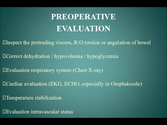
- 14. PREOPERATIVE EVALUATION Inspect the protruding viscera, R/O torsion or angulation of bowel Correct dehydration / hypovolemia
- 15. MANAGEMENT ANESTHETIC MANAGEMENT Airway Maintenance Monitors SURGICAL PROCEDURE Reduction herniated viscera Closure of defect Cardio/respiratory function
- 17. Скачать презентацию
Description of lesion
Preoperative stabilization
Preanesthetic evaluation
Anesthetic management
Postoperative
Description of lesion
Preoperative stabilization
Preanesthetic evaluation
Anesthetic management
Postoperative
GUT DEVELOPMENT
Primitive gut - Divided into 3 regions
Foregut- Pharynx, esophagus and
GUT DEVELOPMENT
Primitive gut - Divided into 3 regions
Foregut- Pharynx, esophagus and
GUT DEVELOPMENT
Week five
Week ten
Week eleven
GUT DEVELOPMENT
Week five
Week ten
Week eleven
OMPHALOCELE
Greek- omphalos-navel, cele- hernia
Absence abdominal wall fascia
Herniation abdominal contents
Eccentric displacement
OMPHALOCELE
Greek- omphalos-navel, cele- hernia
Absence abdominal wall fascia
Herniation abdominal contents
Eccentric displacement
OMPHALOCELE
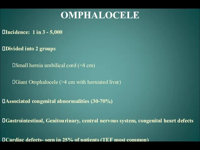
Incidence: 1 in 3 - 5,000
Divided into 2 groups
Small hernia
OMPHALOCELE
Incidence: 1 in 3 - 5,000
Divided into 2 groups
Small hernia
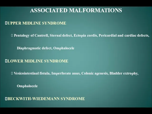
ASSOCIATED MALFORMATIONS
UPPER MIDLINE SYNDROME
Pentalogy of Cantrell, Sternal defect, Ectopia cordis,
ASSOCIATED MALFORMATIONS
UPPER MIDLINE SYNDROME
Pentalogy of Cantrell, Sternal defect, Ectopia cordis,
OMPHALOCELE
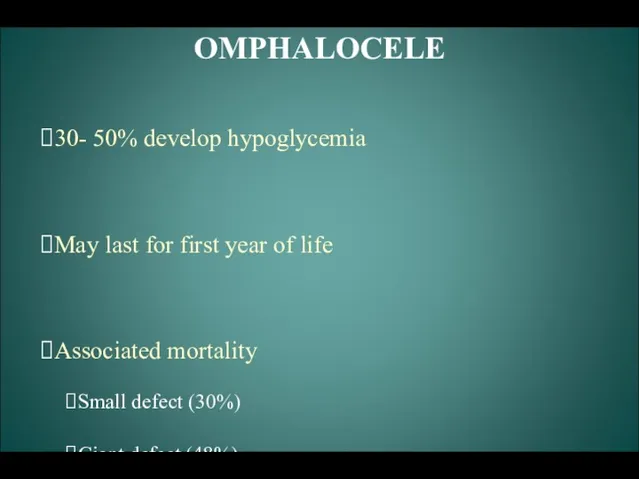
30- 50% develop hypoglycemia
May last for first year of life
Associated
OMPHALOCELE
30- 50% develop hypoglycemia
May last for first year of life
Associated
GASTROSCHISIS
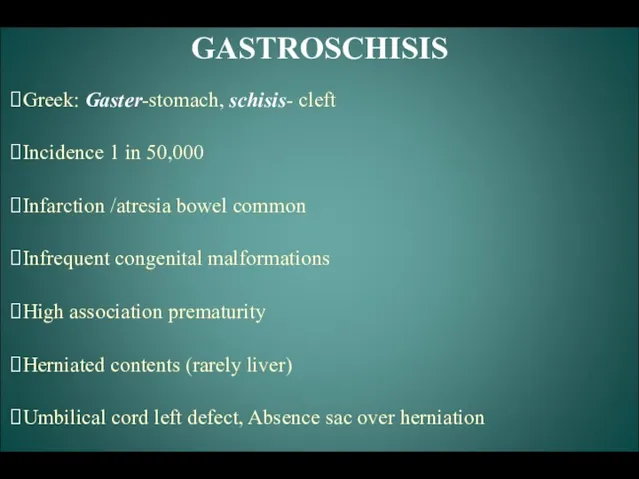
Greek: Gaster-stomach, schisis- cleft
Incidence 1 in 50,000
Infarction /atresia bowel common
Infrequent congenital
GASTROSCHISIS
Greek: Gaster-stomach, schisis- cleft
Incidence 1 in 50,000
Infarction /atresia bowel common
Infrequent congenital
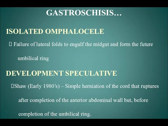
GASTROSCHISIS…
ISOLATED OMPHALOCELE
Failure of lateral folds to engulf the midgut and
GASTROSCHISIS…
ISOLATED OMPHALOCELE
Failure of lateral folds to engulf the midgut and
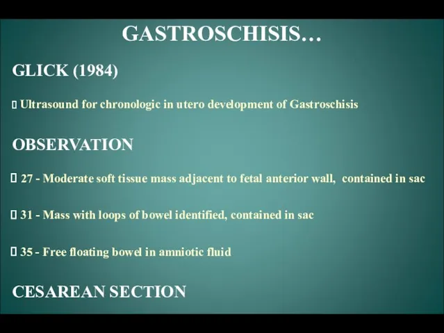
GASTROSCHISIS…
GLICK (1984)
Ultrasound for chronologic in utero development of Gastroschisis
OBSERVATION
GASTROSCHISIS…
GLICK (1984)
Ultrasound for chronologic in utero development of Gastroschisis
OBSERVATION
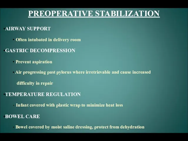
PREOPERATIVE STABILIZATION
AIRWAY SUPPORT
Often intubated in delivery room
GASTRIC DECOMPRESSION
Prevent
PREOPERATIVE STABILIZATION
AIRWAY SUPPORT
Often intubated in delivery room
GASTRIC DECOMPRESSION
Prevent
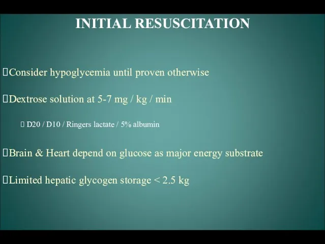
INITIAL RESUSCITATION
Consider hypoglycemia until proven otherwise
Dextrose solution at 5-7 mg /
INITIAL RESUSCITATION
Consider hypoglycemia until proven otherwise
Dextrose solution at 5-7 mg /
PREOPERATIVE EVALUATION
Inspect the protruding viscera, R/O torsion or angulation of bowel
PREOPERATIVE EVALUATION
Inspect the protruding viscera, R/O torsion or angulation of bowel
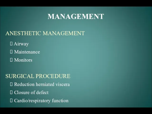
MANAGEMENT
ANESTHETIC MANAGEMENT
Airway
Maintenance
Monitors
SURGICAL PROCEDURE
Reduction herniated viscera
Closure
MANAGEMENT
ANESTHETIC MANAGEMENT
Airway
Maintenance
Monitors
SURGICAL PROCEDURE
Reduction herniated viscera
Closure
 Дифференциальная диагностика пульпитов. Выбор метода лечения пульпитов на основании патогенетических особенностей развития
 Зрение ребёнка - забота родителей
 Гострий діарейний синдром. Холера
 Общая анестезия
 ЭКГ-диагностика ишемической болезни сердца - стенокардии и инфаркта миокарда
 Санитарно-микробиологические и эпидемиологические аспекты биологического терроризма
 Сахарный диабет и беременность
 Хирургическое лечение фибрилляции предсердий
 Гигиена детских и образовательных учреждений
 Инфекционная безопасность
 Әр түрлі туа біткен бүйрек ақаулары кезінде байқалатын бүйрек функциясындағы бейімдеуші өзгерістер
 Современная микроскопия
 Мамандандырылған және арнайы мамандандырылған медициналық көмек
 Антигипертензивная терапия
 Chronic kidney disease
 Қартаюдың биологиялық, медициналық және әлеуметтік негіздері. Қартаю теориялары
 Pulmonary tuberculosis
 Регенерация сердечной мышечной ткани
 Биопсия. Хорион бүрлерінің биопсиясы
 Аллергодерматозы у детей
 Bronchitis. Future in the Past
 Diabetes mellitus. (Subject 8)
 Тірек қимыл аппараты бұзылған балаларға психологиялық педагогикалық көмек көрсету
 Хронический пиелонефрит
 Подвиг медиков в годы Великой Отечественной войны
 Эктопротездер
 Нутриционный статус и его коррекция у пациентов с хронической болезнью почек
 Антиангинальные средства