Слайд 2
Plan
Introduction
History
Etiology
Classification
Epidemiology
Pathogenesis
Clinical features
Diagnosis
Treatment
References
Слайд 3
Introduction
Parainfluenza refers to a group of viruses called human parainfluenza viruses
(HPIVs). There are four viruses in this group. Each one causes different symptoms and illnesses. All forms of HPIV cause an infection in either the upper or lower respiratory area of the body.
Symptoms of HPIVs are like those of the common cold. When cases are mild, the viruses are often misdiagnosed. Most healthy people infected with an HPIV recover with no treatment. A person with a weakened immune system is at risk for developing a life-threatening infection.
Слайд 4
History
Parainfluenza first found in 1952 in Japan. It was look like
influenza, this way renamed parainfluenza in 1959. Disease caused by parainfluenza takes second place in diseases of ARD after influenza: approximately 20 per cent of adults.
Слайд 5
Etiology
Order: Mononegavirales
Family: Paramyxoviridae
Genus: Respirovirus & Rubulavirus
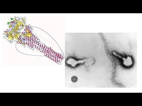
Parainfluenza is RNA virus which includes
Hemagglutinin (H) and Neuraminidase (N). Four types of this virus calls disease in human. Virions are approximately 150-250 nm in size
Слайд 6
Слайд 7
Classification
There are four different types of HPIV. They all cause a
respiratory infection, but the type of infection, symptoms, and location of the infection depend on the type of virus you have. The four types of HPIV can infect anyone.
HPIV-1 is the leading cause of croup in children. Croup is a respiratory illness that manifests as swelling near the vocal cords and in other parts of the upper respiratory system. HPIV-1 is responsible for outbreaks of croup in the autumn. In the United States, the outbreaks tend to be more widespread in odd-numbered years.
HPIV-2 causes croup in children, but doctors detect it much less often than HPIV-1. It’s seen mostly in the autumn but to a lesser degree than HPIV-1.
An HPIV-3 infection is mostly associated with pneumonia and bronchiolitis, which is swelling from inflammation in the smallest airways in the lungs. It often causes infections in the spring and early summer, but it appears in people throughout the year.
HPIV-4 is rarer than the other types. Unlike the other strains of HPIV, there are no known seasonal patterns of HPIV-4.
Слайд 8
Epidemiology
There are several ways you can become infected by an HPIV.
An HPIV can survive on a hard surface for up to 10 hours. If you touch a contaminated surface with your hands and then touch your nose or mouth, you can become infected.
The viruses can also infect you through close contact with an infected person. It usually takes between two and seven days after infection for symptoms to occur.
Слайд 9
Pathogenesis
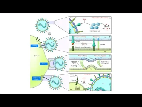
Viral replication is initiated only after successful entry into a cell
by attachment and fusion between the virus and the host cell lipid membrane. Viral RNA is initially associated with nucleoprotein (NP), phosphoprotein (P) and the large protein (L). The hemagglutinin–neuraminidase (HN) is involved with viral attachment and thus hemadsorption and hemagglutination. Furthermore, the fusion (F) protein is important in aiding the fusion of the host and viral cellular membranes, eventually forming syncytia.
Initially the F protein is in an inactive form (F0) but can be cleaved by proteolysis to form its active form, F1 and F2, linked by di-sulphide bonds. Once complete, this is followed by the HPIV nucleocapsid entering the cytoplasm of the cell. Subsequently, genomic transcription occurs using the viruses own 'viral RNA-dependant RNA polymerase' (L protein). The cells own ribosomes are then tasked with translation, forming the viral proteins from the viral mRNA.
Слайд 10
Pathogenesis
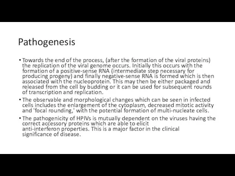
Towards the end of the process, (after the formation of the
viral proteins) the replication of the viral genome occurs. Initially this occurs with the formation of a positive-sense RNA (intermediate step necessary for producing progeny) and finally negative-sense RNA is formed which is then associated with the nucleoprotein. This may then be either packaged and released from the cell by budding or it can be used for subsequent rounds of transcription and replication.
The observable and morphological changes which can be seen in infected cells includes the enlargement of the cytoplasm, decreased mitotic activity and 'focal rounding,' with the potential formation of multi-nucleate cells.
The pathogenicity of HPIVs is mutually dependent on the viruses having the correct accessory proteins which are able to elicit anti-interferon properties. This is a major factor in the clinical significance of disease.
Слайд 11
Слайд 12
Clinical features
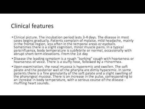
Clinical picture. The incubation period lasts 3-4 days. The disease
in most cases begins gradually. Patients complain of malaise, mild headache, mainly in the frontal region, less often in the temporal areas or eyeballs. Sometimes there is a slight cognition, minor muscle pains. In a typical parainfluenza, body temperature is subfebrile or normal, occasionally with abrupt short-term elevations. From the 1st day
Disease the leading symptom is a rough "barking" cough with hoarseness or hoarseness of voice. There is a stuffy nose, followed by a rhinorrhea.
Upon examination, the nasal mucosa is hyperemic and swollen. The soft palate and the posterior wall of the pharynx are dimly hyperemic. In some patients there is a fine granularity of the soft palate and a slight swelling of the pharyngeal mucosa. There is an increase in the pulse, corresponding to an increase in body temperature, with a serious course of the disease - muffling heart sounds.
Слайд 13
Clinical features
The blood reveals normocytosis or moderate leukopenia. In the period
of convalescence, monocytosis is possible; ESR within normal limits.
Duration of the disease 1-3 weeks.
In persons with chronic diseases of the respiratory system with parainfluenza, the process quickly spreads to the lower respiratory tract. In the early days of the disease, the phenomena of bronchitis are often observed.
Complications. The most frequent complications of parainfluenza are pneumonia caused by a secondary bacterial flora and, as a rule, has a focal character. In children in the first years of life, sometimes cereal occurs as a result of edema and inflammatory infiltration of the laryngeal mucosa, accumulation of secretion in its lumen and reflex muscle spasm.
Parainfluenza leads to an exacerbation of chronic diseases.
Слайд 14
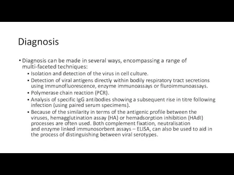
Diagnosis
Diagnosis can be made in several ways, encompassing a range of
multi-faceted techniques:
Isolation and detection of the virus in cell culture.
Detection of viral antigens directly within bodily respiratory tract secretions using immunofluorescence, enzyme immunoassays or fluroimmunoassays.
Polymerase chain reaction (PCR).
Analysis of specific IgG antibodies showing a subsequent rise in titre following infection (using paired serum specimens).
Because of the similarity in terms of the antigenic profile between the viruses, hemagglutination assay (HA) or hemadsorption inhibition (HAdI) processes are often used. Both complement fixation, neutralisation and enzyme linked immunosorbent assays – ELISA, can also be used to aid in the process of distinguishing between viral serotypes.
Слайд 15
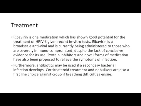
Treatment
Ribavirin is one medication which has shown good potential for the treatment
of HPIV-3 given recent in-vitro tests. Ribavirin is a broadscale anti-viral and is currently being administered to those who are severely immuno-compromised, despite the lack of conclusive evidence for its use. Protein inhibitors and novel forms of medication have also been proposed to relieve the symptoms of infection.
Furthermore, antibiotics may be used if a secondary bacterial infection develops. Corticosteroid treatment and nebulizers are also a first line choice against croup if breathing difficulties ensue.

 Фінанси в системі охорони здоров'я
 Іріңді паротит. Мастит. Парапроктит, лимфангаит, лимфаденит, тромбофлебит
 Urology Clinical Case XL by Slidesgo
 Методы определения подлинности лекарственного растительного сырья
 Жүректің жүрекше мен қарынша гипертрофиясының визуальды диагностикалау әдістері
 Синкопальные состояния
 Дифференциальная диагностика заболеваний с лимфаденитом
 АФО нервной системы и органов чувств ребенка. НПР детей раннего детского возраста
 Анаэробная (инфекционная) энтеротоксемия
 Клиническая терминология
 Сердечная недостаточность. Лечение
 Методы оценки врождённого иммунитета
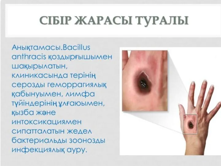 Сібір жарасы туралы
 Доказательная медицина. Систематические обзоры и мета-анализы
 Науки, що вивчають здоров’я людини. Принципи здорового способу життя
 Скорая медицинская помощь при оториноларингологической патологии
 Витаминные и ферментные препараты
 Способы лечения варикозной болезни нижних конечностей
 О коронавирусе детям
 Трихофития
 Тыныс тұншықпасы туралы түсінік
 Ісіктерге қысқаша шолу, ісіктердің пайда болуы
 Микст-патология у больных хроническим описторхозом. Тактика ведения
 План исследования больного при заболеваниях системы дыхания
 Здоровье родителей и здоровье будущего ребенка. Уход за ребенком
 Лечение желудочковых нарушений ритма сердца и профилактика ВСС
 Вакцинация. Современная декларирующая документация
 Passive voice. Modal verbs. Lobular pneumonia