- Creation of radiographs and sonograms for cardiovascular system

Содержание
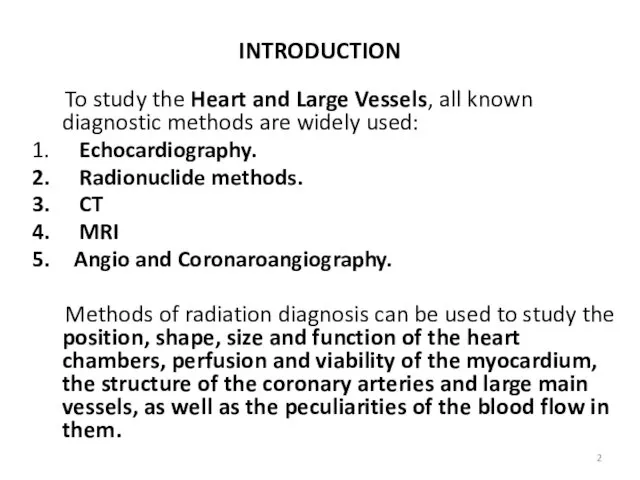
- 2. INTRODUCTION To study the Heart and Large Vessels, all known diagnostic methods are widely used: Echocardiography.
- 3. Roentgenogram of the Chest On the roentgenogram of the chest, most of the middle shadow is
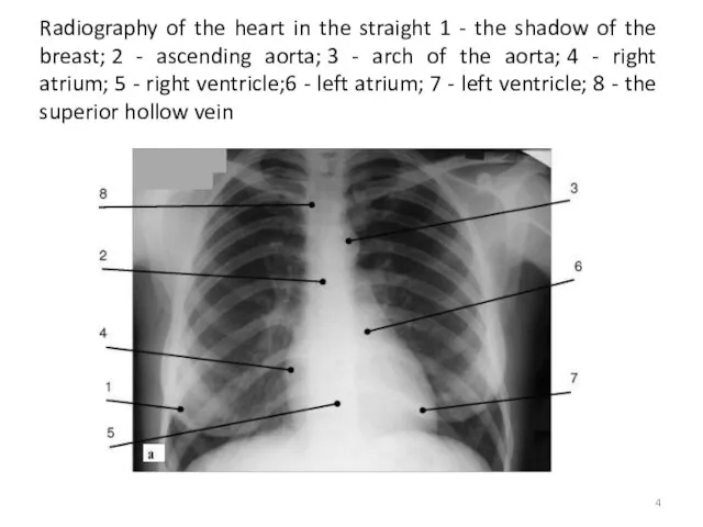
- 4. Radiography of the heart in the straight 1 - the shadow of the breast; 2 -
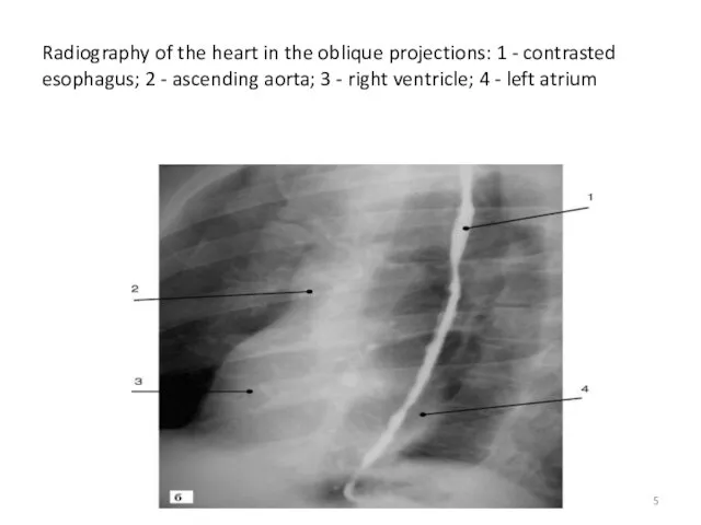
- 5. Radiography of the heart in the oblique projections: 1 - contrasted esophagus; 2 - ascending aorta;
- 6. Echocardiography Echocardiography uses high-frequency ultrasound to evaluate the heart and great vessels. It gives an image
- 7. Indications for Echocardiography Ventricular function Congenital heart disease Valvular heart disease Cardiomyopathy Pericardial effusion Suspected cardiac
- 8. Normal transthoracic echocardiogram from a healthy subject. Views are taken from the left Midparasternal region through
- 9. Transthoracic echocardiogram, left parasternal view, from a patient with a moderate-sized posterior pericardial effusion (PE), visualized
- 10. Angiography Conventional angiography is one of the most commonly performed imaging tests for evaluating the heart
- 11. (A) Coronary arteriogram. Images were obtained from the left lateral projection with contrast injection into the
- 12. (A) Normal aortogram of transverse arch in patient suspected of having traumatic aortic injury. Note the
- 13. CT in Cardiology The use of CT in cardiology allows faster and more accurate diagnosis of
- 14. Normal anatomy at cardiac CT angiography. Ao, aorta; AV, aortic valve; CS, coronary sinus; Diag, diagonal
- 15. 3D volume rendered images in right anterior oblique, left anterior oblique, and cephalad projections (from left
- 16. MRI IN CARDIOLOGY MRI is used for the differential diagnosis of cardiac diseases in complicated cases
- 17. MRI of the heart: a - transverse section at the level of the pulmonary artery trunk;
- 18. Radionuclide Imaging (Nuclear Medicine) Radionuclide Imaging (Nuclear Medicine) Cardiac radionuclide imaging, primarily used for the patient
- 19. Normal myocardial stress/rest study. Stress imaging performed with technetium-99m tetrofosmin following treadmill exercise achieving target heart
- 20. 18F-FDG-PET cardiac study performed after 24-hour fast shows patchy myocardial activity due to cardiac sarcoidosis (arrowheads).
- 21. PATHOLOGICAL RADIOLOGY OF CARDIOVASCULAR SYSYTEM
- 22. Radiograph of the heart with an increase in the left ventricle. Aortic configuration of the heart.
- 23. Radiography of the heart with an increase in the left atrium. Mitral configuration of the heart.
- 24. An increase in the right heart with pulmonary hypertension. Direct projection. Signs of an increase in
- 25. Congenital heart disease without cyanosis Transthoracic echocardiography.Defect of interventricular septum. There is a hole in the
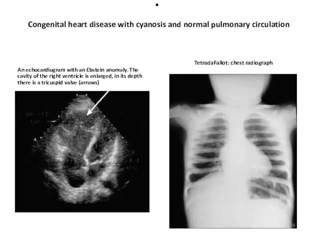
- 26. . Congenital heart disease with cyanosis and normal pulmonary circulation An echocardiogram with an Ebstein anomaly.
- 27. Congenital heart diseases with cyanosis and increased pulmonary circulation angiography with transposition of the main arteries.
- 28. Echocardiography with defects of the mitral valve a - stenosis of the left atrioventricular orifice. Diastole.
- 29. Echodopplerography with aortic valve insufficiency The arrow indicates the retrograde flow of blood through the closed
- 30. DISEASES OF PERICARDIUM Echocardiography with exudative pericarditis.Before the right and behind the left ventricle, a free
- 31. Diagnosis and treatment of arteriovenous fistula of the right kidney: a - on the angiogram, a
- 32. Angiography of the femoral and popliteal arteries: a - a "breakage" of the lumen of the
- 34. Скачать презентацию
INTRODUCTION
To study the Heart and Large Vessels, all known diagnostic
INTRODUCTION
To study the Heart and Large Vessels, all known diagnostic
Roentgenogram of the Chest
On the roentgenogram of the chest, most
Roentgenogram of the Chest
On the roentgenogram of the chest, most
Radiography of the heart in the straight 1 - the shadow
Radiography of the heart in the straight 1 - the shadow
Radiography of the heart in the oblique projections: 1 - contrasted
Radiography of the heart in the oblique projections: 1 - contrasted
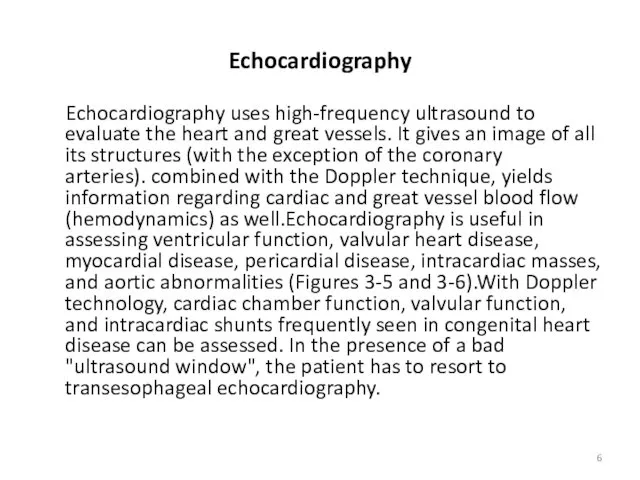
Echocardiography
Echocardiography uses high-frequency ultrasound to evaluate the heart and great
Echocardiography
Echocardiography uses high-frequency ultrasound to evaluate the heart and great
Indications for Echocardiography
Ventricular function
Congenital heart disease
Valvular heart disease
Cardiomyopathy
Pericardial effusion
Suspected cardiac masses
Aortic
Indications for Echocardiography
Ventricular function
Congenital heart disease
Valvular heart disease
Cardiomyopathy
Pericardial effusion
Suspected cardiac masses
Aortic
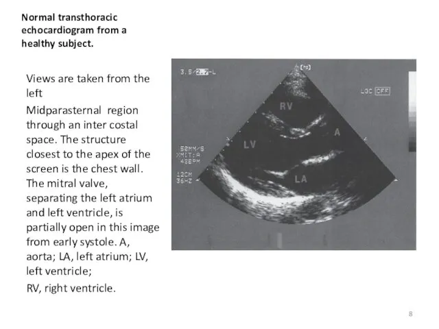
Normal transthoracic
echocardiogram from a healthy subject.
Views are taken from the left
Midparasternal
Normal transthoracic
echocardiogram from a healthy subject.
Views are taken from the left
Midparasternal
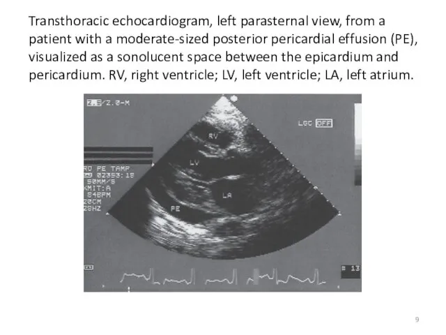
Transthoracic echocardiogram, left parasternal view, from a patient with a moderate-sized
Transthoracic echocardiogram, left parasternal view, from a patient with a moderate-sized
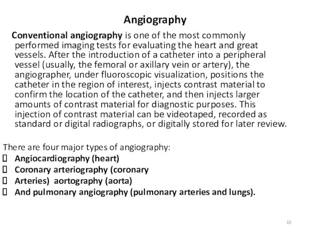
Angiography
Conventional angiography is one of the most commonly performed imaging
Angiography
Conventional angiography is one of the most commonly performed imaging
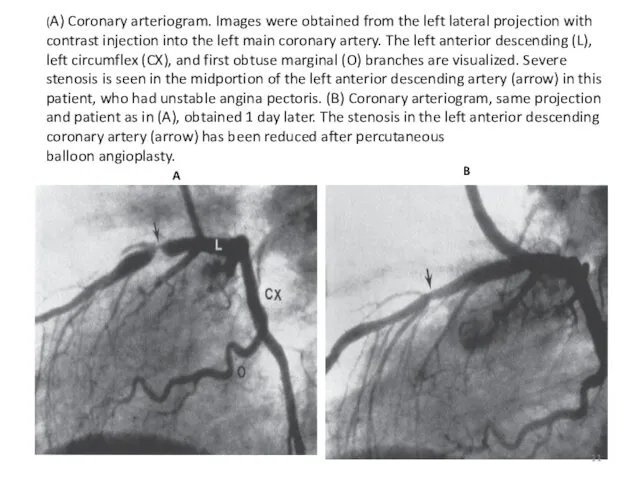
(A) Coronary arteriogram. Images were obtained from the left lateral projection
(A) Coronary arteriogram. Images were obtained from the left lateral projection
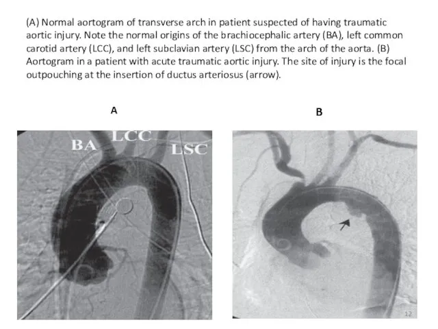
(A) Normal aortogram of transverse arch in patient suspected of having
(A) Normal aortogram of transverse arch in patient suspected of having
CT in Cardiology
The use of CT in cardiology allows faster
CT in Cardiology
The use of CT in cardiology allows faster

Normal anatomy at cardiac CT angiography. Ao, aorta; AV, aortic valve;
Normal anatomy at cardiac CT angiography. Ao, aorta; AV, aortic valve;
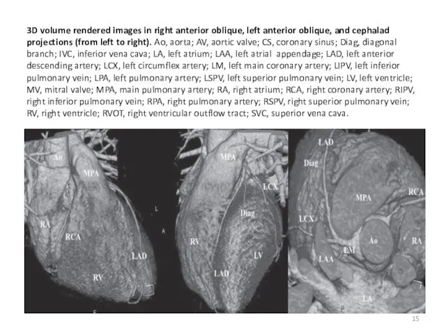
3D volume rendered images in right anterior oblique, left anterior oblique,
3D volume rendered images in right anterior oblique, left anterior oblique,
MRI IN CARDIOLOGY
MRI is used for the differential diagnosis
MRI IN CARDIOLOGY
MRI is used for the differential diagnosis
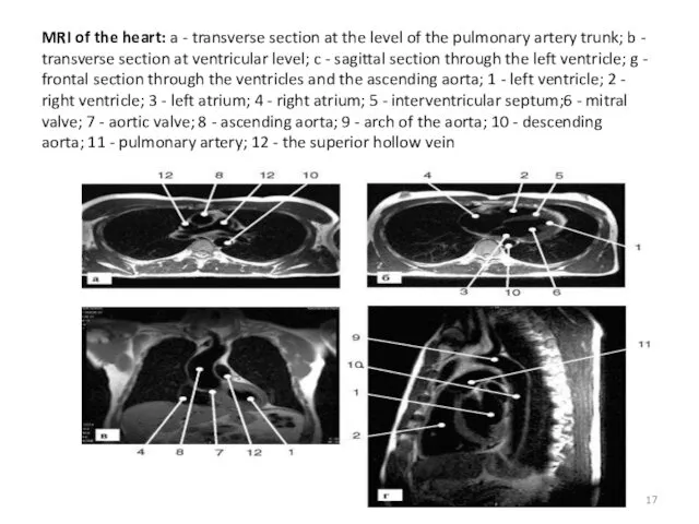
MRI of the heart: a - transverse section at the level
MRI of the heart: a - transverse section at the level
Radionuclide Imaging (Nuclear Medicine)
Radionuclide Imaging (Nuclear Medicine) Cardiac radionuclide imaging,
Radionuclide Imaging (Nuclear Medicine)
Radionuclide Imaging (Nuclear Medicine) Cardiac radionuclide imaging,
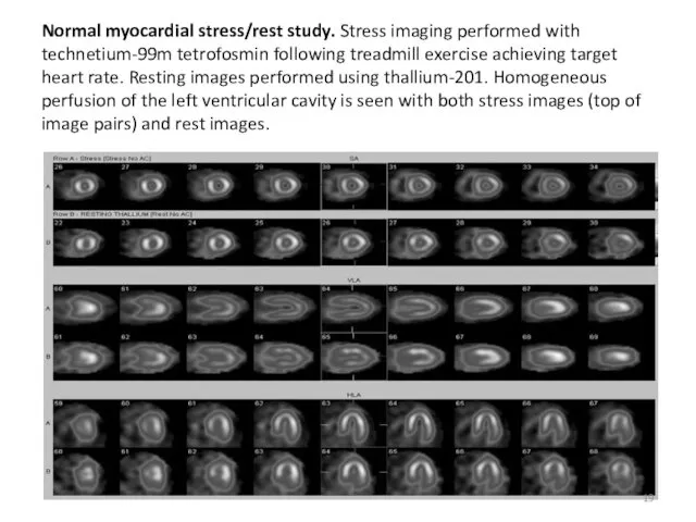
Normal myocardial stress/rest study. Stress imaging performed with technetium-99m tetrofosmin following
Normal myocardial stress/rest study. Stress imaging performed with technetium-99m tetrofosmin following
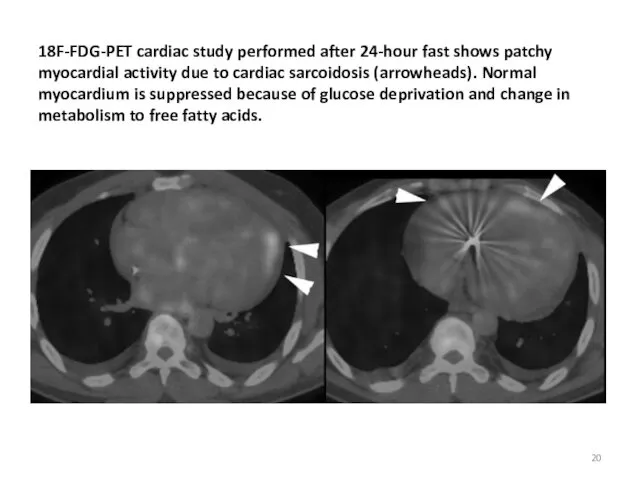
18F-FDG-PET cardiac study performed after 24-hour fast shows patchy myocardial activity
18F-FDG-PET cardiac study performed after 24-hour fast shows patchy myocardial activity
PATHOLOGICAL RADIOLOGY
OF
CARDIOVASCULAR SYSYTEM
PATHOLOGICAL RADIOLOGY
OF
CARDIOVASCULAR SYSYTEM
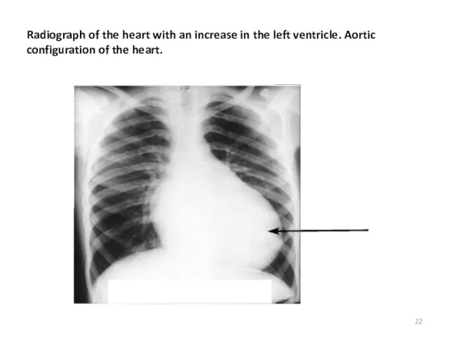
Radiograph of the heart with an increase in the left ventricle. Aortic
Radiograph of the heart with an increase in the left ventricle. Aortic
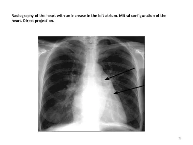
Radiography of the heart with an increase in the left atrium. Mitral
Radiography of the heart with an increase in the left atrium. Mitral
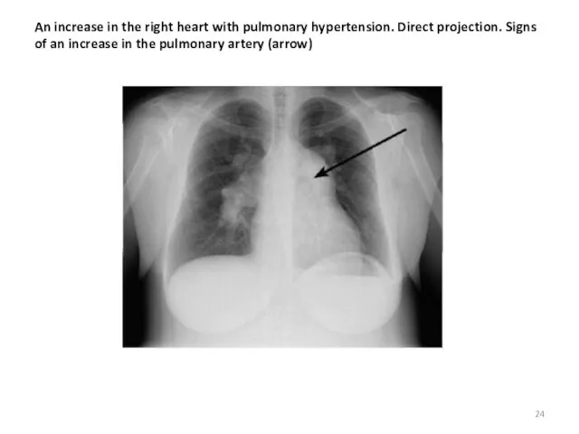
An increase in the right heart with pulmonary hypertension. Direct projection. Signs
An increase in the right heart with pulmonary hypertension. Direct projection. Signs
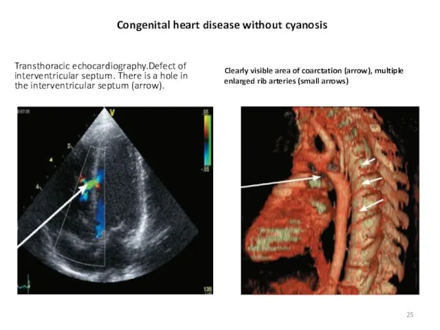
Congenital heart disease without cyanosis
Transthoracic echocardiography.Defect of interventricular septum. There is a
Congenital heart disease without cyanosis
Transthoracic echocardiography.Defect of interventricular septum. There is a
.
Congenital heart disease with cyanosis and normal pulmonary circulation
An echocardiogram with
.
Congenital heart disease with cyanosis and normal pulmonary circulation
An echocardiogram with
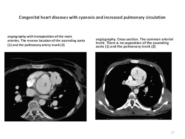
Congenital heart diseases with cyanosis and increased pulmonary circulation
angiography with transposition
Congenital heart diseases with cyanosis and increased pulmonary circulation
angiography with transposition
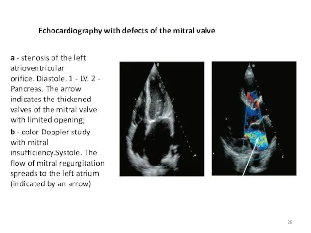
Echocardiography with defects of the mitral valve
a - stenosis of the
Echocardiography with defects of the mitral valve
a - stenosis of the
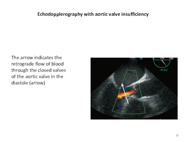
Echodopplerography with aortic valve insufficiency
The arrow indicates the retrograde flow of
Echodopplerography with aortic valve insufficiency
The arrow indicates the retrograde flow of
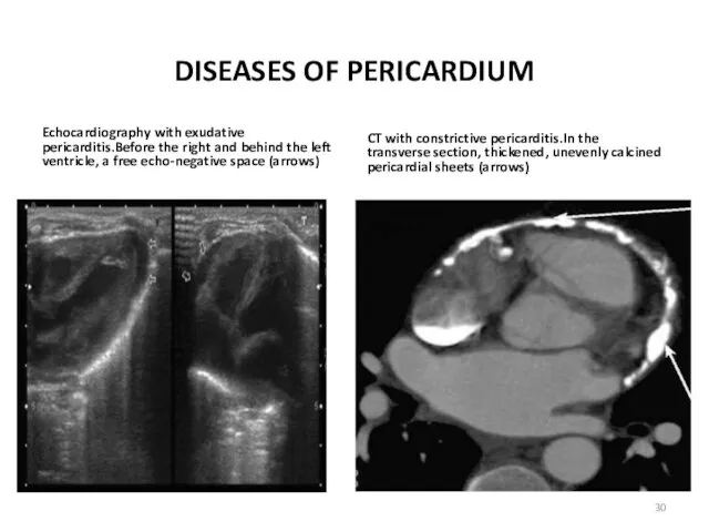
DISEASES OF PERICARDIUM
Echocardiography with exudative pericarditis.Before the right and behind the
DISEASES OF PERICARDIUM
Echocardiography with exudative pericarditis.Before the right and behind the
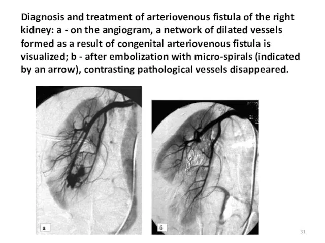
Diagnosis and treatment of arteriovenous fistula of the right kidney: a
Diagnosis and treatment of arteriovenous fistula of the right kidney: a
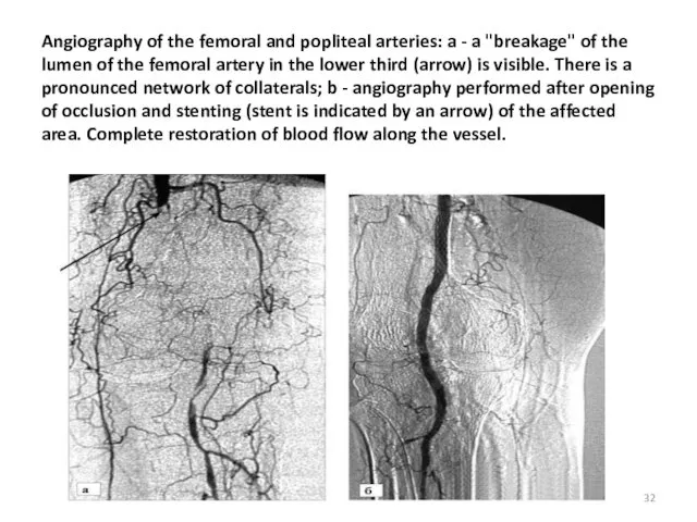
Angiography of the femoral and popliteal arteries: a - a "breakage"
Angiography of the femoral and popliteal arteries: a - a "breakage"
 Профессия врача. Одна из самых нужных и важных профессий
 Орталық жүйке жүйесіне әсер ететін дәрілер
 Диета – здоровье или мода?
 Кашлюк. Етіологія. Клініка. Лікування
 Профилактика ВИЧ-инфекции
 Средства, влияющие на систему крови
 СПИД. Пути передачи вируса
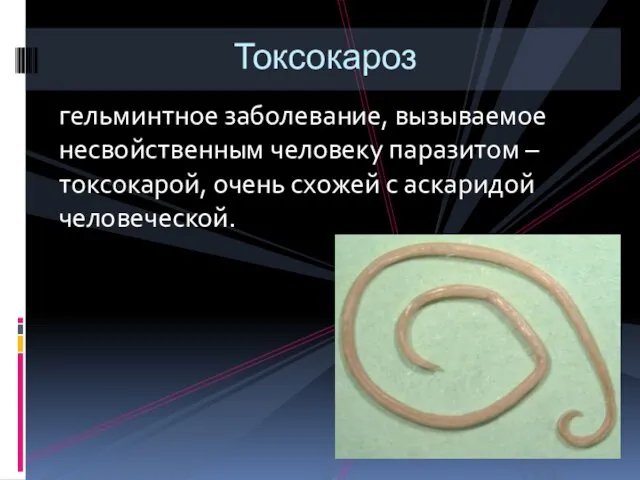 Токсокароз. Жизненный цикл токсокар
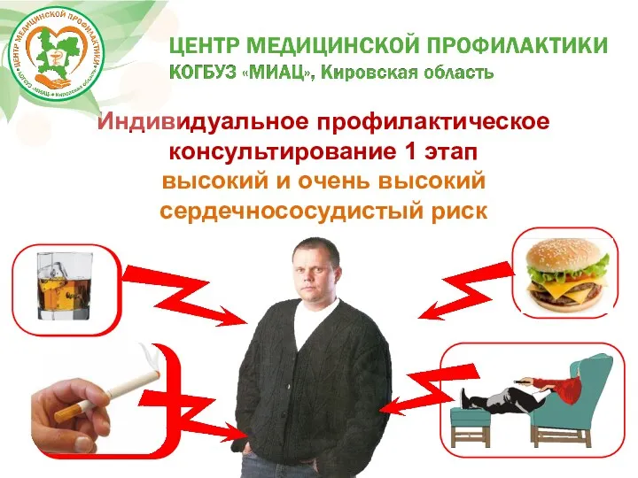 Высокий и очень высокий сердечно-сосудистый риск. Индивидуальное профилактическое консультирование
 Основні методи дослідження в психіатрії
 Мочекаменная болезнь
 Гіпертонічна хвороба
 Принципы и современные методы лечения переломов. Несросшиеся переломы, ложные суставы
 Дыхательная гимнастика для школьников по методике Стрельниковой А.Н
 Гомеопатия и ноотропные препараты от головокружения
 Физиология мозжечка и переднего мозга, их участие в регуляции мышечного тонуса и движения
 Основні переваги грудного вигодовування малят
 Паразитарлы аурулар
 Некроз. Апоптоз
 Догляд за хворими з хірургічними захворюваннями прямої кишки
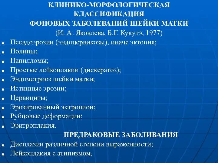 Клинико-морфологическая классификация фоновых заболеваний шейки матки
 Тематикалық аурулардан анамнез жинау ерекшеліктері
 Здоровый образ жизни. Личная гигиена
 Disorders of metabolism. (Subject 9)
 Патогенные и условно патогенные микроорганизмы
 Лечебная физкультура при гинекологических заболеваниях
 Первичные иммунодефициты. Этиология, патогенез, клинические проявления. Принципы диагностики. (Лекция 8)
 Кардиология. ИБС. Классификация. Стенокардия