- Pathogenetic peculiarities and pathoanatomical changes in bronchial pneumonia of calves

Содержание
- 2. Currently, it revealed a clear tendency to increase the number of patients suffering from diseases of
- 3. The aim of research was to study the incidence and prevalence of pneumonia in young cattle
- 4. The experimental part of the work is carried out at the Department of “Clinical Veterinary Medicine”
- 5. Fixation material for electronic microscope studies were performed in 2.5%-gluteraldehyde on collodion buffer with post fixation
- 6. Results of studies have shown that the structural organization of the respiratory system in clinically healthy
- 7. The structural organization of the alveoli and bronchioles in the lungs of healthy calves: a)the bronchial
- 8. The structural organization of the alveoli and bronchioles in the lungs of healthy calves: b)the terminal
- 9. The structural organization of the alveoli and bronchioles in the lungs of healthy calves: c)Interalveolar partition
- 10. The structural organization of the alveoli and bronchioles in the lungs of healthy calves: d) The
- 11. Thus, the results of their own research and analysis of data in the literature suggest that
- 13. Скачать презентацию
Currently, it revealed a clear tendency to increase the number
Currently, it revealed a clear tendency to increase the number
The aim of research was to study the incidence
The aim of research was to study the incidence
The experimental part of the work is carried out at
The experimental part of the work is carried out at
Fixation material for electronic microscope studies were performed in 2.5%-gluteraldehyde
Fixation material for electronic microscope studies were performed in 2.5%-gluteraldehyde
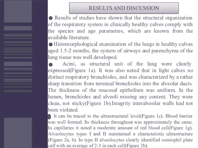
Results of studies have shown that the structural organization of
Results of studies have shown that the structural organization of
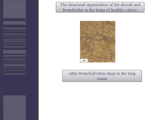
The structural organization of the alveoli and bronchioles in the lungs
The structural organization of the alveoli and bronchioles in the lungs
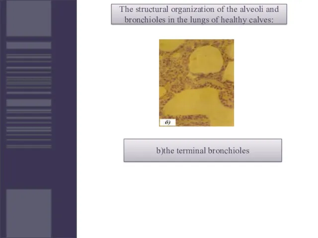
The structural organization of the alveoli and bronchioles in the lungs
The structural organization of the alveoli and bronchioles in the lungs
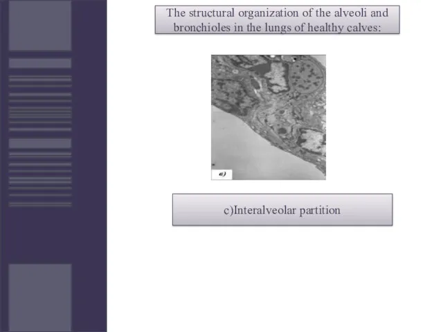
The structural organization of the alveoli and bronchioles in the lungs
The structural organization of the alveoli and bronchioles in the lungs

The structural organization of the alveoli and bronchioles in the lungs
The structural organization of the alveoli and bronchioles in the lungs
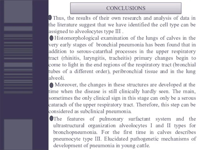
Thus, the results of their own research and analysis of
Thus, the results of their own research and analysis of
 Врожденный и приобретенный иммунитет. Клеточные и гуморальные механизмы
 Спортивна медицина
 Трихинеллёз. Возбудитель Трихинеллёза
 Инфекция. Классификация инфекций и инвазий человека
 Болезни почек
 Эндокриндік жүйенің бұзылыстары
 Геморрагический инсульт (спонтанное субарахноидальное кровоизлияние – САК) у детей
 Диагностика рака щитовидной железы
 Инструментальные и функциональные методы исследования сердца
 Стоматологическая служба в системе охраны материнства и детства
 Мочеполовая система
 General course of syphilis. Primary syphilis secondary syphslis
 Острый одонтогенный, гематогенный, хронический остеомиелит
 Противовоспалительные лекарственные средства (ПВЛС)
 Оборотные средства аптечной организации. (Тема 21)
 Первая помощь при растяжении связок, вывихах суставов, переломах костей
 Общая семиотика экстрапирамидных нарушений
 Остеопороз и его связь со стоматологическими заболеваниями
 Өлім жақындағанда адамның бойында не болады
 Гомеостаз зуба после прорезывания. Резистентность эмали зуба к кариозному поражению. Влияние на процессы формирования
 Сердечная недостаточность у детей
 Нравственная и правовая ответственность врача перед пациентом
 Сальмонеллез. Основные факторы патогенности
 Бронхит. Классификация по течению болезни
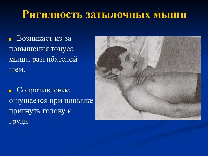 Ригидность затылочных мышц
 Ғылыми жұмыстағы әдістер мен материалдар
 Склера. Заболевания склеры
 Артериальная гипертензия у беременных в терапии и гинекологии: два взгляда – одно решение