- Pelvic аnatomy

Содержание
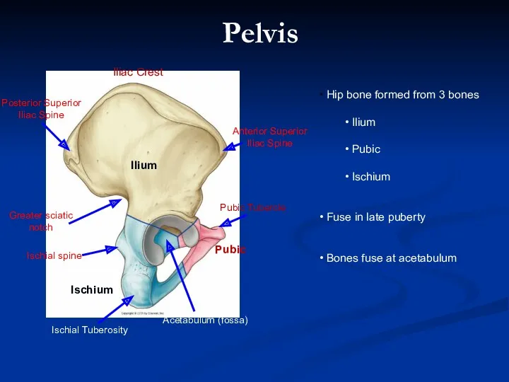
- 2. Ischium Pubic Ilium Hip bone formed from 3 bones Ilium Pubic Ischium Fuse in late puberty
- 3. L4 Ischial spine Ischial tuberosity 48 Sacral promontory Left sacro-iliac joint Iliopectineal line Sacrospinous ligament Sacrotuberous
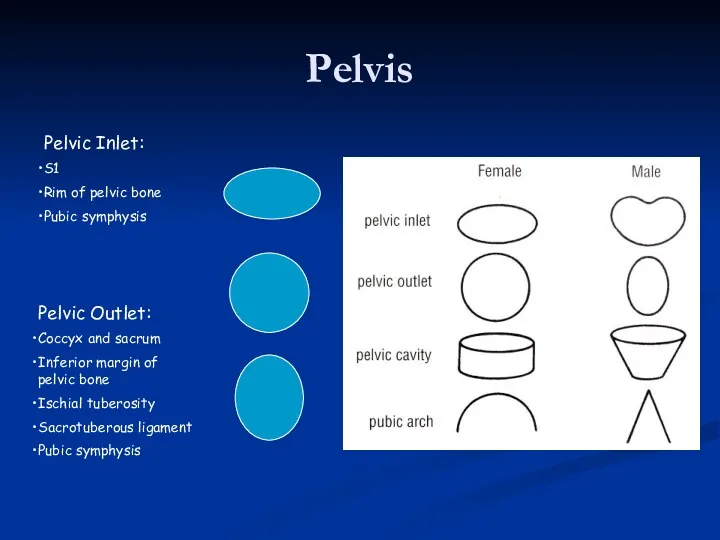
- 5. Pelvis Pelvic Inlet: S1 Rim of pelvic bone Pubic symphysis Pelvic Outlet: Coccyx and sacrum Inferior
- 6. Frolich, Human Anatomy, Pelvis I REVIEW OF PELVIS I Pelvic brim, inlet Pelvic outlet True pelvis--viscera
- 7. Female Male Cavity is broad, shallow Pelvic inlet oval + outlet round Bones are lighter, thinner
- 8. Pelvis Sacrospinous Sacrotuberous Sacrotuberous Apex: medial ischial tuberosity Base: PSIS to sacrum to coccyx to Sacrospinous
- 9. Pelvic Foramen Obturator Canal: Obturator nerve and vessels. Greater Sciatic Foramen: Above piriformis: - superior gluteal
- 10. The Pelvic Floor Musculotendinous hammock or sling Termination of the pelvic outlet Muscles of the pelvis
- 11. The Function of Pelvic Floor Support pelvic and abdominal organs during stress of increased abdominal pressure
- 12. PERINEUM Diamond shape area It is bouded: Anteriorly: lower edge of symphysis pubis Posteriorly: coccyx Lateraly:
- 13. Both the male and female anal triangles are similar so we will just describe one. Starting
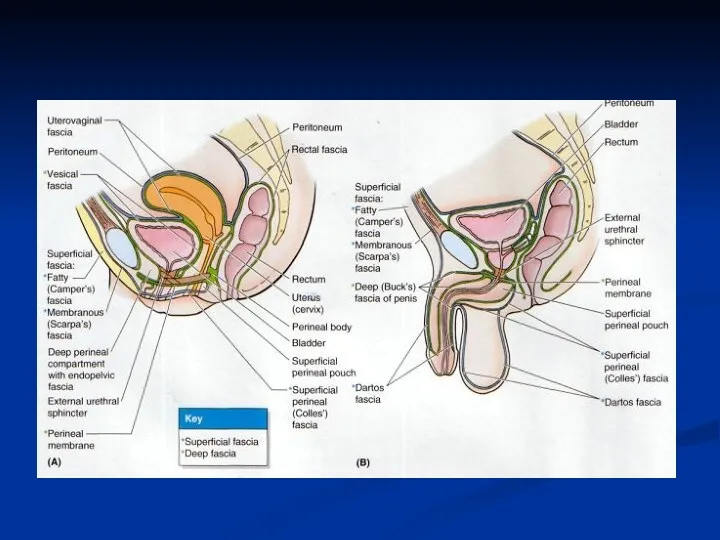
- 14. Compartments of the urogenital traingle Skin Colles Fascia Perineal Membrane Superior layer of fascia of UGD
- 15. 1. Structures forming the 3 roots of the penis (2 crura and 1 bulb) covered by
- 16. Structures forming the roots of clitoris: Two crura of the clitoris, two vestibular bulbs, and their
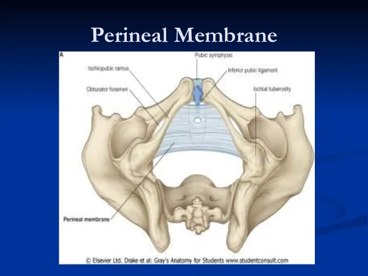
- 17. Perineal Membrane
- 18. 2. INFERIOR LAYER of fascia of UGD= called also perineal membrane The closed space between 2
- 19. Deep Perineal Pouch Male contents: The membranous urethra Sphincter urethrae muscle Deep transverse perineal muscles Bulbourethral
- 20. Pelvic diaphragm = levator ani and coccygeus muscles. Pelvic floor= pelvic diaphragm, perineal membrane and muscles
- 21. Levator Ani Muscles Pubococcygeus Iliococcygeus Puborectalis Ischiococcygeus
- 22. Puborectalis U-shaped, medial most located levator ani muscle Pulls the anorectal junction anteriorly, forming the anorectal
- 23. The puborectalis muscle (Inferior fibers of pubococcygeus)
- 24. Functional Anatomy Puborectalis and the anorectal angle allow for gross fecal continence Relieves pressure from the
- 25. PERFORATIONS OF PELVIC DIAPHRAGM 1. Anteriorly: urethral and vaginal opening (the bulb of the penis in
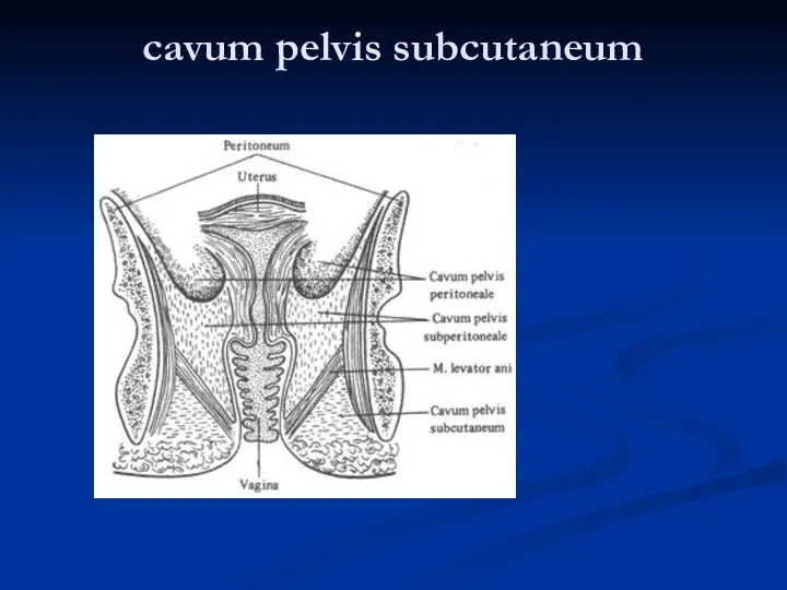
- 26. 1 – cavum pelvis peritoneale 2 – cavum pelvis subperitoneale 3 –cavum pelvis subcutaneum
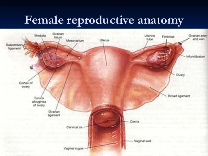
- 28. Female reproductive anatomy
- 29. Cavum pelvis subperitoneale
- 30. cavum pelvis subcutaneum
- 31. Female reproductive anatomy
- 32. Uterine Support Uterine support thought to be by: Ligaments: - from the uterus to the pelvic
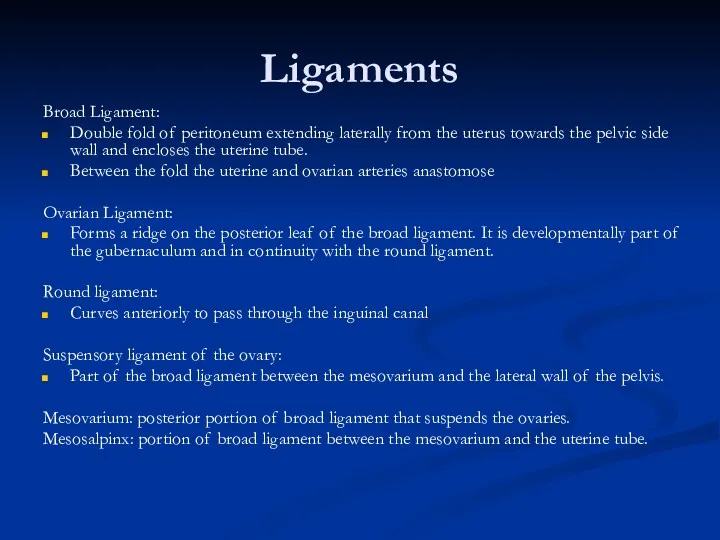
- 33. Ligaments Broad Ligament: Double fold of peritoneum extending laterally from the uterus towards the pelvic side
- 34. Nerve supply Pudendal nerve Left hypogastric nerve Sacral splanchnic nerve Superior hypogastric plexus Inferior hypogastric plexus
- 35. Greater TSN (T5-9) Lesser TSN (T10-11) Least TSN (T12) Lumbar SN (L1/2) Sacral SN (L1/2) Coeliac
- 37. Frolich, Human Anatomy, Pelvis I
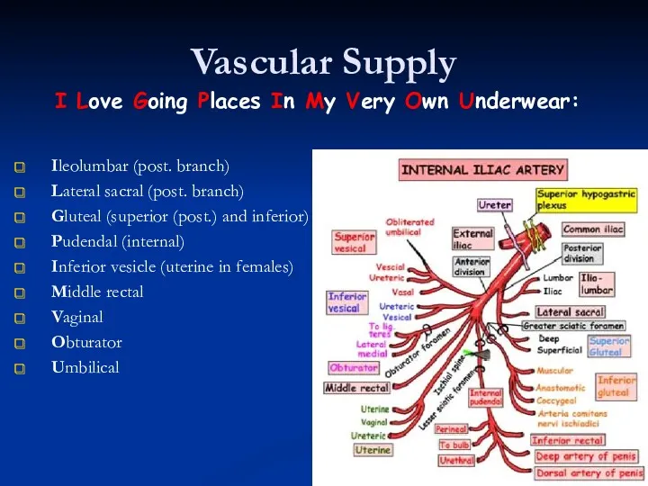
- 38. Vascular Supply Ileolumbar (post. branch) Lateral sacral (post. branch) Gluteal (superior (post.) and inferior) Pudendal (internal)
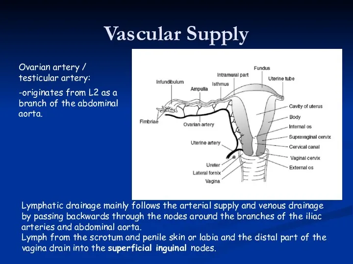
- 39. Vascular Supply Ovarian artery / testicular artery: -originates from L2 as a branch of the abdominal
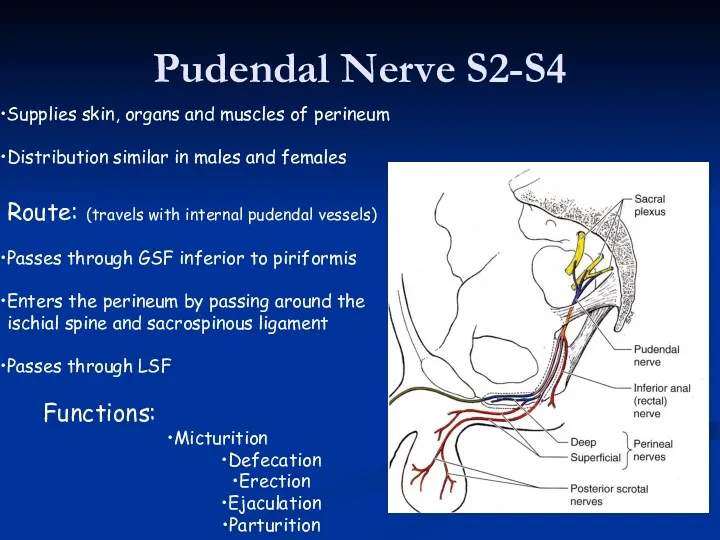
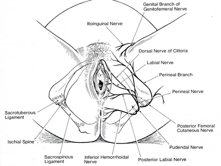
- 40. Pudendal Nerve S2-S4 Supplies skin, organs and muscles of perineum Distribution similar in males and females
- 42. Pudendal Nerve Blockade Medial to ishial tuberosity at sacrospinous ligament Transvaginal
- 43. Spinal: Into subarachnoid space Dense block for 2-4 hours Can cause hypotension Needle goes through: The
- 44. PR exam
- 46. Скачать презентацию
Ischium
Pubic
Ilium
Hip bone formed from 3 bones
Ilium
Pubic
Ischium
Fuse in
Ischium
Pubic
Ilium
Hip bone formed from 3 bones
Ilium
Pubic
Ischium
Fuse in
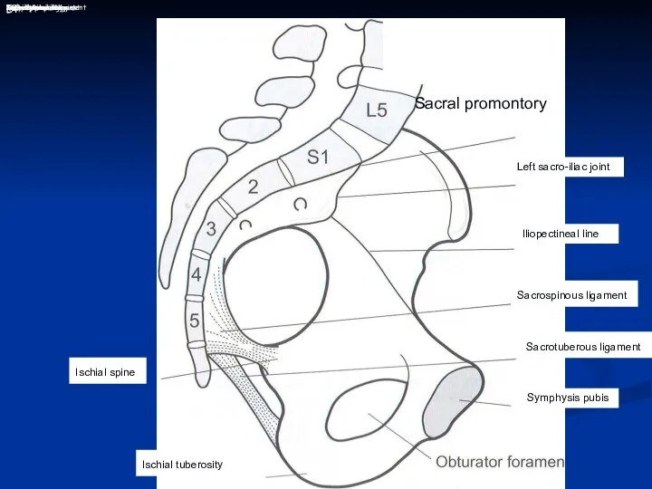
L4
Ischial spine
Ischial tuberosity
48
Sacral promontory
Left sacro-iliac joint
Iliopectineal line
Sacrospinous ligament
Sacrotuberous ligament
Symphysis pubis
Sacral promontory
L4
Ischial spine
Ischial tuberosity
48
Sacral promontory
Left sacro-iliac joint
Iliopectineal line
Sacrospinous ligament
Sacrotuberous ligament
Symphysis pubis
Sacral promontory
Pelvis
Pelvic Inlet:
S1
Rim of pelvic bone
Pubic symphysis
Pelvic Outlet:
Coccyx and sacrum
Inferior margin of
Pelvis
Pelvic Inlet:
S1
Rim of pelvic bone
Pubic symphysis
Pelvic Outlet:
Coccyx and sacrum
Inferior margin of
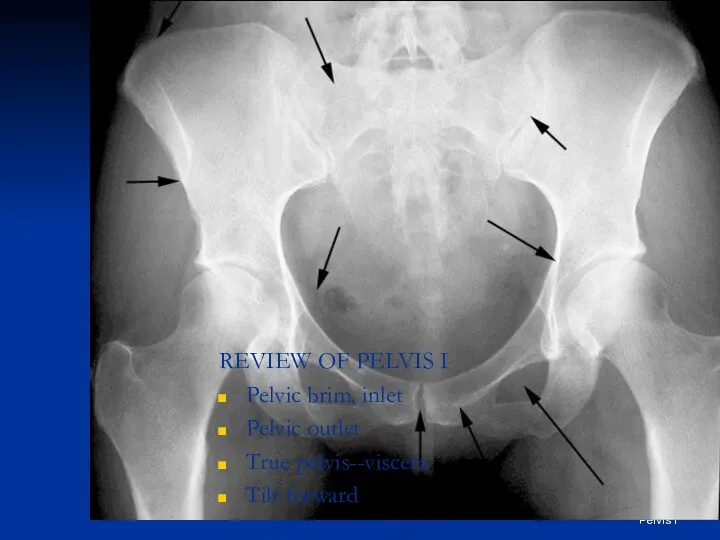
Frolich, Human Anatomy, Pelvis I
REVIEW OF PELVIS I
Pelvic brim, inlet
Pelvic outlet
True
Frolich, Human Anatomy, Pelvis I
REVIEW OF PELVIS I
Pelvic brim, inlet
Pelvic outlet
True
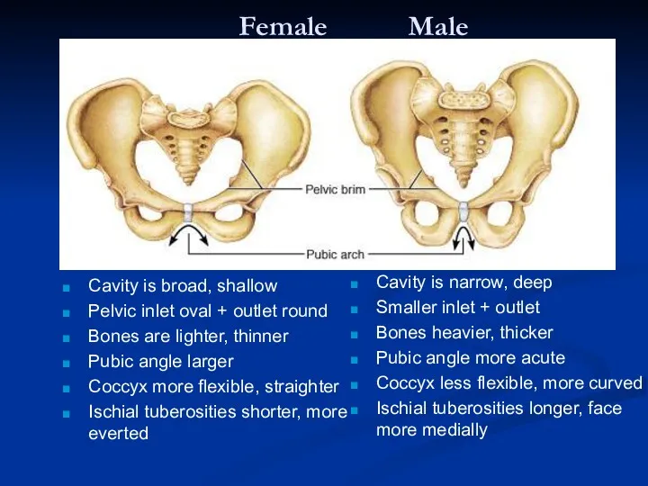
Female Male
Cavity is broad, shallow
Pelvic inlet oval + outlet round
Bones are
Female Male
Cavity is broad, shallow
Pelvic inlet oval + outlet round
Bones are
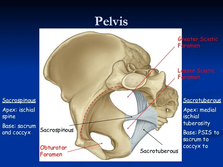
Pelvis
Sacrospinous
Sacrotuberous
Sacrotuberous
Apex: medial ischial tuberosity
Base: PSIS to sacrum to coccyx to
Sacrospinous
Apex: ischial
Pelvis
Sacrospinous
Sacrotuberous
Sacrotuberous
Apex: medial ischial tuberosity
Base: PSIS to sacrum to coccyx to
Sacrospinous
Apex: ischial
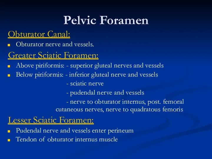
Pelvic Foramen
Obturator Canal:
Obturator nerve and vessels.
Greater Sciatic Foramen:
Above piriformis: - superior
Pelvic Foramen
Obturator Canal:
Obturator nerve and vessels.
Greater Sciatic Foramen:
Above piriformis: - superior
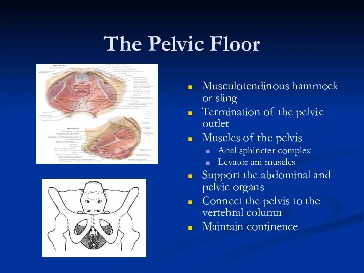
The Pelvic Floor
Musculotendinous hammock or sling
Termination of the pelvic outlet
Muscles of
The Pelvic Floor
Musculotendinous hammock or sling
Termination of the pelvic outlet
Muscles of
The Function of Pelvic Floor
Support pelvic and abdominal organs during stress
The Function of Pelvic Floor
Support pelvic and abdominal organs during stress
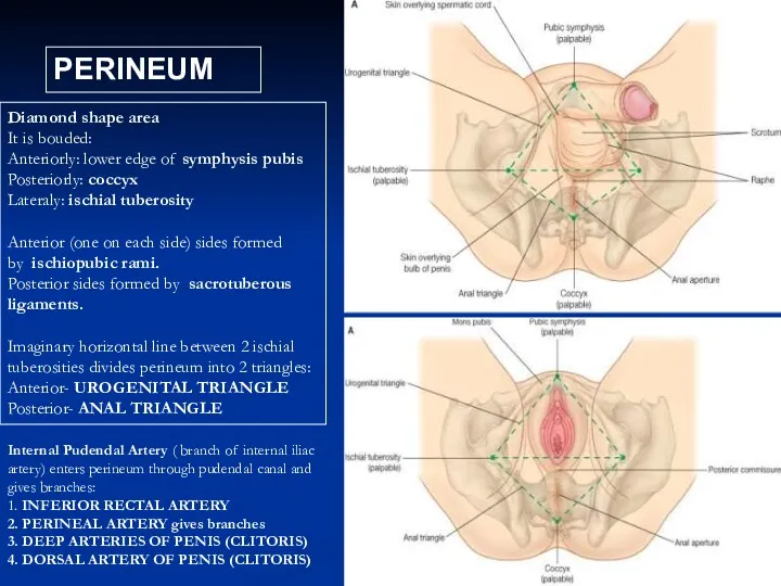
PERINEUM
Diamond shape area
It is bouded:
Anteriorly: lower edge of symphysis pubis
Posteriorly: coccyx
Lateraly:
PERINEUM
Diamond shape area
It is bouded:
Anteriorly: lower edge of symphysis pubis
Posteriorly: coccyx
Lateraly:
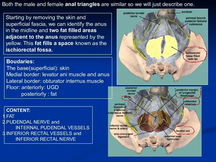
Both the male and female anal triangles are similar so we
Both the male and female anal triangles are similar so we
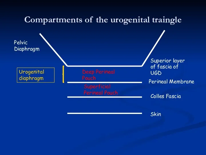
Compartments of the urogenital traingle
Skin
Colles Fascia
Perineal Membrane
Superior layer of fascia of
Compartments of the urogenital traingle
Skin
Colles Fascia
Perineal Membrane
Superior layer of fascia of
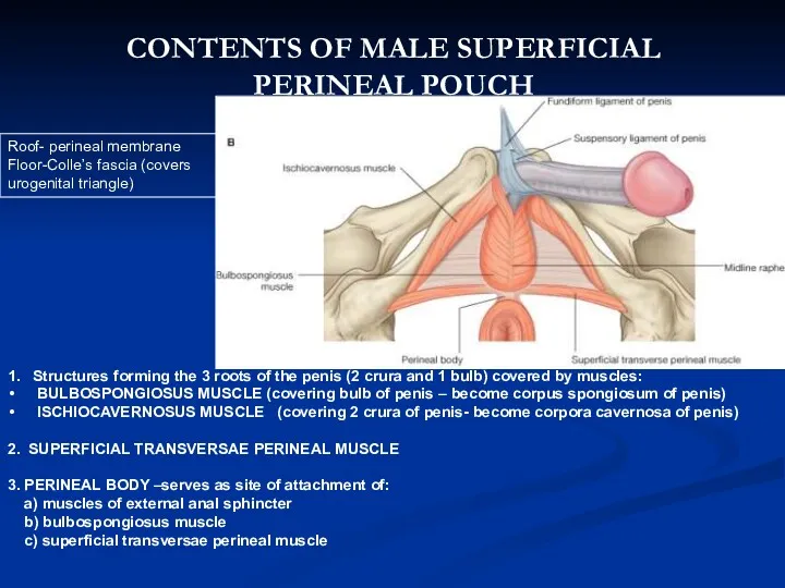
1. Structures forming the 3 roots of the penis (2 crura
1. Structures forming the 3 roots of the penis (2 crura
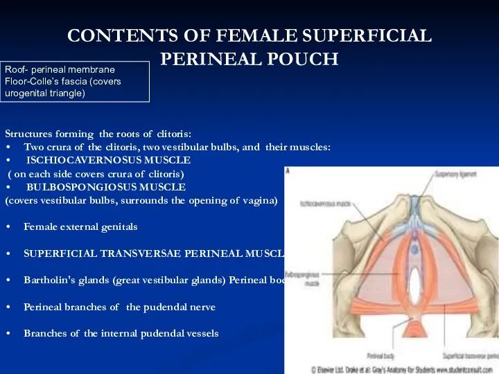
Structures forming the roots of clitoris:
Two crura of the clitoris, two
Structures forming the roots of clitoris:
Two crura of the clitoris, two
Perineal Membrane
Perineal Membrane
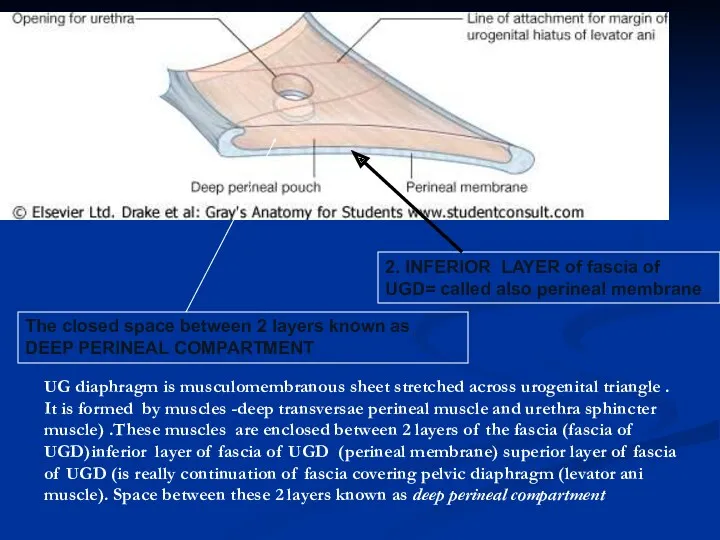
2. INFERIOR LAYER of fascia of UGD= called also perineal membrane
2. INFERIOR LAYER of fascia of UGD= called also perineal membrane
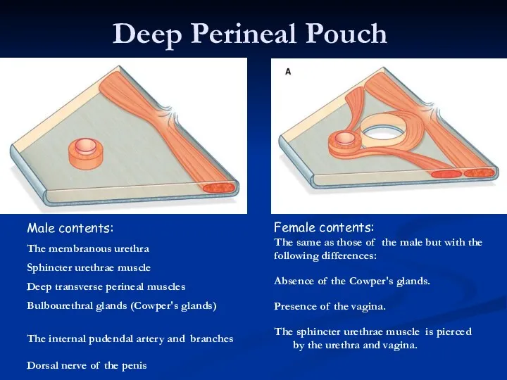
Deep Perineal Pouch
Male contents:
The membranous urethra
Sphincter urethrae muscle
Deep transverse perineal
Deep Perineal Pouch
Male contents:
The membranous urethra
Sphincter urethrae muscle
Deep transverse perineal
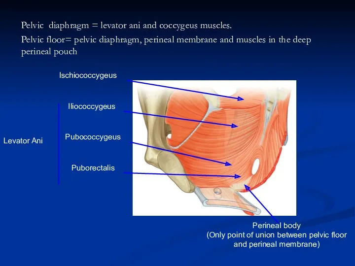
Pelvic diaphragm = levator ani and coccygeus muscles.
Pelvic floor= pelvic diaphragm,
Pelvic diaphragm = levator ani and coccygeus muscles. Pelvic floor= pelvic diaphragm,
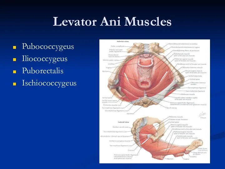
Levator Ani Muscles
Pubococcygeus
Iliococcygeus
Puborectalis
Ischiococcygeus
Levator Ani Muscles
Pubococcygeus
Iliococcygeus
Puborectalis
Ischiococcygeus
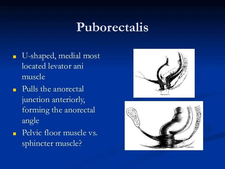
Puborectalis
U-shaped, medial most located levator ani muscle
Pulls the anorectal junction anteriorly,
Puborectalis
U-shaped, medial most located levator ani muscle
Pulls the anorectal junction anteriorly,
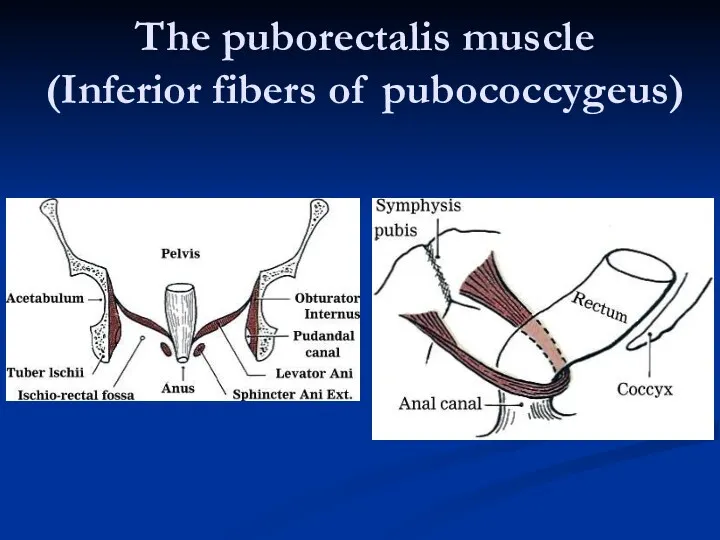
The puborectalis muscle
(Inferior fibers of pubococcygeus)
The puborectalis muscle
(Inferior fibers of pubococcygeus)
Functional Anatomy
Puborectalis and the anorectal angle allow for gross fecal continence
Relieves
Functional Anatomy
Puborectalis and the anorectal angle allow for gross fecal continence
Relieves
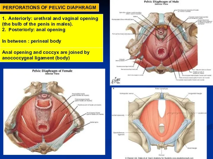
PERFORATIONS OF PELVIC DIAPHRAGM
1. Anteriorly: urethral and vaginal opening
(the bulb of
PERFORATIONS OF PELVIC DIAPHRAGM
1. Anteriorly: urethral and vaginal opening
(the bulb of
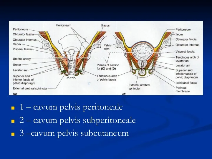
1 – cavum pelvis peritoneale
2 – cavum pelvis subperitoneale
3 –cavum pelvis
1 – cavum pelvis peritoneale
2 – cavum pelvis subperitoneale
3 –cavum pelvis
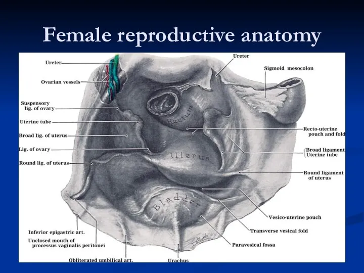
Female reproductive anatomy
Female reproductive anatomy
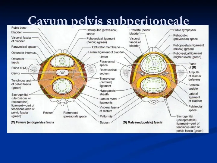
Cavum pelvis subperitoneale
Cavum pelvis subperitoneale
cavum pelvis subcutaneum
cavum pelvis subcutaneum
Female reproductive anatomy
Female reproductive anatomy
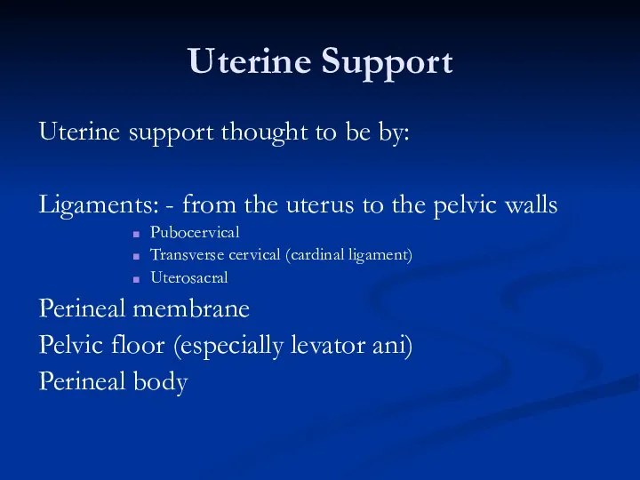
Uterine Support
Uterine support thought to be by:
Ligaments: - from the uterus
Uterine Support
Uterine support thought to be by:
Ligaments: - from the uterus
Ligaments
Broad Ligament:
Double fold of peritoneum extending laterally from the uterus towards
Ligaments
Broad Ligament:
Double fold of peritoneum extending laterally from the uterus towards
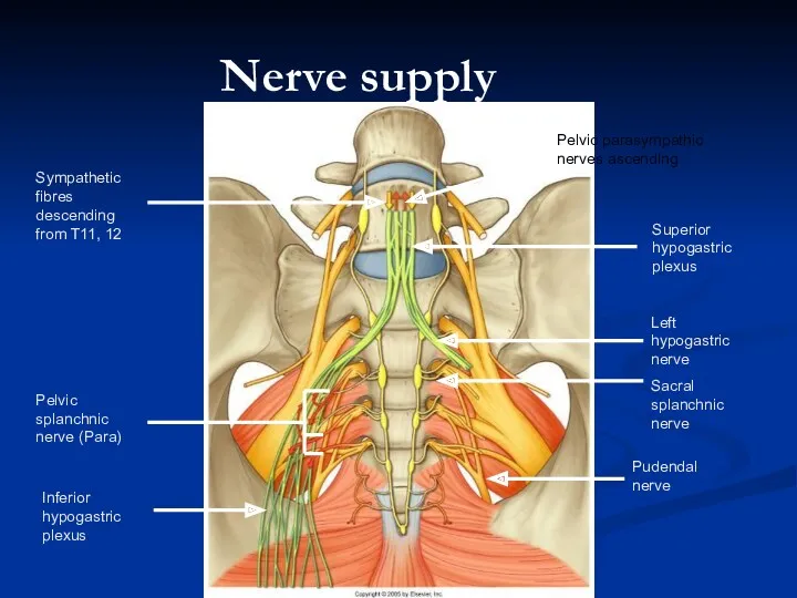
Nerve supply
Pudendal nerve
Left hypogastric nerve
Sacral splanchnic nerve
Superior hypogastric plexus
Inferior hypogastric plexus
Pelvic
Nerve supply
Pudendal nerve
Left hypogastric nerve
Sacral splanchnic nerve
Superior hypogastric plexus
Inferior hypogastric plexus
Pelvic
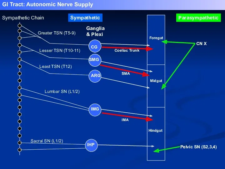
Greater TSN (T5-9)
Lesser TSN (T10-11)
Least TSN (T12)
Lumbar SN (L1/2)
Sacral SN (L1/2)
Coeliac
Greater TSN (T5-9)
Lesser TSN (T10-11)
Least TSN (T12)
Lumbar SN (L1/2)
Sacral SN (L1/2)
Coeliac
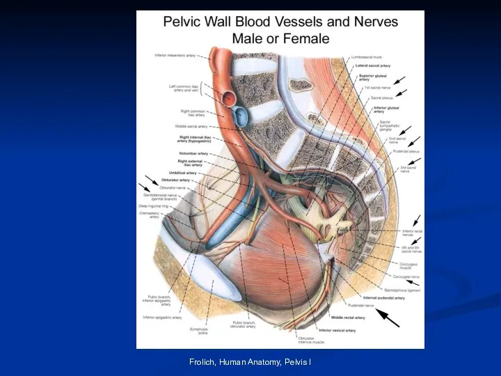
Frolich, Human Anatomy, Pelvis I
Frolich, Human Anatomy, Pelvis I
Vascular Supply
Ileolumbar (post. branch)
Lateral sacral (post. branch)
Gluteal (superior (post.) and inferior)
Pudendal
Vascular Supply
Ileolumbar (post. branch)
Lateral sacral (post. branch)
Gluteal (superior (post.) and inferior)
Pudendal
Vascular Supply
Ovarian artery / testicular artery:
-originates from L2 as a branch
Vascular Supply
Ovarian artery / testicular artery:
-originates from L2 as a branch
Pudendal Nerve S2-S4
Supplies skin, organs and muscles of perineum
Distribution similar in
Pudendal Nerve S2-S4
Supplies skin, organs and muscles of perineum
Distribution similar in
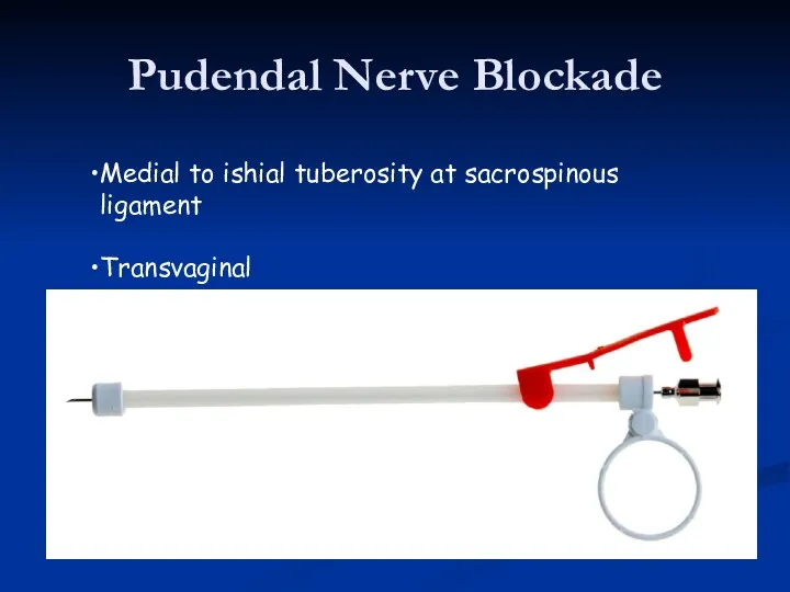
Pudendal Nerve Blockade
Medial to ishial tuberosity at sacrospinous ligament
Transvaginal
Pudendal Nerve Blockade
Medial to ishial tuberosity at sacrospinous ligament
Transvaginal
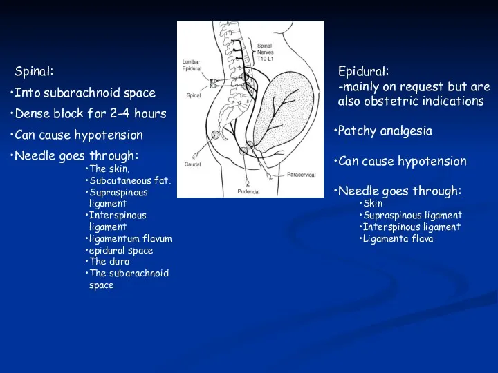
Spinal:
Into subarachnoid space
Dense block for 2-4 hours
Can cause hypotension
Needle goes through:
The
Spinal:
Into subarachnoid space
Dense block for 2-4 hours
Can cause hypotension
Needle goes through:
The
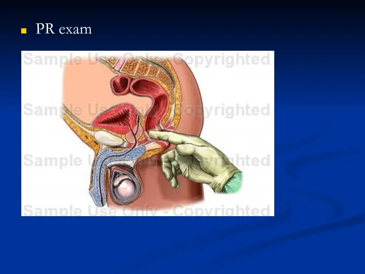
PR exam
PR exam
 Патология терморегуляции. Лекция № 8
 Шетелдегі инклюзивті білім беру
 Планирование научного исследования в медицине
 Спинальная мышечная атрофия тип lll. Болезнь Кугельберга-Веландера
 Prezentatsia_po_biologii_na_temu_Znachenie_pischi_i_eyo_sostav__8_klass
 Анемія. Етіологія і патогенез
 Борьба с вирусными заболеваниями (СПИД и другие)
 Заманауи гепатопротекторлы заттар
 Ортопедиялық стоматология
 Комплексная программа организации летнего отдыха и оздоровления детей и подростков Беломорская волна
 Дети с особыми образовательными потребностями
 Непрерывное медицинское образование. Периодическая аккредитация
 Гравидограмма интерпритациясы
 Экстрагенитальная патология и беременность
 Фізіологічні механізми та закономірності формування рухових навичок
 Физиология плода. Физиология беременности
 Асфиксии новорождённых
 Герпетическая инфекция
 Риккетсии. Хламидии
 Общая и специальная подготовка полости рта перед протезированием
 Босанғаннан кейінгі ерте кезеңдегі қан кетудің себептері: травма,тромбин
 Антибиотики и химиотерапия. Химиотерапевтические препараты
 Первая помощь при синдроме длительного сдавления (СДС) или тяжелая компрессионная травма
 Мышцы верхних и нижних конечностей человека
 Синдром диабетической стопы
 Острый аппендицит. История учения об аппендиците. Анатомо-физиологические особенности
 Холера
 Острые респираторные вирусные инфекции