- Sexually transmitted bacterial diseases

Содержание
- 2. PLAN Morphology Culture Antigenic structure Virulence factors Pathogenesis Immunity Clinical syndromes Epidemiology Laboratory diagnosis Treatment Prevention
- 3. A 28-year-old hair dresser complained of a painless small ulcer on the penis during the last
- 4. TREPONEMA PALLIDUM: MORPHOLOGY
- 5. TREPONEMA PALLIDUM : ANTIGENIC STRUCTURE Cardiolipin antigen T. pallidum group-specific antigen T. pallidum species-specific antigen
- 6. TREPONEMA PALLIDUM : VIRULENCE FACTORS
- 7. TREPONEMA PALLIDUM : PATHOGENESIS
- 8. TREPONEMA PALLIDUM : CLINICAL SYNDROMES Venereal syphilis (transmitted by sexual contact) Nonvenereal syphilis (congenital syphilis and
- 9. TREPONEMA PALLIDUM : EPIDEMIOLOGY
- 10. TREPONEMA PALLIDUM : LABORATORY DIAGNOSIS Microscopy Direct antigen detection Serodiagnosis: Nontreponemal tests (standard tests of syphilis:
- 11. TREPONEMA PALLIDUM : LABORATORY DIAGNOSIS
- 12. A 6-year-old boy attended the Ophthalmology OPD with symptoms of conjunctivitis of the right eye. Examination
- 13. CHLAMYDIA TRACHOMATIS: MORPHOLOGY
- 14. CHLAMYDIA TRACHOMATIS: CULTURE
- 15. CHLAMYDIA TRACHOMATIS: ANTIGENIC STRUCTURE Genus-specific antigen Species-specific antigen Serotype-specific antigen Typing of species trachoma biovar causing
- 16. CHLAMYDIA TRACHOMATIS: VIRULENCE FACTORS The ability to multiply intracellularly in the infected cell is the key
- 17. CHLAMYDIA TRACHOMATIS: PATHOGENESIS
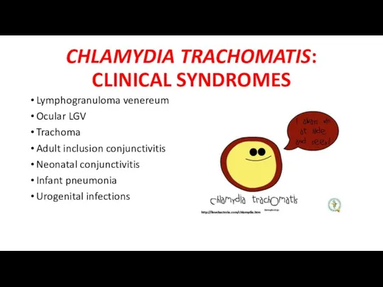
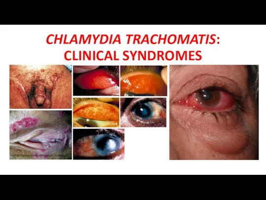
- 18. CHLAMYDIA TRACHOMATIS: CLINICAL SYNDROMES Lymphogranuloma venereum Ocular LGV Trachoma Adult inclusion conjunctivitis Neonatal conjunctivitis Infant pneumonia
- 19. CHLAMYDIA TRACHOMATIS: CLINICAL SYNDROMES
- 20. CHLAMYDIA TRACHOMATIS: LABORATORY DIAGNOSIS Microscopy Culture Antigen detection Serodiagnosis Frei’s skin test
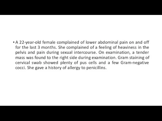
- 21. A 22-year-old female complained of lower abdominal pain on and off for the last 3 months.
- 22. NEISSERIA GONORRHOEAE: MORPHOLOGY
- 23. NEISSERIA GONORRHOEAE: CULTURE
- 24. NEISSERIA GONORRHOEAE: VIRULENCE FACTORS
- 25. NEISSERIA GONORRHOEAE: PATHOGENESIS
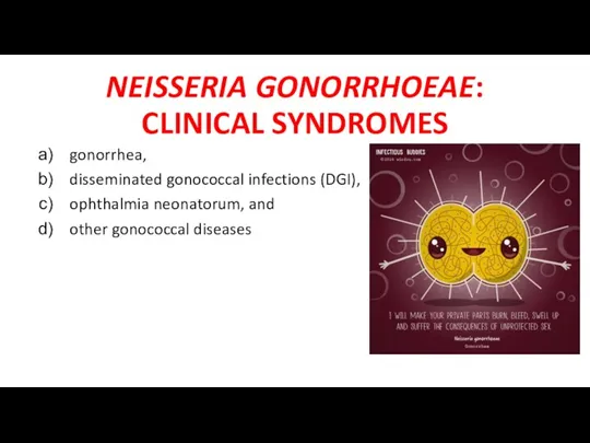
- 26. NEISSERIA GONORRHOEAE: CLINICAL SYNDROMES gonorrhea, disseminated gonococcal infections (DGI), ophthalmia neonatorum, and other gonococcal diseases
- 27. NEISSERIA GONORRHOEAE: LABORATORY DIAGNOSIS Microscopy Culture Antigen detection SerodiagnosiS
- 28. NEISSERIA GONORRHOEAE: CLINICAL SYNDROMES gonorrhea, disseminated gonococcal infections (DGI), ophthalmia neonatorum, and other gonococcal diseases
- 30. Скачать презентацию
PLAN
Morphology
Culture
Antigenic structure
Virulence factors
Pathogenesis
Immunity
Clinical syndromes
Epidemiology
Laboratory diagnosis
Treatment
Prevention
PLAN
Morphology
Culture
Antigenic structure
Virulence factors
Pathogenesis
Immunity
Clinical syndromes
Epidemiology
Laboratory diagnosis
Treatment
Prevention
A 28-year-old hair dresser complained of a painless small ulcer on
A 28-year-old hair dresser complained of a painless small ulcer on
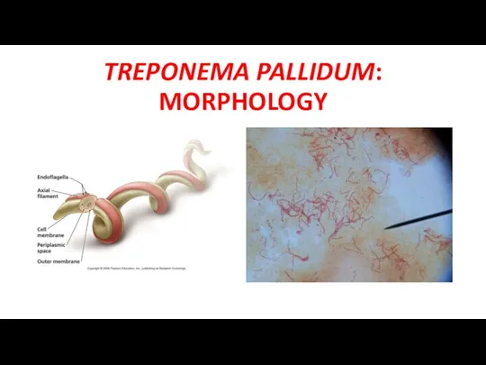
TREPONEMA PALLIDUM: MORPHOLOGY
TREPONEMA PALLIDUM: MORPHOLOGY
TREPONEMA PALLIDUM : ANTIGENIC STRUCTURE
Cardiolipin antigen
T. pallidum group-specific antigen
T.
TREPONEMA PALLIDUM : ANTIGENIC STRUCTURE
Cardiolipin antigen
T. pallidum group-specific antigen
T.
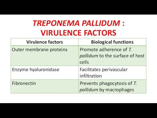
TREPONEMA PALLIDUM : VIRULENCE FACTORS
TREPONEMA PALLIDUM : VIRULENCE FACTORS
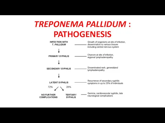
TREPONEMA PALLIDUM : PATHOGENESIS
TREPONEMA PALLIDUM : PATHOGENESIS
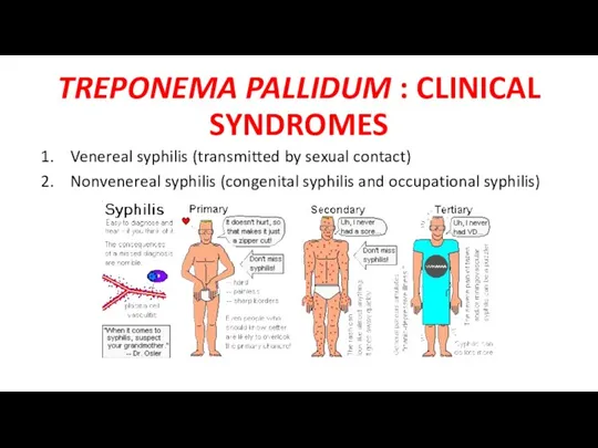
TREPONEMA PALLIDUM : CLINICAL SYNDROMES
Venereal syphilis (transmitted by sexual contact)
Nonvenereal syphilis
TREPONEMA PALLIDUM : CLINICAL SYNDROMES
Venereal syphilis (transmitted by sexual contact)
Nonvenereal syphilis
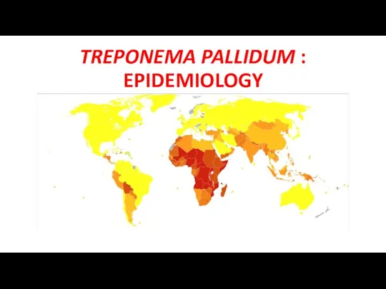
TREPONEMA PALLIDUM : EPIDEMIOLOGY
TREPONEMA PALLIDUM : EPIDEMIOLOGY
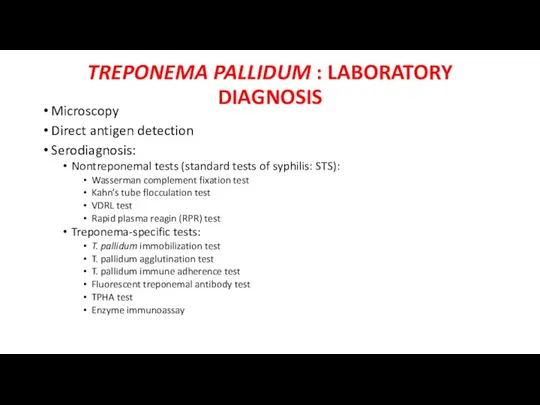
TREPONEMA PALLIDUM : LABORATORY DIAGNOSIS
Microscopy
Direct antigen detection
Serodiagnosis:
Nontreponemal tests (standard tests of
TREPONEMA PALLIDUM : LABORATORY DIAGNOSIS
Microscopy
Direct antigen detection
Serodiagnosis:
Nontreponemal tests (standard tests of
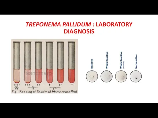
TREPONEMA PALLIDUM : LABORATORY DIAGNOSIS
TREPONEMA PALLIDUM : LABORATORY DIAGNOSIS
A 6-year-old boy attended the Ophthalmology OPD with symptoms of conjunctivitis
A 6-year-old boy attended the Ophthalmology OPD with symptoms of conjunctivitis
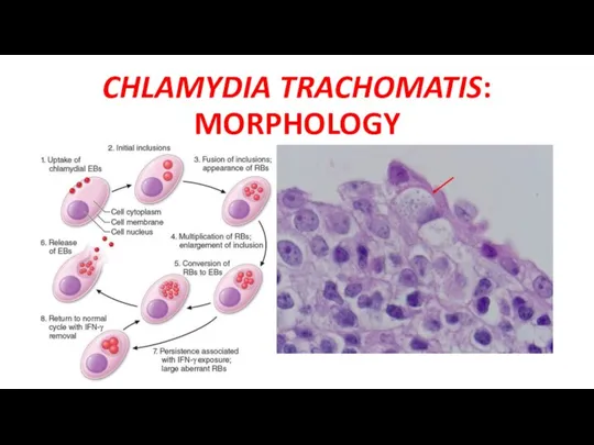
CHLAMYDIA TRACHOMATIS: MORPHOLOGY
CHLAMYDIA TRACHOMATIS: MORPHOLOGY
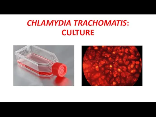
CHLAMYDIA TRACHOMATIS: CULTURE
CHLAMYDIA TRACHOMATIS: CULTURE
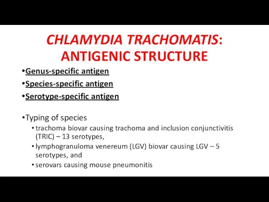
CHLAMYDIA TRACHOMATIS: ANTIGENIC STRUCTURE
Genus-specific antigen
Species-specific antigen
Serotype-specific antigen
Typing of species
trachoma biovar causing
CHLAMYDIA TRACHOMATIS: ANTIGENIC STRUCTURE
Genus-specific antigen
Species-specific antigen
Serotype-specific antigen
Typing of species
trachoma biovar causing
CHLAMYDIA TRACHOMATIS: VIRULENCE FACTORS
The ability to multiply intracellularly in the infected
CHLAMYDIA TRACHOMATIS: VIRULENCE FACTORS
The ability to multiply intracellularly in the infected
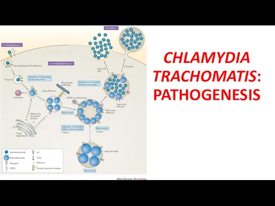
CHLAMYDIA TRACHOMATIS: PATHOGENESIS
CHLAMYDIA TRACHOMATIS: PATHOGENESIS
CHLAMYDIA TRACHOMATIS: CLINICAL SYNDROMES
Lymphogranuloma venereum
Ocular LGV
Trachoma
Adult inclusion conjunctivitis
Neonatal conjunctivitis
Infant pneumonia
Urogenital infections
CHLAMYDIA TRACHOMATIS: CLINICAL SYNDROMES
Lymphogranuloma venereum
Ocular LGV
Trachoma
Adult inclusion conjunctivitis
Neonatal conjunctivitis
Infant pneumonia
Urogenital infections
CHLAMYDIA TRACHOMATIS: CLINICAL SYNDROMES
CHLAMYDIA TRACHOMATIS: CLINICAL SYNDROMES
CHLAMYDIA TRACHOMATIS: LABORATORY DIAGNOSIS
Microscopy
Culture
Antigen detection
Serodiagnosis
Frei’s skin test
CHLAMYDIA TRACHOMATIS: LABORATORY DIAGNOSIS
Microscopy
Culture
Antigen detection
Serodiagnosis
Frei’s skin test
A 22-year-old female complained of lower abdominal pain on and off
A 22-year-old female complained of lower abdominal pain on and off
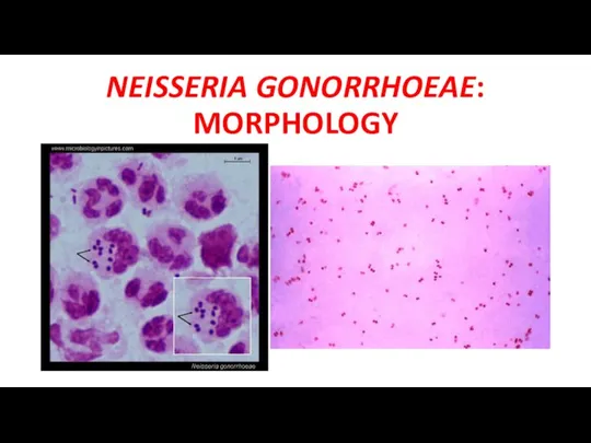
NEISSERIA GONORRHOEAE: MORPHOLOGY
NEISSERIA GONORRHOEAE: MORPHOLOGY
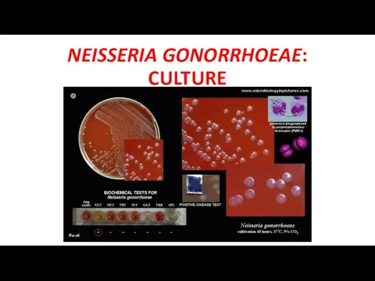
NEISSERIA GONORRHOEAE: CULTURE
NEISSERIA GONORRHOEAE: CULTURE
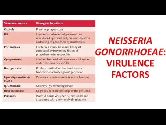
NEISSERIA GONORRHOEAE: VIRULENCE FACTORS
NEISSERIA GONORRHOEAE: VIRULENCE FACTORS
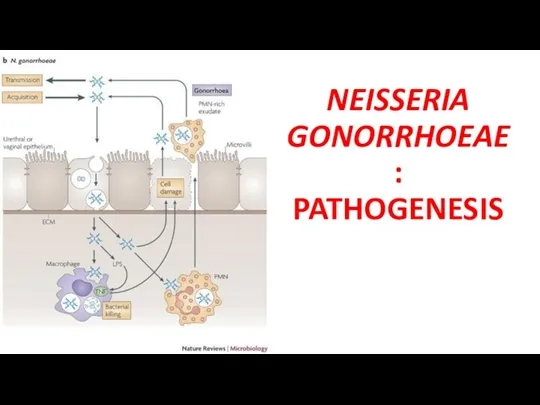
NEISSERIA GONORRHOEAE: PATHOGENESIS
NEISSERIA GONORRHOEAE: PATHOGENESIS
NEISSERIA GONORRHOEAE: CLINICAL SYNDROMES
gonorrhea,
disseminated gonococcal infections (DGI),
ophthalmia neonatorum, and
NEISSERIA GONORRHOEAE: CLINICAL SYNDROMES
gonorrhea,
disseminated gonococcal infections (DGI),
ophthalmia neonatorum, and
NEISSERIA GONORRHOEAE: LABORATORY DIAGNOSIS
Microscopy
Culture
Antigen detection
SerodiagnosiS
NEISSERIA GONORRHOEAE: LABORATORY DIAGNOSIS
Microscopy
Culture
Antigen detection
SerodiagnosiS

NEISSERIA GONORRHOEAE: CLINICAL SYNDROMES
gonorrhea,
disseminated gonococcal infections (DGI),
ophthalmia neonatorum, and
NEISSERIA GONORRHOEAE: CLINICAL SYNDROMES
gonorrhea,
disseminated gonococcal infections (DGI),
ophthalmia neonatorum, and
 Переломы шейки бедра
 Гипертоническая болезнь (ГБ). Ишемическая болезнь сердца (ИБС). Цереброваскулярные болезни (ЦВБ)
 Неотложные состояния в эндокринологии
 Микобактерии. Туберкулез
 Здоровье как показатель эффективности медико - профилактической деятельности
 Медицина Древнего Китая
 Созылмалы аурулары бар науқастардағы тіс жұлу операциясы
 Күйік
Күйік Сенсорні системи
 Проблема старения в современном мире
 Планирование стоматологической помощи населению
 Электрокардиография при нарушениях ритма сердца
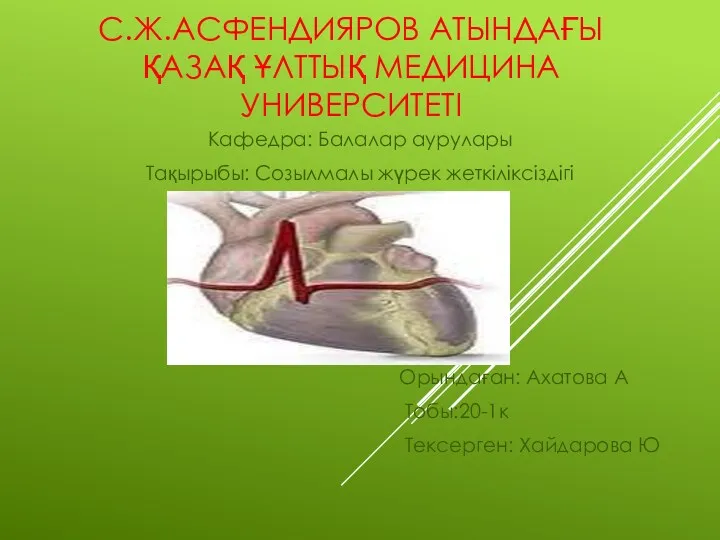 Созылмалы жүрек жеткіліксіздігі
 Травма шейного отдела позвоночника
 Общие принципы лечения и профилактики наследственных болезней
 Физиология высшей нервной деятельности. Методы исследования головного мозга. Фазы сна. Виды памяти (Лекция 2)
 Иммунопрофилактика инфекционных заболеваний
 Внематочная беременность
 Влияние болезней кровообращения на течение беременности
 Введение в венерологию. История развития венерологии. Инфекции, передающиеся половым путем
 Методи дослідження та діагностики захворювань органів дихання та серцево-судинної системи
 Болезни склеры. Клиника, диагностика, лечение
 Общая и частная миология
 Основы иммунопрофилактики и иммунотерапии инфекционных заболеваний
 Қабыну. Анықтамасы,мәні мен биологиялық маңызы,даму заңдылықтары
 Организация гигиенического воспитания населения в условиях поликлиники
 Организация противоэпидемических мероприятий при чрезвычайных ситуациях
 Биологиялық ұлпалардағы жоғары интенсивті лазерлік сәуле әсерінің механизмі