- The Alimentary Tract

Содержание
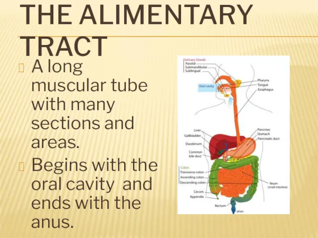
- 2. THE ALIMENTARY TRACT A long muscular tube with many sections and areas. Begins with the oral
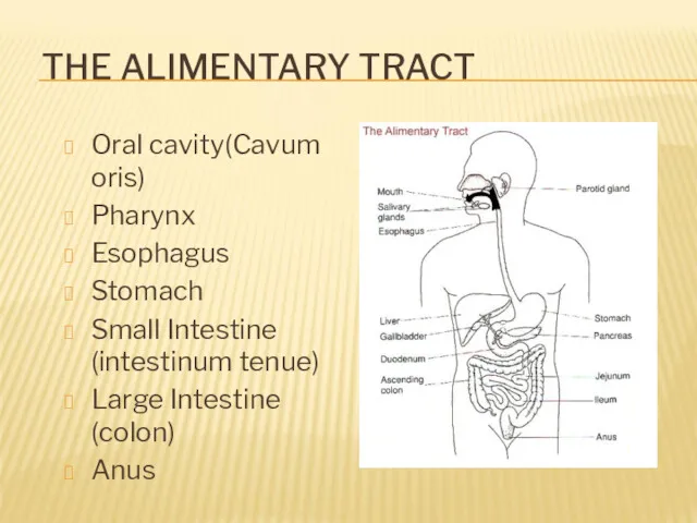
- 3. THE ALIMENTARY TRACT Oral cavity(Cavum oris) Pharynx Esophagus Stomach Small Intestine (intestinum tenue) Large Intestine (colon)
- 4. ACCESSORY PARTS Organs that are not in the Alimentary tract but helps in the digestion Teeth
- 5. ORAL CAVITY (CAVUM ORIS) Functions: Food enters in the mouth or oral cavity Tasting Mechanical breakdown
- 6. MOUTH Structures in the mouth that aids digestion: Teeth – cut, tear, crush and grind food.
- 7. MECHANISM OF SWALLOWING Swallowing is a coordinated activity of the tongue, soft palate, pharynx and esophagus.
- 8. ESOPHAGUS A straight muscular tube that is about 25 cm long which connects the mouth with
- 9. STOMACH J-shaped muscular sac Has inner folds (rugae) that increases the surface area of the stomach.
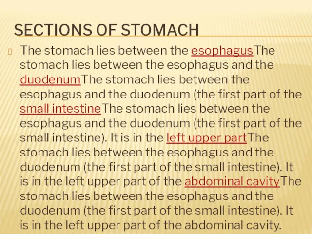
- 10. SECTIONS OF STOMACH The stomach lies between the esophagusThe stomach lies between the esophagus and the
- 11. SECTIONS OF STOMACH
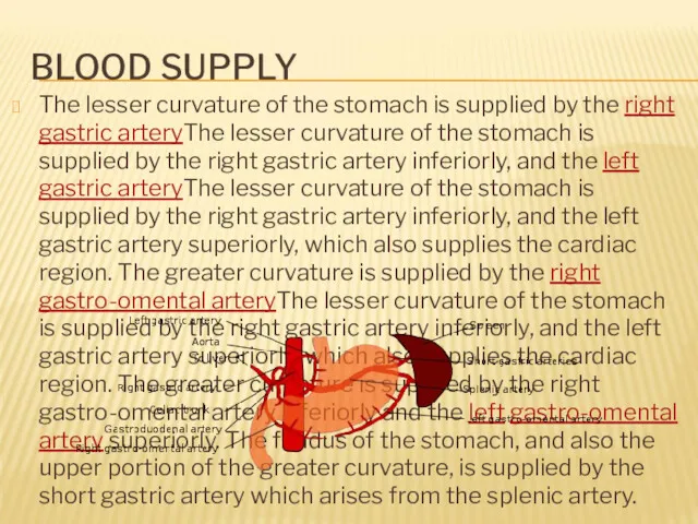
- 12. BLOOD SUPPLY The lesser curvature of the stomach is supplied by the right gastric arteryThe lesser
- 13. MOVEMENTS IN STOMACH
- 14. SMALL INTESTINE Long (5-6m), coiled tube beneath the stomach. Has three parts: Duodenum – upper part;
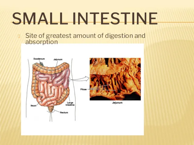
- 15. SMALL INTESTINE Site of greatest amount of digestion and absorption
- 16. DUODENUM The duodenum is a short structure (about 20–25 cm long) continuous with the stomach and
- 17. JEJENUM The jejunum is the midsection of the small intestine, connecting the duodenum to the ileum.
- 18. ILEUM The ileum: The final section of the small intestine. It is about 3 m long,
- 19. MOVEMENT IN SMALL INTESTINE: Mixing: Segmental contraction that occurs in small intestine Secretion: Lubricate, liquefy, digest
- 20. FUNCTIONS OF SMALL INTESTINE DIGESTION ABSORBTION IMMUNOLOGICAL
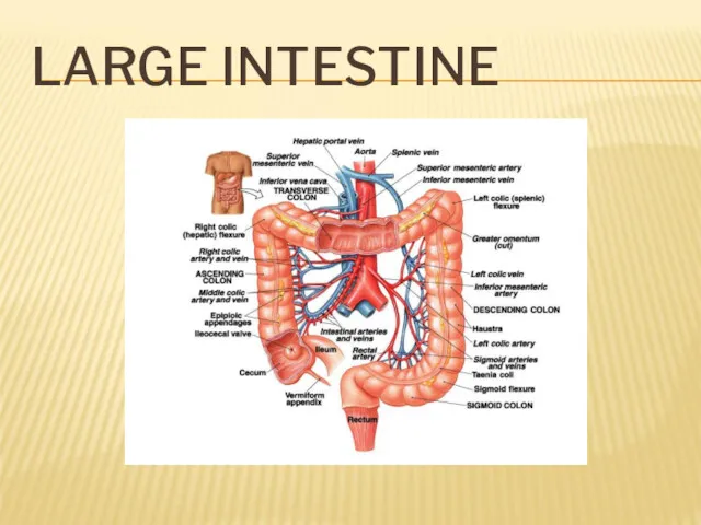
- 21. LARGE INTESTINE a.k.a. Colon larger diameter, but shorter (1-1,5м) Water is absorbed from the undigested food
- 22. LARGE INTESTINE
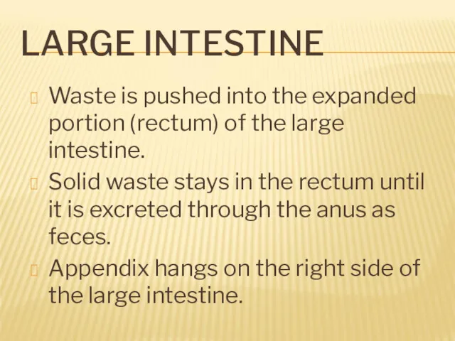
- 23. LARGE INTESTINE Waste is pushed into the expanded portion (rectum) of the large intestine. Solid waste
- 24. ACCESSORY ORGANS Produce or store enzymes that helps in digestion. Liver Largest gland of the body
- 26. Скачать презентацию
THE ALIMENTARY TRACT
A long muscular tube with many sections and areas.
Begins
THE ALIMENTARY TRACT
A long muscular tube with many sections and areas.
Begins
THE ALIMENTARY TRACT
Oral cavity(Cavum oris)
Pharynx
Esophagus
Stomach
Small Intestine (intestinum tenue)
Large Intestine (colon)
Anus
THE ALIMENTARY TRACT
Oral cavity(Cavum oris)
Pharynx
Esophagus
Stomach
Small Intestine (intestinum tenue)
Large Intestine (colon)
Anus
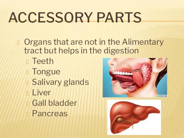
ACCESSORY PARTS
Organs that are not in the Alimentary tract but helps
ACCESSORY PARTS
Organs that are not in the Alimentary tract but helps
ORAL CAVITY (CAVUM ORIS)
Functions:
Food enters in the mouth or oral
ORAL CAVITY (CAVUM ORIS)
Functions:
Food enters in the mouth or oral
MOUTH
Structures in the mouth that aids digestion:
Teeth – cut, tear,
MOUTH
Structures in the mouth that aids digestion:
Teeth – cut, tear,
MECHANISM OF SWALLOWING
Swallowing is a coordinated activity of the tongue, soft
MECHANISM OF SWALLOWING
Swallowing is a coordinated activity of the tongue, soft
ESOPHAGUS
A straight muscular tube that is about 25 cm long which
ESOPHAGUS
A straight muscular tube that is about 25 cm long which
STOMACH
J-shaped muscular sac
Has inner folds (rugae) that increases the surface area
STOMACH
J-shaped muscular sac
Has inner folds (rugae) that increases the surface area
SECTIONS OF STOMACH
The stomach lies between the esophagusThe stomach lies between
SECTIONS OF STOMACH
The stomach lies between the esophagusThe stomach lies between
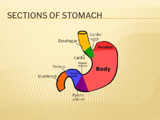
SECTIONS OF STOMACH
SECTIONS OF STOMACH
BLOOD SUPPLY
The lesser curvature of the stomach is supplied by the
BLOOD SUPPLY
The lesser curvature of the stomach is supplied by the
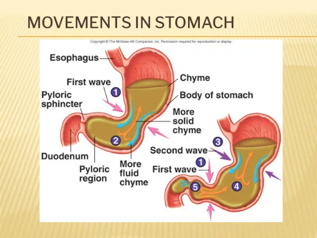
MOVEMENTS IN STOMACH
MOVEMENTS IN STOMACH
SMALL INTESTINE
Long (5-6m), coiled tube beneath the stomach.
Has three parts:
Duodenum –
SMALL INTESTINE
Long (5-6m), coiled tube beneath the stomach.
Has three parts:
Duodenum –
SMALL INTESTINE
Site of greatest amount of digestion and absorption
SMALL INTESTINE
Site of greatest amount of digestion and absorption
DUODENUM
The duodenum is a short structure (about 20–25 cm long) continuous with
DUODENUM
The duodenum is a short structure (about 20–25 cm long) continuous with
JEJENUM
The jejunum is the midsection of the small intestine, connecting the
JEJENUM
The jejunum is the midsection of the small intestine, connecting the
ILEUM
The ileum: The final section of the small intestine. It is
ILEUM
The ileum: The final section of the small intestine. It is
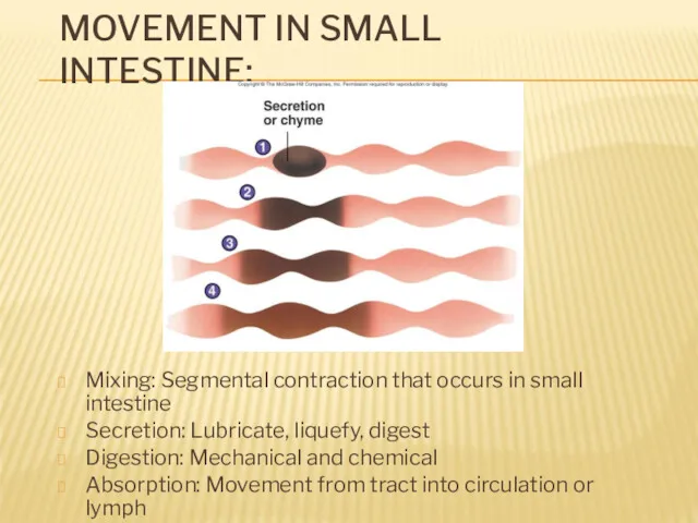
MOVEMENT IN SMALL INTESTINE:
Mixing: Segmental contraction that occurs in small intestine
Secretion:
MOVEMENT IN SMALL INTESTINE:
Mixing: Segmental contraction that occurs in small intestine
Secretion:
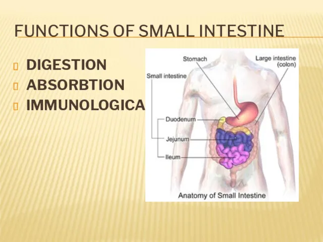
FUNCTIONS OF SMALL INTESTINE
DIGESTION
ABSORBTION
IMMUNOLOGICAL
FUNCTIONS OF SMALL INTESTINE
DIGESTION
ABSORBTION
IMMUNOLOGICAL
LARGE INTESTINE
a.k.a. Colon
larger diameter, but shorter (1-1,5м)
Water is absorbed from the
LARGE INTESTINE
a.k.a. Colon
larger diameter, but shorter (1-1,5м)
Water is absorbed from the
LARGE INTESTINE
LARGE INTESTINE
LARGE INTESTINE
Waste is pushed into the expanded portion (rectum) of the
LARGE INTESTINE
Waste is pushed into the expanded portion (rectum) of the
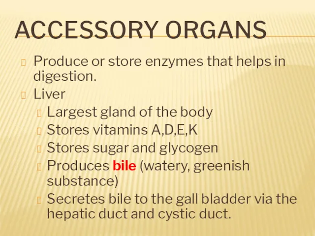
ACCESSORY ORGANS
Produce or store enzymes that helps in digestion.
Liver
Largest
ACCESSORY ORGANS
Produce or store enzymes that helps in digestion.
Liver
Largest
 Вступление в систему непревыного медицинского и фармацевтического образования: пошаговая инструкция
 Гигиена детей и подростков
 Экзогенный аллергический альвеолит
 Операции на кишечнике
 Повреждения органов брюшной полости
 Сучасні перинатальні технології та спостереження за новонародженим в перші 72 години життя
 Первичные бактериальные менингиты. Диагностика и лечение
 Профессиональная БА
 Захворювання периферичної нервової системи
 Применение лабораторных методов при судебно-медицинской экспертизе
 Дисфункциональные маточные кровотечения
 Никотиновая зависимость и как от нее избавится
 Антибиотикотерапия у детей
 Современные способы обработки рук хирурга и операционного поля
 Подагра. Стадии развития подагры
 Види кровотеч
 Background and precancerous diseases of female genital. Malignant neoplasms of female genital organs
 Сладкая жизнь. Опасность сахарного диабета
 Гемофилии А, В, С
 Средства гигиены
 Пластика стебельчатым лоскутом. Лекция для студентов стоматологического факультета
 Chronic hepatitis
 Общая физиология ЦНС
 Хронические воспалительные заболевания кишечника. Лекция № 13
 Крымская-конго геморрагическая лихорадка (ККГЛ)
 Рахит. Диагностика и лечение
 Женское здоровье. Продукты компании TianDe
 Копинг-стратегии и соматическая патология- есть ли связь