Background and precancerous diseases of female genital. Malignant neoplasms of female genital organs презентация
- Background and precancerous diseases of female genital. Malignant neoplasms of female genital organs

Содержание
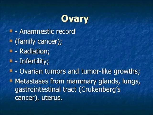
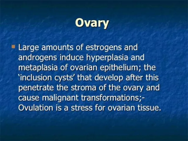
- 2. In benign tumors of the external genitals include fibroma, leiomyoma, lipoma (adipose tumor), myxoma (mucous tumor),
- 3. Fibroma – this tumor is mainly localized inside the labia majora or in a thin submucous
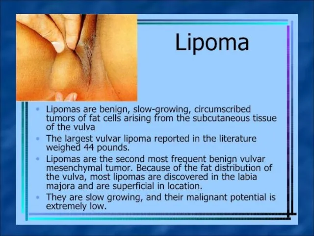
- 4. Lipoma – a tumor that formed mature fatty tissue with connective tissue fibers in the pubic
- 6. Hemangioma– a tumor that arises due to atelectasis vessels of the skin (as node) and mucous
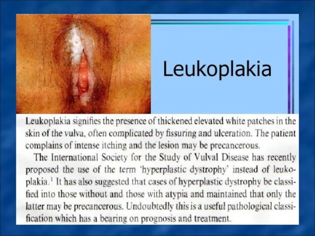
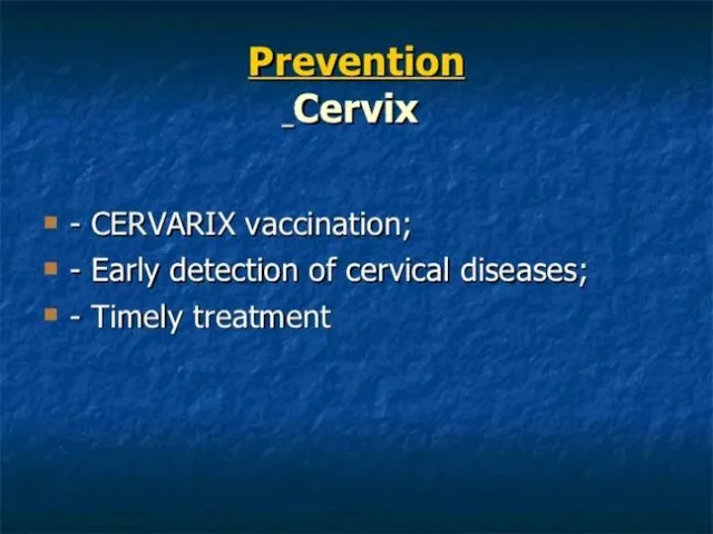
- 9. In precancerous diseases of external genitalia include leukoplakia, kraurosis and Bowen’s and Paget disease.
- 10. Vulvar leukoplakia developing mainly in the perimenopausal period (probably due to hormonal disorders and immune status),
- 12. Vulvar kraurosis develops mostly in the perimenopausal period is characterized by papillary atrophy and mesh layers
- 13. Vulvar kraurosis
- 14. Bowen’s and Paget disease characterized by hyperkeratosis and acanthosis of the external genital organs, shows bright-red,
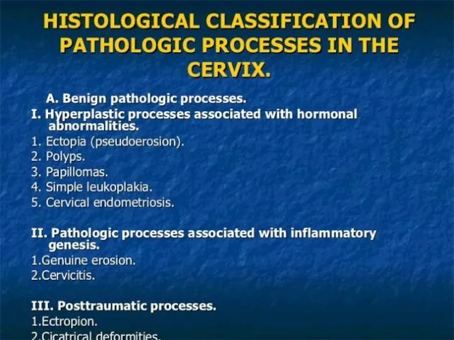
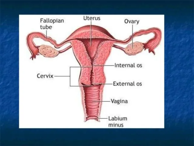
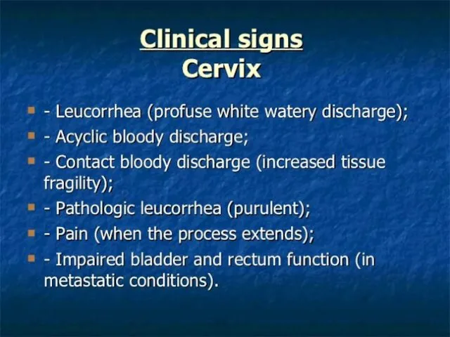
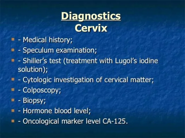
- 15. In benign (background) cervical diseases include such pathological processes in which the epithelium remains normoplaziya -
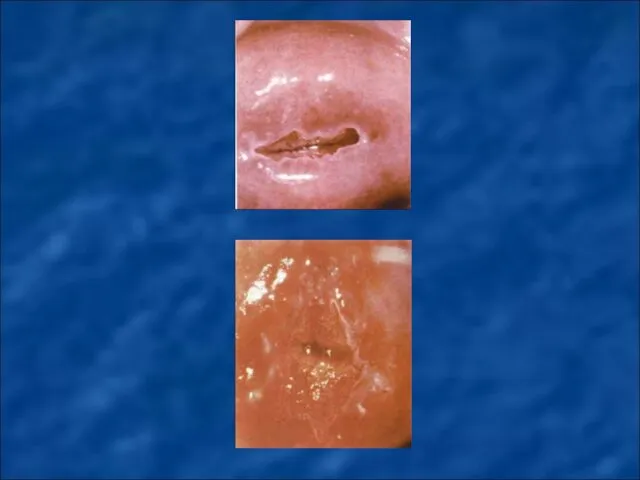
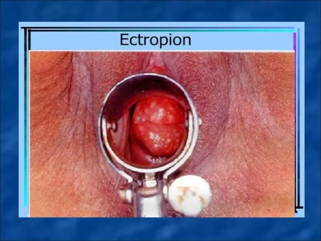
- 21. Ectopia of columnar epithelium (dishormonal, inflammatory, posttraumatic - ektropion) - move the cervical mucous membrane (columnar
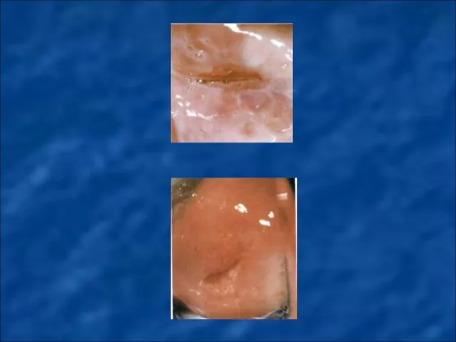
- 22. Cervicitis (endo-and exocervicitis) - inflammatory processes in the area of vaginal mucous membrane of the cervix
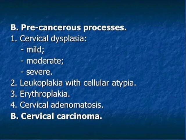
- 28. Precancerous cervical deseases (dysplasia, CIN, cervical intraepithelial neoplasia) - is the proliferation of cervical tissue with
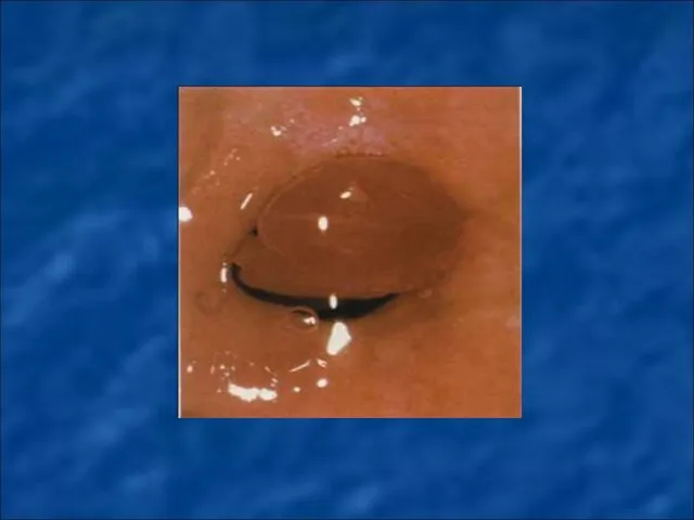
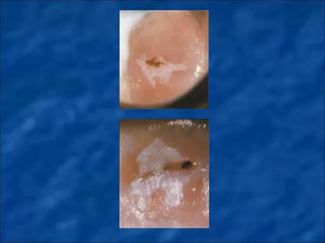
- 29. Simple leukoplakia - during colposcopic examination shown in the form of white spots with a smooth
- 36. Methods of treating precancerous cervical deseasesl are determined by the nature and degree of dysplasia and
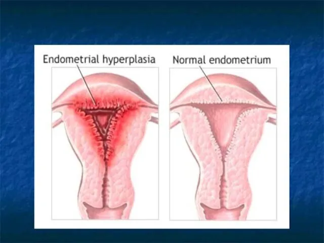
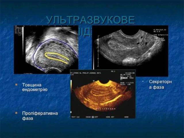
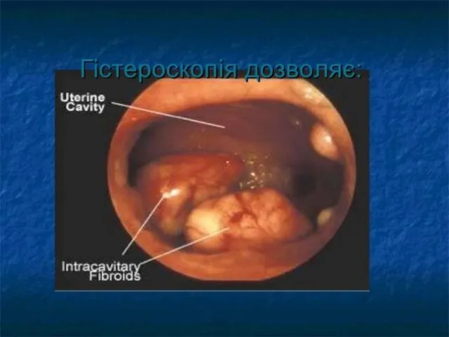
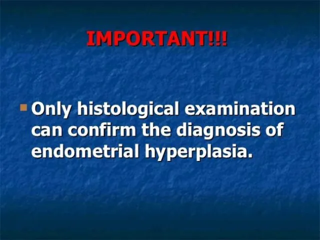
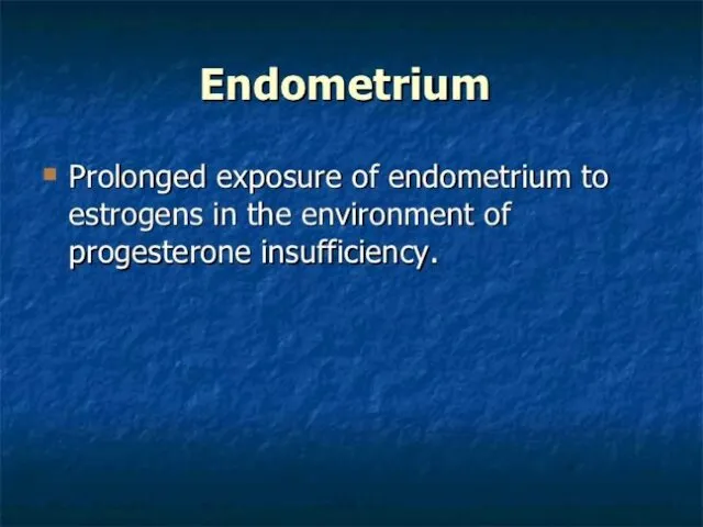
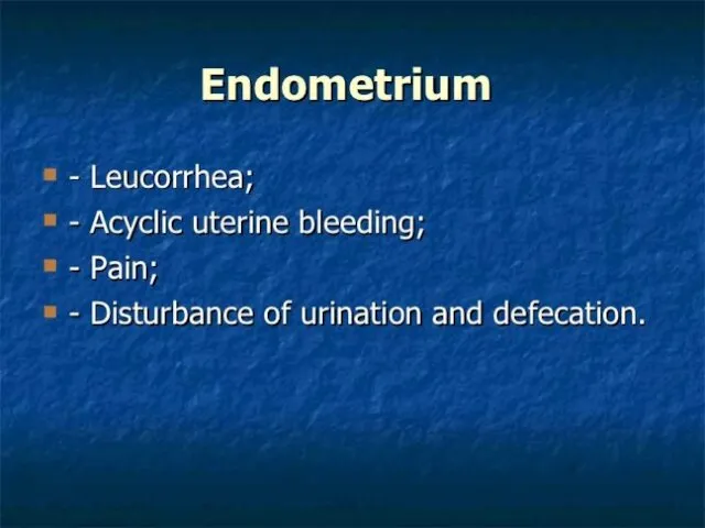
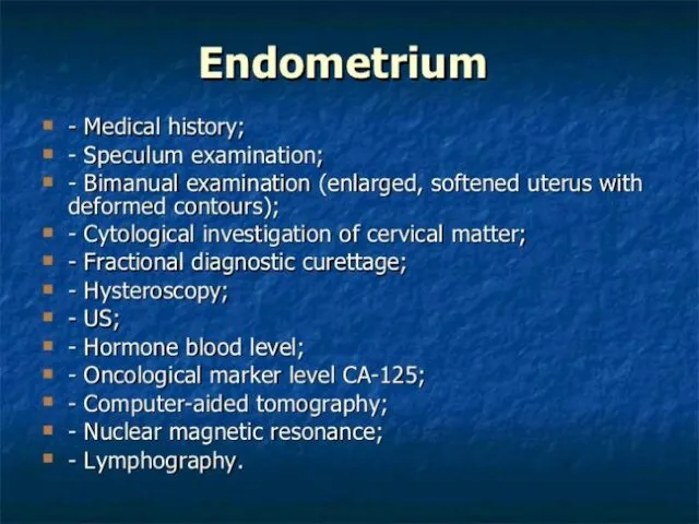
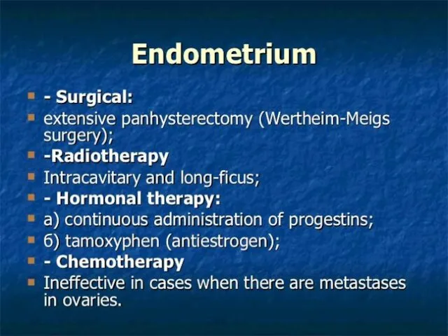
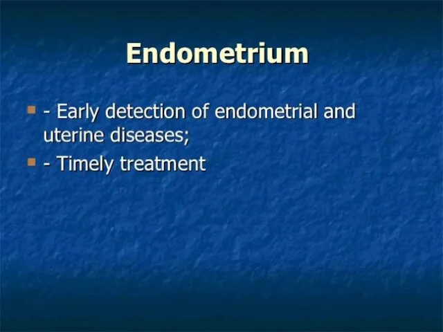
- 38. Endometrial hyperplasia - benign pathology of mucous membrane of uterine and a pathological proliferation of endometrium
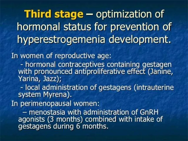
- 41. Classification of endometrial hyperplasia (developed by International Society Pathologist-gynecologists and approved by WHO, 1994): - simple
- 42. Clinical and morphological classification of endometrial hyperplasia [Y. Bohman, 1985]: 1. Background processes: glandular and glandular-cystic
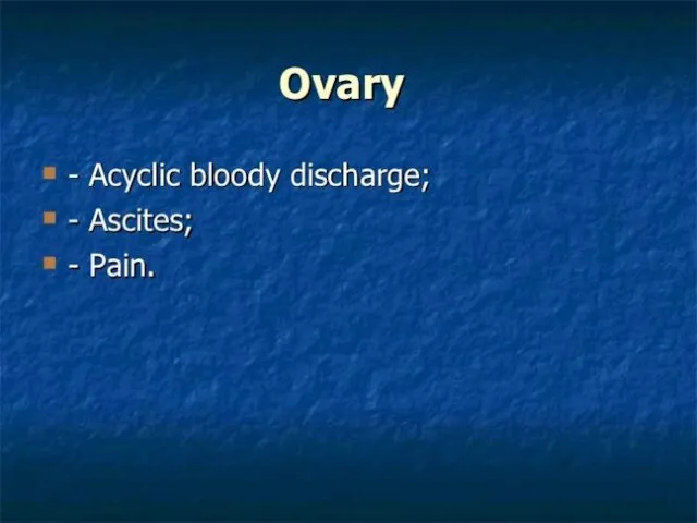
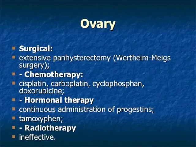
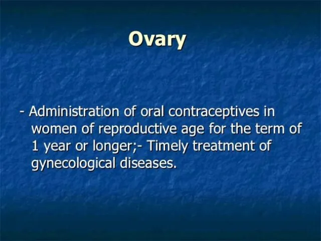
- 58. MALIGNANT NEOPLASMS OF FEMALE GENITAL ORGANS.
- 85. Скачать презентацию
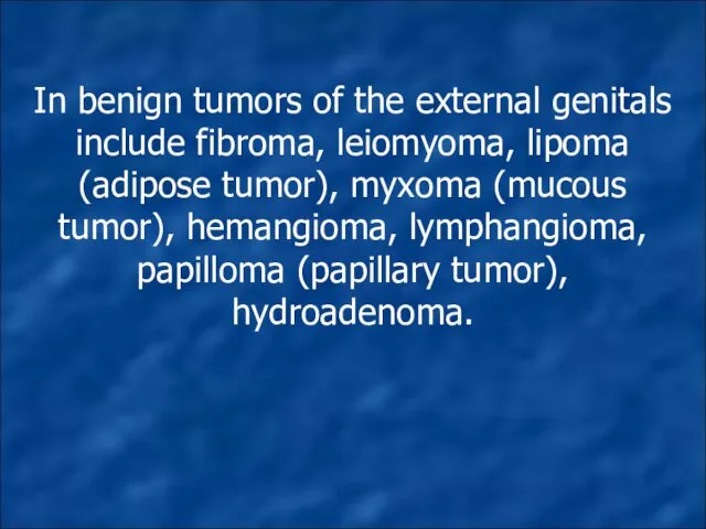
In benign tumors of the external genitals include fibroma, leiomyoma, lipoma
In benign tumors of the external genitals include fibroma, leiomyoma, lipoma
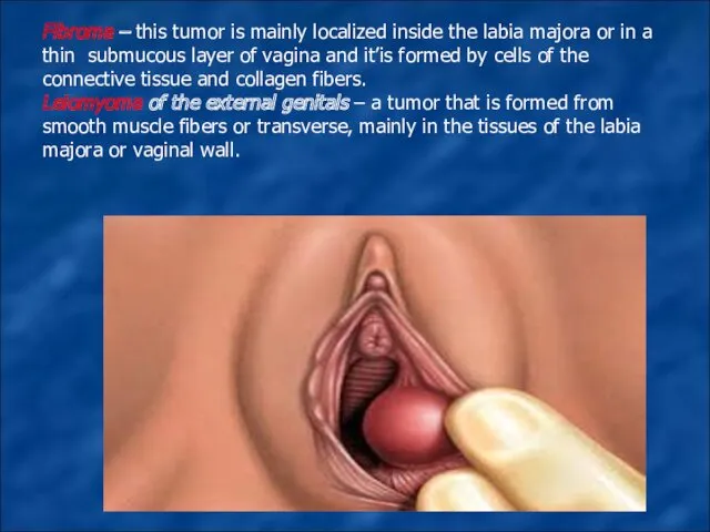
Fibroma – this tumor is mainly localized inside the labia majora
Fibroma – this tumor is mainly localized inside the labia majora
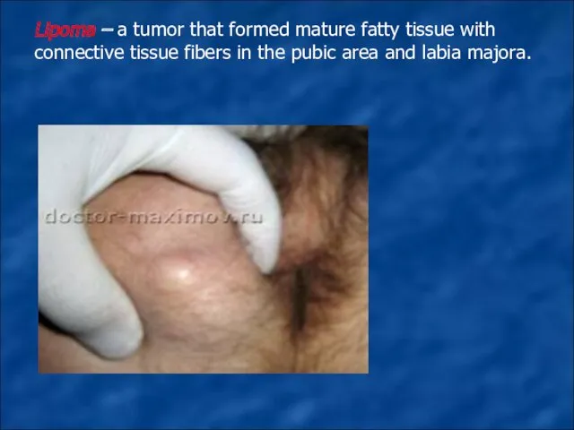
Lipoma – a tumor that formed mature fatty tissue with connective
Lipoma – a tumor that formed mature fatty tissue with connective

Hemangioma– a tumor that arises due to atelectasis vessels of the
Hemangioma– a tumor that arises due to atelectasis vessels of the
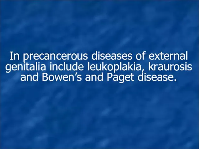
In precancerous diseases of external genitalia include leukoplakia, kraurosis and Bowen’s
In precancerous diseases of external genitalia include leukoplakia, kraurosis and Bowen’s
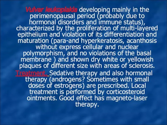
Vulvar leukoplakia developing mainly in the perimenopausal period (probably due
Vulvar leukoplakia developing mainly in the perimenopausal period (probably due
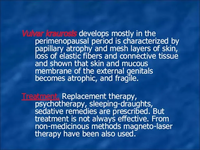
Vulvar kraurosis develops mostly in the perimenopausal period is characterized by
Vulvar kraurosis develops mostly in the perimenopausal period is characterized by
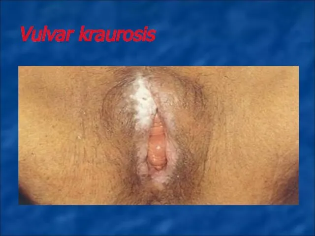
Vulvar kraurosis
Vulvar kraurosis
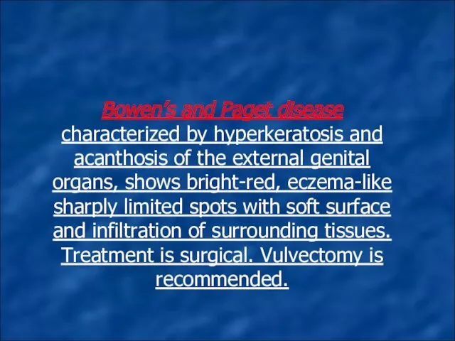
Bowen’s and Paget disease characterized by hyperkeratosis and acanthosis of the
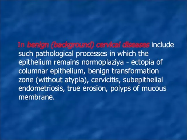
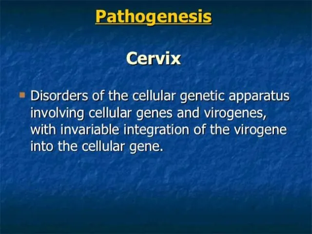
In benign (background) cervical diseases include such pathological processes in which
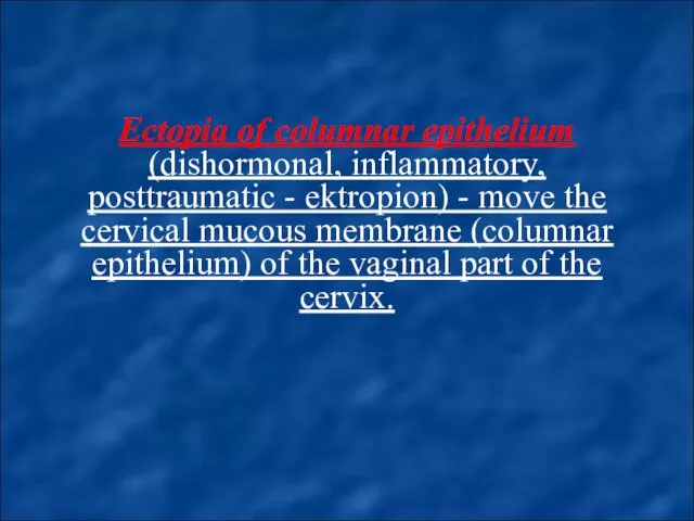
Ectopia of columnar epithelium (dishormonal, inflammatory, posttraumatic - ektropion) - move
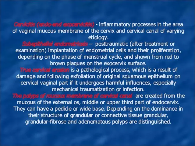
Cervicitis (endo-and exocervicitis) - inflammatory processes in the area of vaginal
Cervicitis (endo-and exocervicitis) - inflammatory processes in the area of vaginal

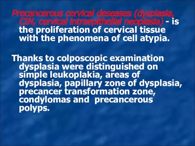
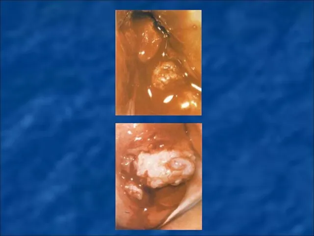
Precancerous cervical deseases (dysplasia, CIN, cervical intraepithelial neoplasia) - is the
Precancerous cervical deseases (dysplasia, CIN, cervical intraepithelial neoplasia) - is the
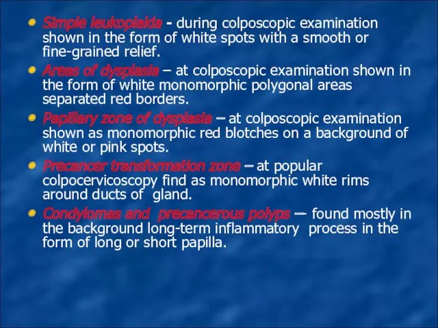
Simple leukoplakia - during colposcopic examination shown in the form of
Simple leukoplakia - during colposcopic examination shown in the form of

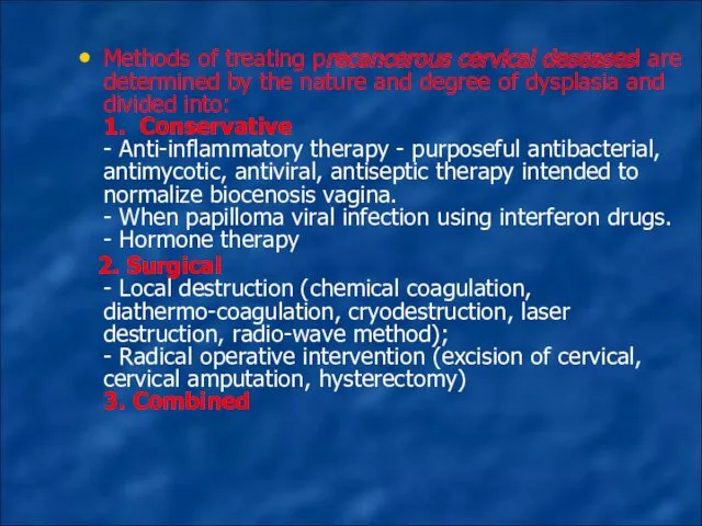
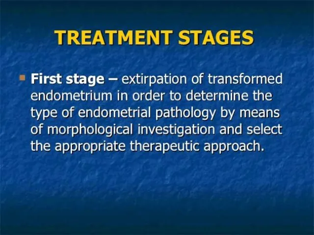
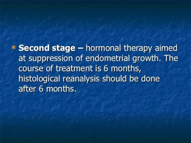
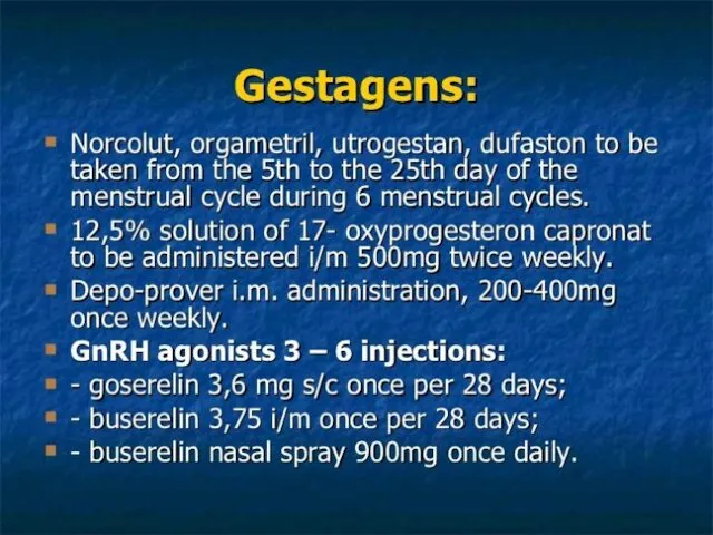
Methods of treating precancerous cervical deseasesl are determined by the nature
Methods of treating precancerous cervical deseasesl are determined by the nature
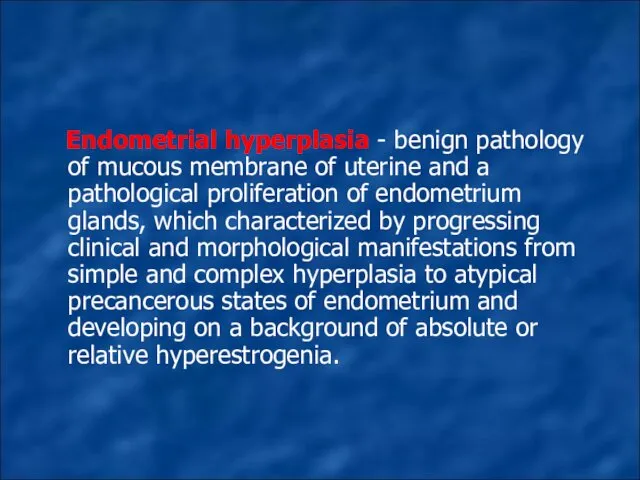
Endometrial hyperplasia - benign pathology of mucous membrane of uterine
Endometrial hyperplasia - benign pathology of mucous membrane of uterine
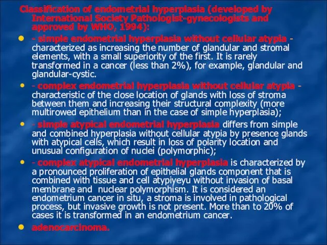
Classification of endometrial hyperplasia (developed by International Society Pathologist-gynecologists and approved
Classification of endometrial hyperplasia (developed by International Society Pathologist-gynecologists and approved
![Clinical and morphological classification of endometrial hyperplasia [Y. Bohman, 1985]:](/_ipx/f_webp&q_80&fit_contain&s_1440x1080/imagesDir/jpg/19591/slide-41.jpg)
Clinical and morphological classification of endometrial hyperplasia [Y. Bohman, 1985]:
1.
Clinical and morphological classification of endometrial hyperplasia [Y. Bohman, 1985]:
1.
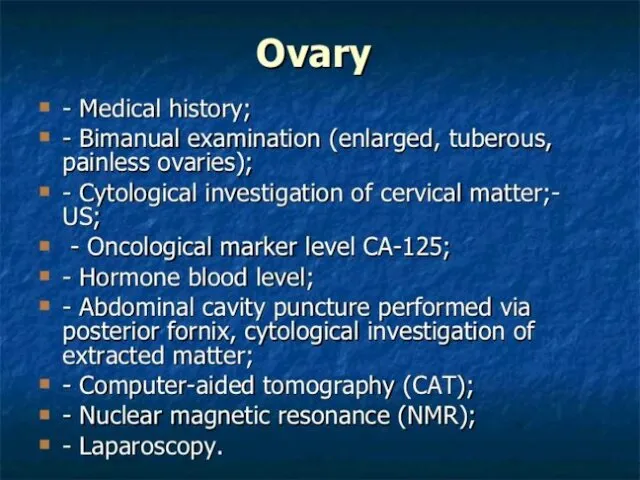
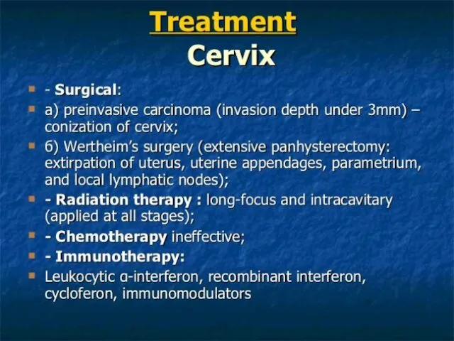
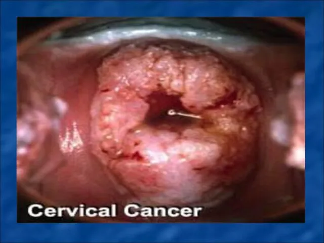
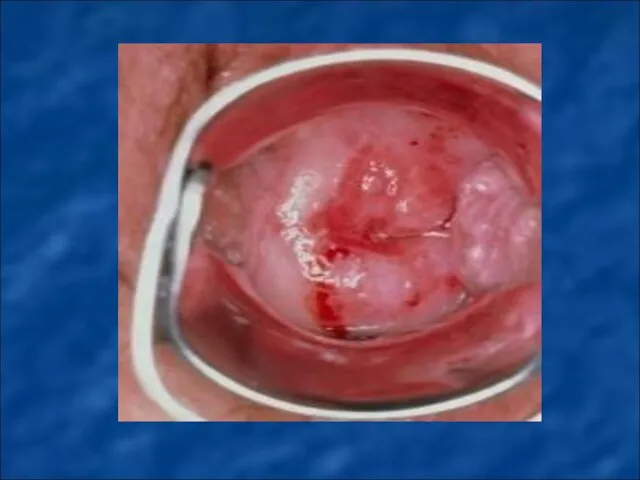
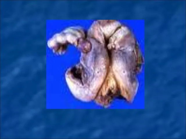
MALIGNANT NEOPLASMS OF FEMALE GENITAL ORGANS.
MALIGNANT NEOPLASMS OF FEMALE GENITAL ORGANS.

 Кардиогенный шок и его причины
 Rickettsioses
 Фармакология системы крови
 Гинекология детей и подростков
 Хронические тонзиллиты
 ПХО ран шеи.Типичные разрезы при абсцессах и флегмонах шеи
 Организация акушерскогинекологической помощи в Российской Федерации. Основные показатели родовспоможения
 Уильям Гарвей (1578 – 1657)
 Психометаболические стимуляторы (ноотропные препараты)
 Эндоваскулярные операции на коронарных артериях. Транслюминальная баллонная ангиопластика и стентирование
 Профилактика неинфекционных заболеваний
 Дезинфекция. Виды дезинфекции
 Злоякісні пухлини жіночих статевих органів
 Нарушения ритма и проводимости сердца
 Хронические воспалительные заболевания гортани
 Dermatologiya fanidan
 Медицина катастроф
 Пошкодження ока та його додаткового апарату, клініка, невідкладна допомога, профілактика, диспансеризація
 Резекционная трепанация черепа
 Хронический пиелонефрит у пациентов пожилого и старческого возраста
 Ayurveda doctor
 Плазменное звено системы гемостаза
 Заболевания губ у детей
 Недоношенный ребенок. Причины преждевременных родов. Классификация. Дифференциальная диагностика незрелости, недоношенности
 Оценка тяжести пациента
 Оториноларингологиялық аурулардың қазіргі заманға сай диагностикасы мен емдеу әдістері
 Лечение заболеваний крови у детей. Железодефицитная анемия. Лейкозы. Геморрагические диатезы
 Хирургическая анатомия позвоночника и шеи