- The hormonal regulation of the body

Содержание
- 2. Neural and humoral regulation of body functions
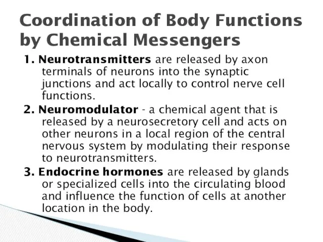
- 3. 1. Neurotransmitters are released by axon terminals of neurons into the synaptic junctions and act locally
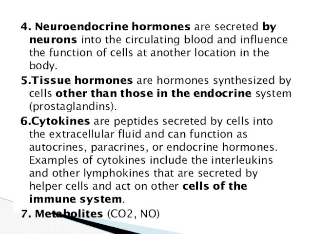
- 4. 4. Neuroendocrine hormones are secreted by neurons into the circulating blood and influence the function of
- 5. Endocrine hormones are released by glands or specialized cells into the circulating blood and influence the
- 6. The locations for the different types of hormone receptors are generally the following: 1. In or
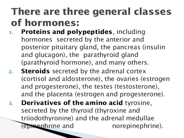
- 7. Proteins and polypeptides, including hormones secreted by the anterior and posterior pituitary gland, the pancreas (insulin
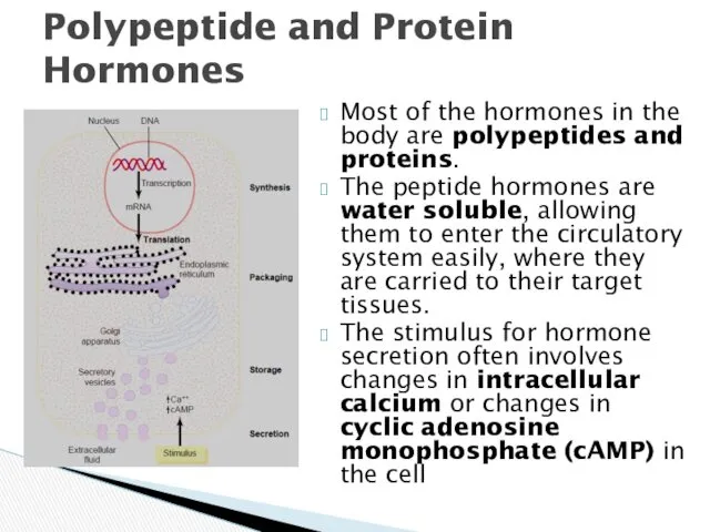
- 8. Most of the hormones in the body are polypeptides and proteins. The peptide hormones are water
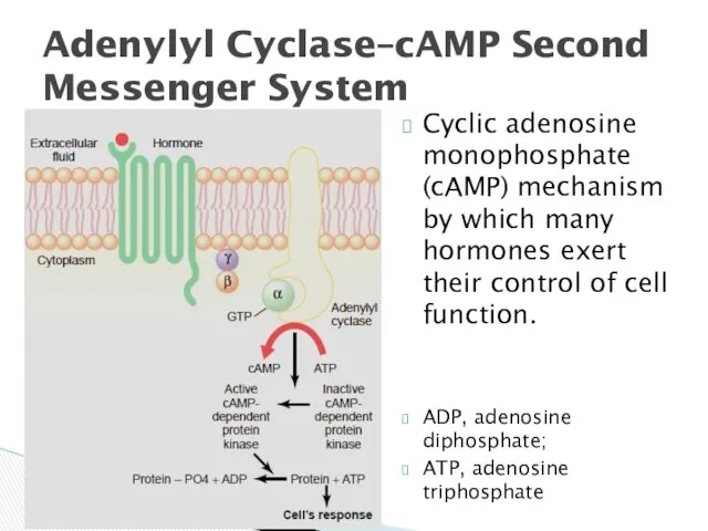
- 9. Cyclic adenosine monophosphate (cAMP) mechanism by which many hormones exert their control of cell function. ADP,
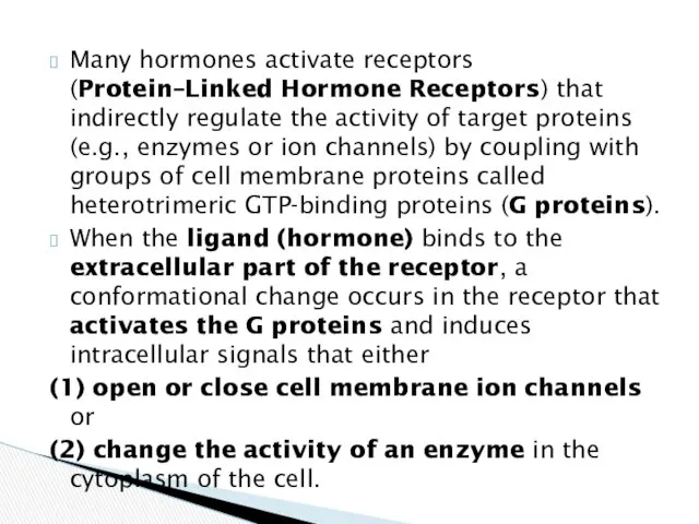
- 10. Many hormones activate receptors (Protein–Linked Hormone Receptors) that indirectly regulate the activity of target proteins (e.g.,
- 11. cAMP is not the only second messenger used by the different hormones. Two other especially important
- 12. Enzyme-linked receptors have their hormone-binding site on the outside of the cell membrane and their catalytic
- 13. The chemical structure of steroid hormones is similar to that of cholesterol. Because the steroids are
- 14. After the hormone binds to the receptor in the cytoplasm or in the nucleus, the hormone-receptor
- 15. 1. The steroid hormone diffuses across the cell membrane and enters the cytoplasm of the cell,
- 16. Amine hormones are derived from tyrosine (the thyroid and the adrenal medullary hormones). Contrary to common
- 17. 1. Metabolism and tissue maturation. The endocrine system regulates the rate of metabolism and influences the
- 18. 5. Heart rate and blood pressure regulation. The endocrine system helps regulate the heart rate and
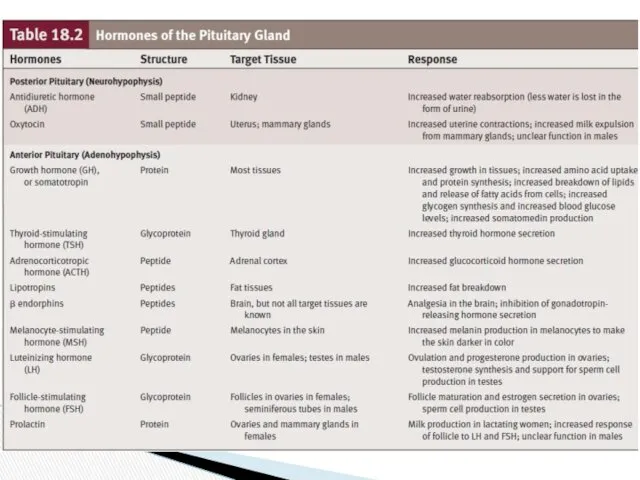
- 19. The pituitary gland, or hypophysis, secretes nine major hormones that regulate numerous body functions and the
- 20. Relationship Among the Hypothalamus, Posterior Pituitary, and Target Tissues
- 21. Antidiuretic Hormone is so named because it prevents the output of large amounts of urine (diuresis).
- 22. Control of Antidiuretic Hormone (ADH) Secretion
- 23. When blood osmolality increases, the frequency of action potentials in the osmoreceptors increases, resulting in a
- 24. A lack of ADH secretion is one cause of diabetes insipidus and leads to the production
- 25. Oxytocin is synthesized by neuron cell bodies in the paraventricular nuclei of the hypothalamus and then
- 26. Action potentials are carried by sensory neurons from the uterus and from the nipples to the
- 27. Portal vessels are blood vessels that begin and end in a capillary network. Neurohormones, produced and
- 28. Relationship Among the Hypothalamus, Anterior Pituitary, and Target Tissues
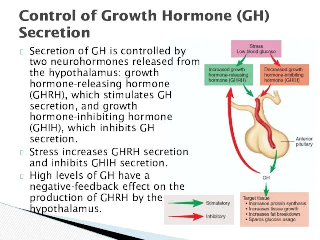
- 31. Control of Growth Hormone (GH) Secretion Secretion of GH is controlled by two neurohormones released from
- 32. Chronic hyposecretion of GH in infants and children leads to dwarfism, or short stature due to
- 33. Regulation of Thyroid Hormone (T3 and T4) Secretion
- 34. An abnormal enlargement of the thyroid gland is called a goiter. Goiters can result from conditions
- 35. Hormones of the Adrenal Gland
- 36. Regulation of Adrenal Medullary Secretions
- 37. The adrenal cortex secretes three hormone types: mineralocorticoids, glucocorticoids, and androgens. All are similar in structure
- 38. Target Tissues and Their Responses to Glucocorticoid Hormones
- 39. Regulation of Cortisol Secretion
- 40. The major secretory products of the zona glomerulosa are the mineralocorticoids. Aldosterone is produced in the
- 41. Aldosterone increases K excretion into the urine by the kidneys, thereby decreasing blood levels of K.
- 42. Symptoms of Hyposecretion and Hypersecretion of Adrenal Cortex Hormones
- 44. Скачать презентацию
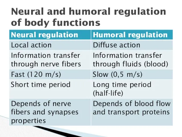
Neural and humoral regulation of body functions
Neural and humoral regulation of body functions
1. Neurotransmitters are released by axon terminals of neurons into the
1. Neurotransmitters are released by axon terminals of neurons into the
4. Neuroendocrine hormones are secreted by neurons into the circulating blood
4. Neuroendocrine hormones are secreted by neurons into the circulating blood
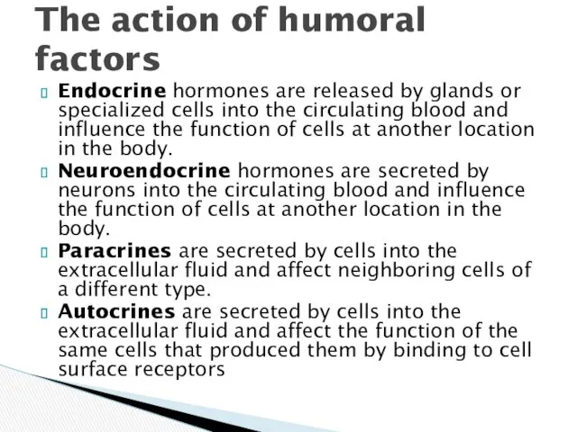
Endocrine hormones are released by glands or specialized cells into the
Endocrine hormones are released by glands or specialized cells into the

The locations for the different types of hormone receptors are generally
The locations for the different types of hormone receptors are generally
Proteins and polypeptides, including hormones secreted by the anterior and posterior
Proteins and polypeptides, including hormones secreted by the anterior and posterior
Most of the hormones in the body are polypeptides and proteins.
The
Most of the hormones in the body are polypeptides and proteins.
The
Cyclic adenosine monophosphate (cAMP) mechanism by which many hormones exert their
Cyclic adenosine monophosphate (cAMP) mechanism by which many hormones exert their
Many hormones activate receptors (Protein–Linked Hormone Receptors) that indirectly regulate the
Many hormones activate receptors (Protein–Linked Hormone Receptors) that indirectly regulate the
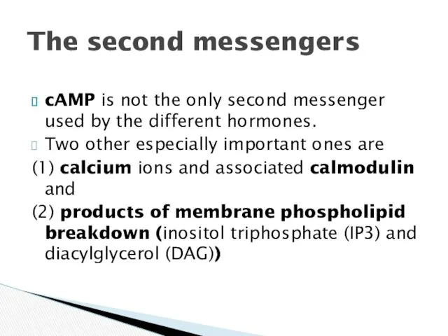
cAMP is not the only second messenger used by the different
cAMP is not the only second messenger used by the different
Enzyme-linked receptors have their hormone-binding site on the outside of the
Enzyme-linked receptors have their hormone-binding site on the outside of the
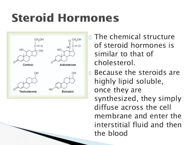
The chemical structure of steroid hormones is similar to that of
The chemical structure of steroid hormones is similar to that of
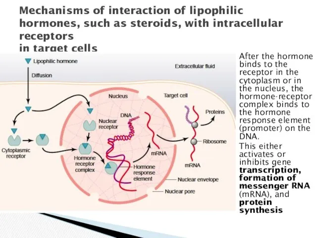
After the hormone binds to the receptor in the cytoplasm or
After the hormone binds to the receptor in the cytoplasm or
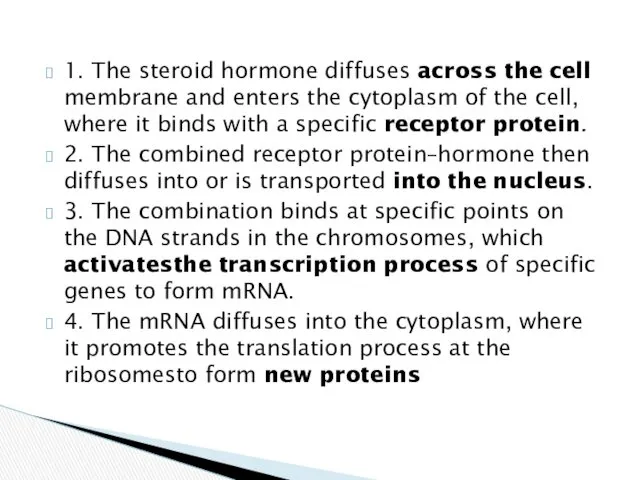
1. The steroid hormone diffuses across the cell membrane and enters
1. The steroid hormone diffuses across the cell membrane and enters
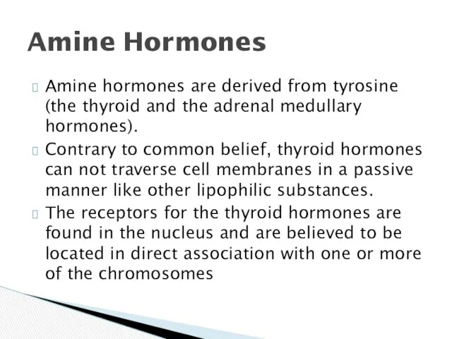
Amine hormones are derived from tyrosine (the thyroid and the adrenal
Amine hormones are derived from tyrosine (the thyroid and the adrenal
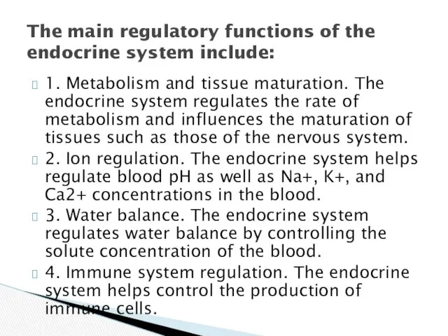
1. Metabolism and tissue maturation. The endocrine system regulates the rate
1. Metabolism and tissue maturation. The endocrine system regulates the rate
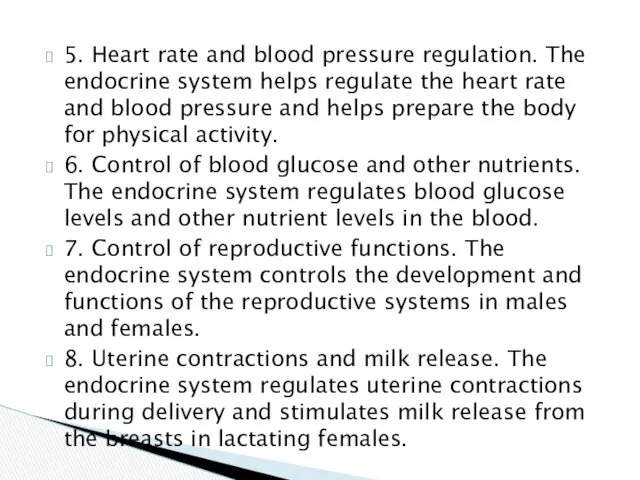
5. Heart rate and blood pressure regulation. The endocrine system helps
5. Heart rate and blood pressure regulation. The endocrine system helps
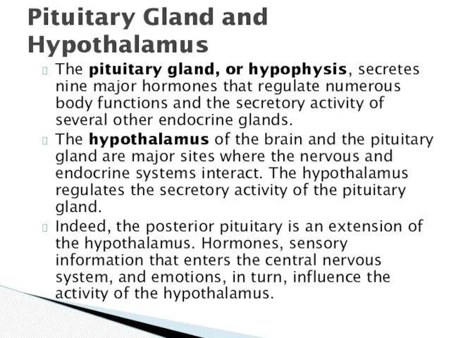
The pituitary gland, or hypophysis, secretes nine major hormones that regulate
The pituitary gland, or hypophysis, secretes nine major hormones that regulate
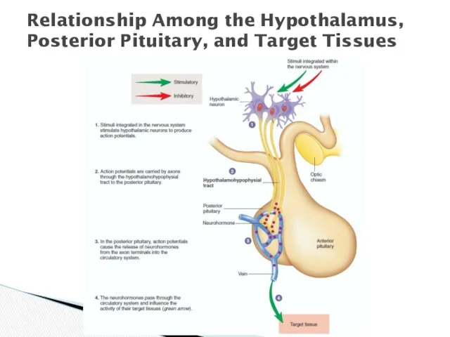
Relationship Among the Hypothalamus, Posterior Pituitary, and Target Tissues
Relationship Among the Hypothalamus, Posterior Pituitary, and Target Tissues
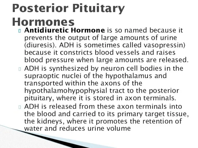
Antidiuretic Hormone is so named because it prevents the output of
Antidiuretic Hormone is so named because it prevents the output of
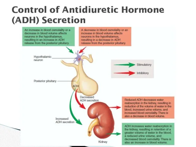
Control of Antidiuretic Hormone (ADH) Secretion
Control of Antidiuretic Hormone (ADH) Secretion
When blood osmolality increases, the frequency of action potentials in the
When blood osmolality increases, the frequency of action potentials in the
A lack of ADH secretion is one cause of diabetes insipidus
A lack of ADH secretion is one cause of diabetes insipidus
Oxytocin is synthesized by neuron cell bodies in the paraventricular nuclei
Oxytocin is synthesized by neuron cell bodies in the paraventricular nuclei
Action potentials are carried by sensory neurons from the uterus and
Action potentials are carried by sensory neurons from the uterus and
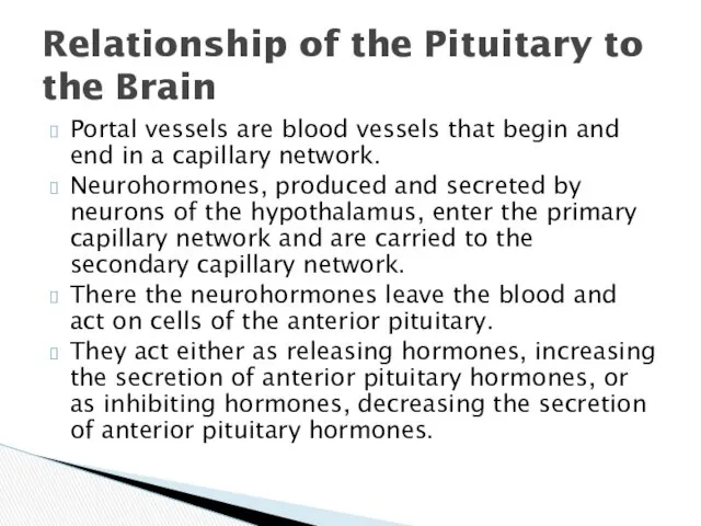
Portal vessels are blood vessels that begin and end in a
Portal vessels are blood vessels that begin and end in a
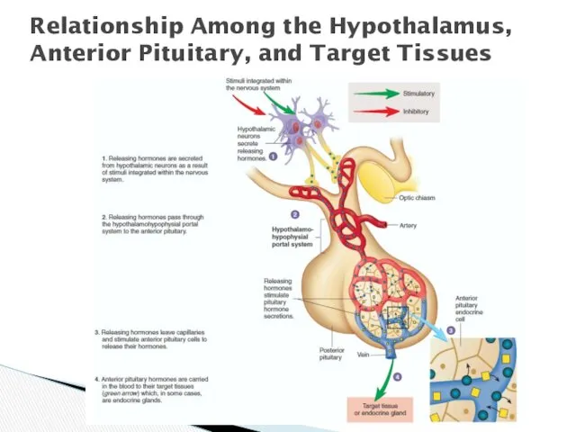
Relationship Among the Hypothalamus, Anterior Pituitary, and Target Tissues
Relationship Among the Hypothalamus, Anterior Pituitary, and Target Tissues
Control of Growth Hormone (GH) Secretion
Secretion of GH is controlled by
Control of Growth Hormone (GH) Secretion
Secretion of GH is controlled by
Chronic hyposecretion of GH in infants and children leads to dwarfism,
Chronic hyposecretion of GH in infants and children leads to dwarfism,
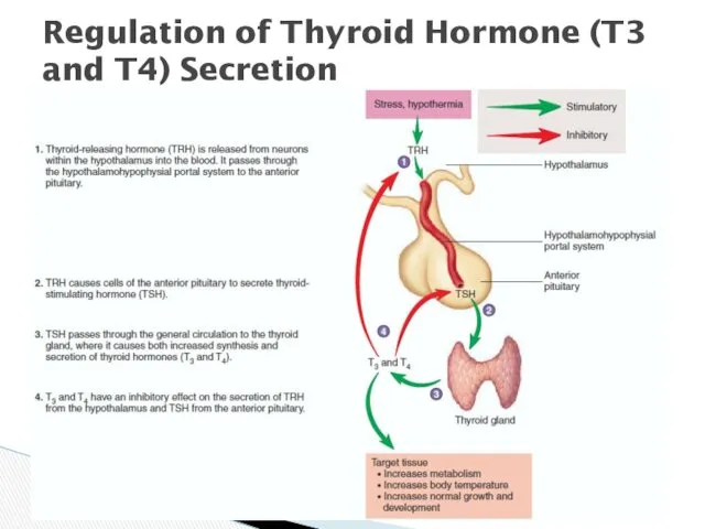
Regulation of Thyroid Hormone (T3 and T4) Secretion
Regulation of Thyroid Hormone (T3 and T4) Secretion
An abnormal enlargement of the thyroid gland is called a goiter.
An abnormal enlargement of the thyroid gland is called a goiter.
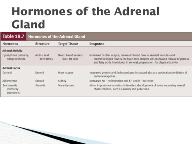
Hormones of the Adrenal Gland
Hormones of the Adrenal Gland
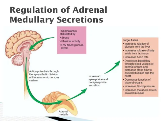
Regulation of Adrenal Medullary Secretions
Regulation of Adrenal Medullary Secretions
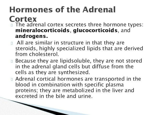
The adrenal cortex secretes three hormone types: mineralocorticoids, glucocorticoids, and androgens.
The adrenal cortex secretes three hormone types: mineralocorticoids, glucocorticoids, and androgens.
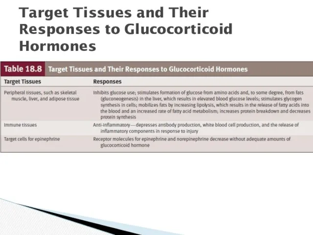
Target Tissues and Their Responses to Glucocorticoid Hormones
Target Tissues and Their Responses to Glucocorticoid Hormones
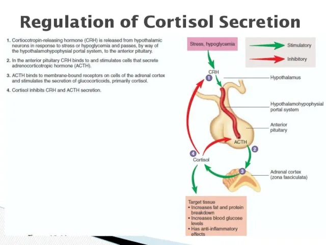
Regulation of Cortisol Secretion
Regulation of Cortisol Secretion
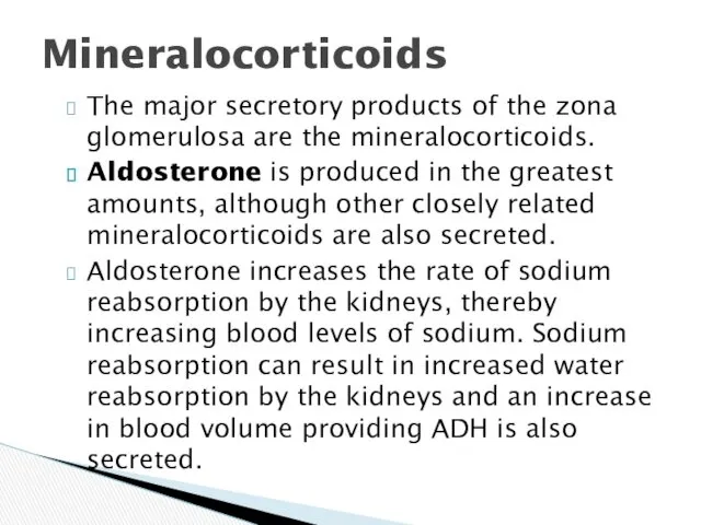
The major secretory products of the zona glomerulosa are the mineralocorticoids.
The major secretory products of the zona glomerulosa are the mineralocorticoids.
Aldosterone increases K excretion into the urine by the kidneys, thereby
Aldosterone increases K excretion into the urine by the kidneys, thereby
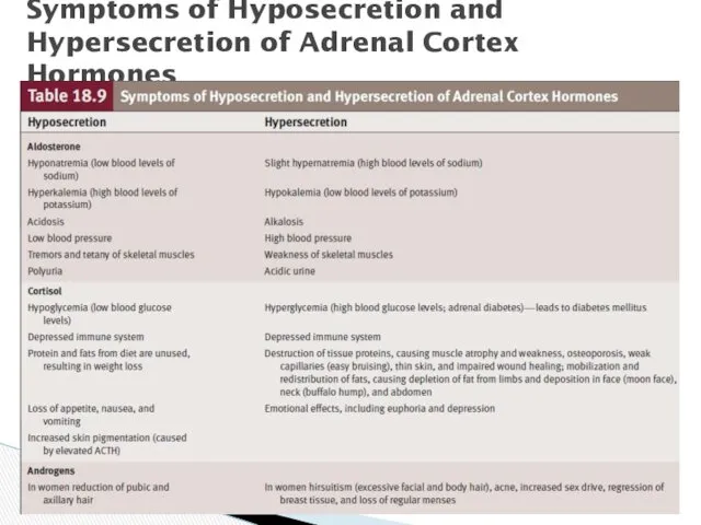
Symptoms of Hyposecretion and Hypersecretion of Adrenal Cortex Hormones
Symptoms of Hyposecretion and Hypersecretion of Adrenal Cortex Hormones
 Миастения. Никотиновый ацетилхолиновый рецептор
 Негізгі психопатологиялық синдромдар
 Соединительно-тканные дисплазии
 Стерилизация тиімділігін бақылау әдістері
 Післяпологові септичні ускладнення
 Очищение организма – обязательная мера для крепкого здоровья и стойкого иммунитета
 Rubella (краснуха)
 Клинический протокол диагностики и лечения послеродового кровотечения
 Саранск БУ. Анонимные Наркоманы
 Работа в очаге туберкулёзной инфекции
 Ультрафиолетовое облучение (УФО)
 Центр медико-социального притяжения Вектор здоровья
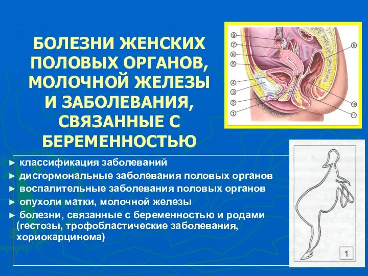 Болезни женских половых органов, молочной железы и заболевания, связанные с беременностью
 Болезни органов пищеварения лошадей
 Несахарный диабет
 2 типті қант диабеті дамуының факторлары
 Основные нейропсихологические синдромы в детском возрасте
 Дені сау нәрестеге үйде патронаж жасап амбулаториялық оқу картасын толтыру
 Эндокринология естественной и хирургической менопаузы
 Почему важно грудное вскармливание
 Оказание первой медицинской помощи
 Зоонозы. Туляремия
 Микоздық қоздырғыштар. Саңырауқұлақтардың морфологиялық, дақылдық қасиеттері
 Здоров’я населення як медико-соціальна проблема. Стратегії охорони здоров’я
 Денсаулық сақтауға арналған ҚР бюджет жүйесі
 Желтухи у новорожденных
 Еңбекті қорғау
 Современные лабораторные технологии в трансплантологии