- Tissue repair. Regeneration and Reparation

Содержание
- 2. Tissue Repair may start early after tissue damage regeneration by parenchymal cells of the same type
- 3. Regeneration and Reparation regeneration restoration of normal structure and function persistence of supportive „tissue skeleton“ necessary
- 4. Tissue types permanent nonproliferative in postnatal life neurons (?), cardiomyocytes (?) stable regeneration as response to
- 5. Cell-ECM interactions not only cells! EMC plays important role in healing interstitial matrix – by fibroblasts
- 6. Cell-ECM interactions ECM function mechanical support determination of cell polarity control of cell growth maintenance of
- 7. Replacement of necrotic tissue resorption by macrophages dissolution by enzymes replacement by granulation tissue uniform mechanism
- 8. Granulation tissue new-formed connective tissue, apparent from 3rd day thin-walled capillary vessels fibroblasts loose extracellular matrix
- 9. Granulation tissue pink soft granular appearance richly vascularized highly cellular myxoid matrix inflammatory cells e.g. surface
- 10. Angiogenesis neovascularization x vasculogenesis (embryonic process only) highly complex phenomenon angiogenic factors (FGF, VEGF) antiangiogenic factors
- 11. Fibrosis and Remodeling scar formation fibroblasts myofibroblasts spindle cells of both fibroblastic and smooth muscle phenotype
- 12. Fibrosis and Remodeling abundant collagen fibres bridging the defect devoid of inflammatory cells reepithelization of surface
- 14. Скачать презентацию
Tissue Repair
may start early after tissue damage
regeneration
by parenchymal cells of the
Tissue Repair
may start early after tissue damage
regeneration
by parenchymal cells of the
Regeneration and Reparation
regeneration
restoration of normal structure and function
persistence of supportive „tissue
Regeneration and Reparation
regeneration
restoration of normal structure and function
persistence of supportive „tissue
Tissue types
permanent
nonproliferative in postnatal life
neurons (?), cardiomyocytes (?)
stable
regeneration as response
Tissue types
permanent
nonproliferative in postnatal life
neurons (?), cardiomyocytes (?)
stable
regeneration as response
Cell-ECM interactions
not only cells!
EMC plays important role in healing
interstitial matrix –
Cell-ECM interactions
not only cells!
EMC plays important role in healing
interstitial matrix –
Cell-ECM interactions
ECM function
mechanical support
determination of cell polarity
control of cell growth
maintenance of
Cell-ECM interactions
ECM function
mechanical support
determination of cell polarity
control of cell growth
maintenance of
Replacement of necrotic tissue
resorption by macrophages
dissolution by enzymes
replacement by granulation tissue
uniform
Replacement of necrotic tissue
resorption by macrophages
dissolution by enzymes
replacement by granulation tissue
uniform
Granulation tissue
new-formed connective tissue, apparent from 3rd day
thin-walled capillary vessels
fibroblasts
loose extracellular
Granulation tissue
new-formed connective tissue, apparent from 3rd day
thin-walled capillary vessels
fibroblasts
loose extracellular
Granulation tissue
pink soft granular appearance
richly vascularized
highly cellular
myxoid matrix
inflammatory cells
e.g. surface of
Granulation tissue
pink soft granular appearance
richly vascularized
highly cellular
myxoid matrix
inflammatory cells
e.g. surface of
Angiogenesis
neovascularization
x vasculogenesis (embryonic process only)
highly complex phenomenon
angiogenic factors (FGF, VEGF)
antiangiogenic factors
Angiogenesis
neovascularization
x vasculogenesis (embryonic process only)
highly complex phenomenon
angiogenic factors (FGF, VEGF)
antiangiogenic factors
Fibrosis and Remodeling
scar formation
fibroblasts
myofibroblasts
spindle cells of both fibroblastic and smooth muscle
Fibrosis and Remodeling
scar formation
fibroblasts
myofibroblasts
spindle cells of both fibroblastic and smooth muscle
Fibrosis and Remodeling
abundant collagen fibres bridging the defect
devoid of inflammatory cells
reepithelization
Fibrosis and Remodeling
abundant collagen fibres bridging the defect
devoid of inflammatory cells
reepithelization
 HELLP синдромы
 Бронхоэктатическая болезнь
 Адаптация организма к различным условиям среды. (Лекция 7)
 Методы выявления возбудителей туберкулеза и микобактериоза
 Геморрагическая лихорадка с почечным синдромом
 Часто болеющие дети. Тактика ведения
 Appendectomy By Mohan Krishna Redlapalle
 Менингококковая инфекция у детей
 Гарднереллалар туыстастығы
 Лекарственное растительное сырье, применяемое при заболеваниях органов пищеварения
 Астматический статус. Тяжелые формы БА
 Профилактика ОРВИ и гриппа
 Гепаторенальный синдром
 Вирус гриппа и его влияние на организм
 7 и 8 меридианы в рефлексотерапии. Инь-Ян
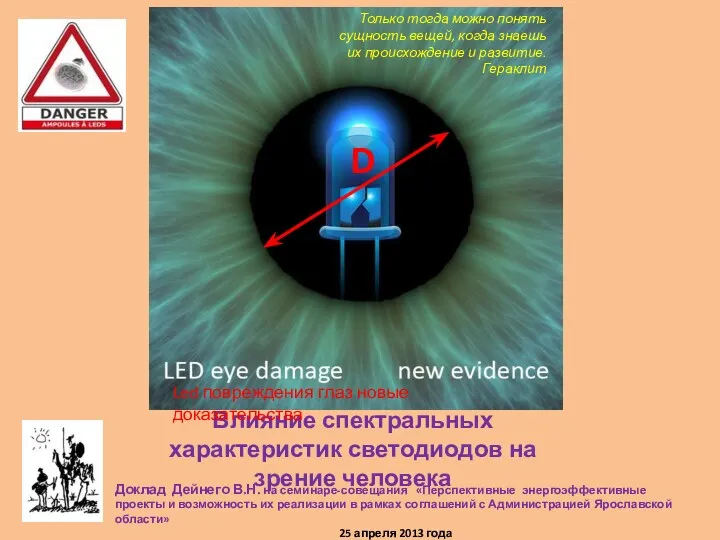 Влияние спектральных характеристик светодиодов на зрение человека
 Основные аллергены молока
 Кольцевидные тени. Рентгенопульмонология
 Бет-жақсүйек аймағының флегмоналар кезіндегі дренаждау (кәріздеу) тәсілдері. Көз аймағы
 Организация общей врачебной практики в городах и в сельской местности
 Острая дыхательная недостаточность
 Гломерулонефрит. Патогенез. Классификация
 Гигиена рук медицинского персонала
 Санитарно-гигиеническое обучение
 Когнитивно–поведенческая психотерапия
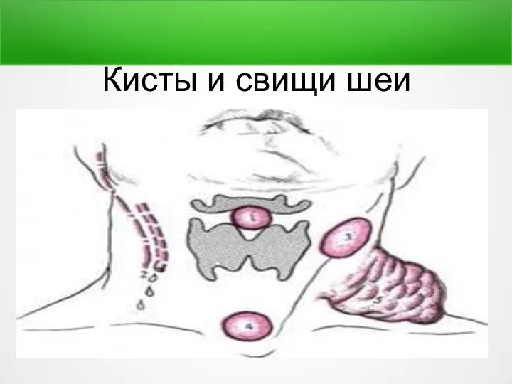 Кисты и свищи шеи
 Микроциркуляция. Транскапиллярный обмен. Лимфообращение
 Бактериальный бронхит