- general_myology

Содержание
- 2. Myology is the study of the muscular system, including the study of the structure, function and
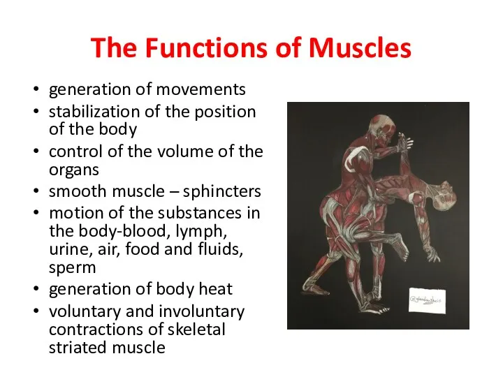
- 3. The Functions of Muscles generation of movements stabilization of the position of the body control of
- 4. 3 types of muscle tissue Smooth muscle tissue (textus muscularis levis) Striated muscle tissue (textus muscularis
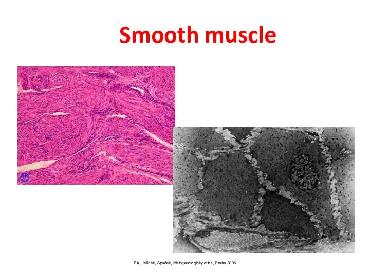
- 5. Eis, Jelínek, Špaček, Histopatologický atlas, Praha 2006 Smooth muscle
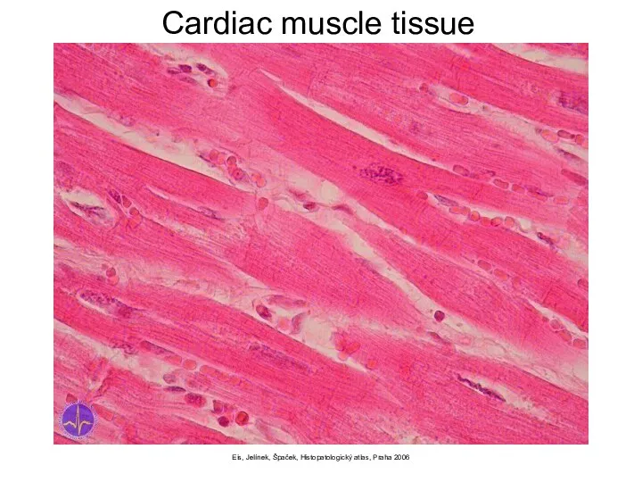
- 6. Eis, Jelínek, Špaček, Histopatologický atlas, Praha 2006 Cardiac muscle tissue
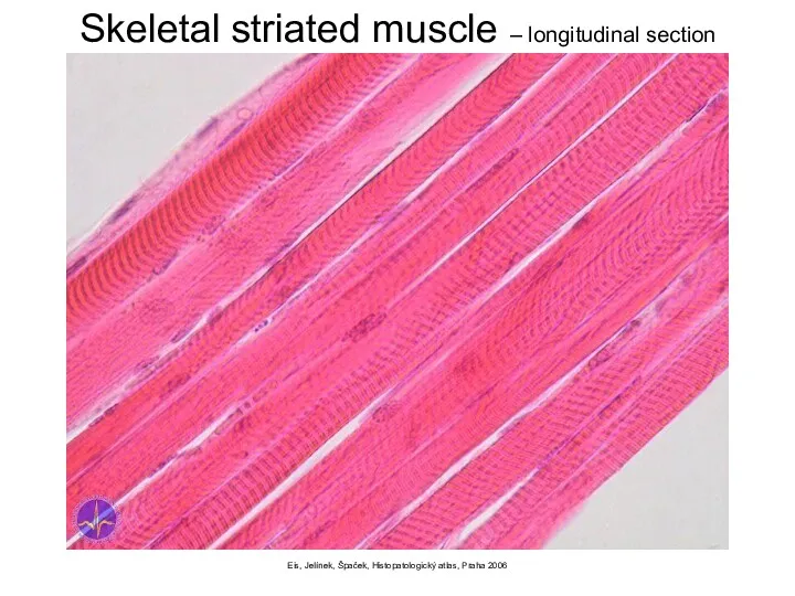
- 7. Eis, Jelínek, Špaček, Histopatologický atlas, Praha 2006 Skeletal striated muscle – longitudinal section
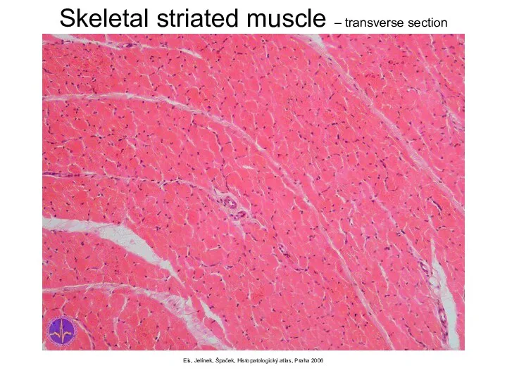
- 8. Eis, Jelínek, Špaček, Histopatologický atlas, Praha 2006 Skeletal striated muscle – transverse section
- 9. Skeletal striated muscle Myoglobin (pigment causing red colouring) Fibres Quick quickly fatigued light (white) in superficial
- 10. Skeletal striated muscle myofibre (myofibra) Elementary structural unit Multinucleated thickness: 10–100 µm length: mm – cm
- 11. http://www.baileybio.com/plogger/?level=picture&id=264 http://www.bms.ed.ac.uk/research/others/smaciver/Myosin%20II.htm
- 12. Functions of skeletal muscle Movement of animal body 2. Control of body openings and passages "maintain
- 13. Basic muscle structure striated muscle fibres special muscle structures primary muscle bundle 10-100 fibres connected and
- 14. Basic muscle structure fibrous tissue endomysium (perimysium internum) covers myofibres and bundles epimysium (perimysium externum) =
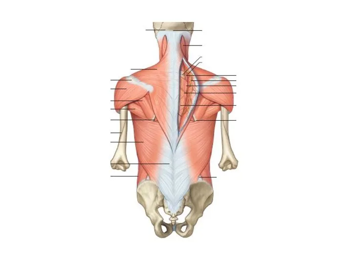
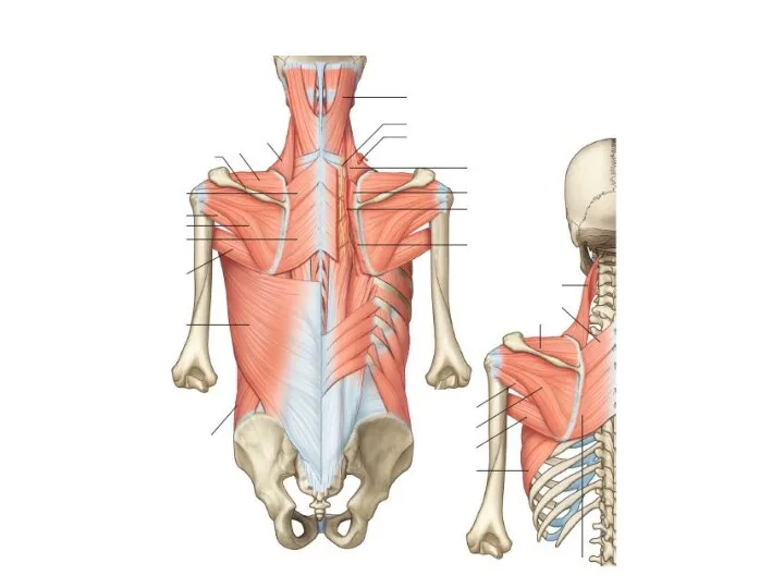
- 15. The parts of muscles origin (origo) mobile end (punctum fixum) head (caput musculi) belly (venter musculi)
- 16. Classification of skeletal muscles by form
- 17. Classification of skeletal muscles by movement agonists in the same direction acting muscles antagonists counteracting muscles
- 18. Classification of skeletal muscles by number of joints one-jointed muscles they´re causing the movement only in
- 19. Classification of skeletal muscles by the direction of movement flexor (m. flexor) makes the angle in
- 20. The work of muscles Dynamic-work in which muscles move parts of a person’s body, and the
- 21. Punctum fixum is a point, which is not moving during a contraction of a muscle, ie
- 22. Special muscle structures fascia (= perimysium externum) fibrous envelope of muscle or muscle group barrier for
- 23. Fasciotomy http://lifeinthefastlane.com/ortho-library/compartment-syndrome/
- 24. Special muscle structures tendon (tendo) strip of tough fibrous connective tissue composed of bundles of collagenous
- 25. Special muscle structures bursae mucosae pouches in the vincinity of the joints, tendons and muscles lined
- 35. EMG (electromyography) detection of the superficial muscle or the intramuscular activity detects the change of electrical
- 36. Functional muscle test informs us about the muscle strength helps to assess the extent and location
- 37. spasm – involuntary contraction of one muscle cramp – painful spasm tetanus – multiple spasms of
- 39. Скачать презентацию
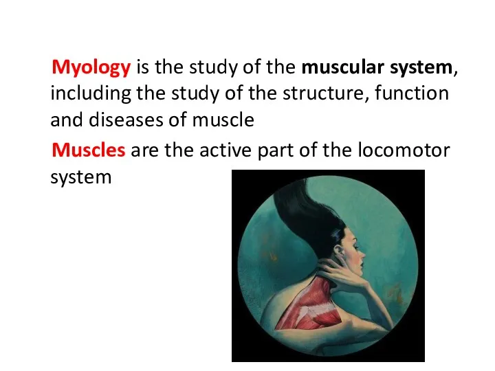
Myology is the study of the muscular system, including the study of
Myology is the study of the muscular system, including the study of
The Functions of Muscles
generation of movements
stabilization of the position of the
The Functions of Muscles
generation of movements
stabilization of the position of the
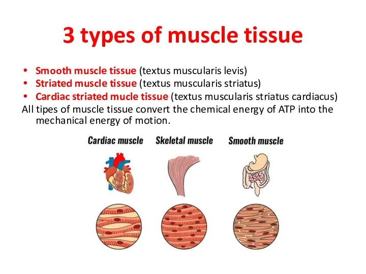
3 types of muscle tissue
Smooth muscle tissue (textus muscularis levis)
Striated muscle
3 types of muscle tissue
Smooth muscle tissue (textus muscularis levis)
Striated muscle
Eis, Jelínek, Špaček, Histopatologický atlas, Praha 2006
Smooth muscle
Eis, Jelínek, Špaček, Histopatologický atlas, Praha 2006
Smooth muscle
Eis, Jelínek, Špaček, Histopatologický atlas, Praha 2006
Cardiac muscle tissue
Eis, Jelínek, Špaček, Histopatologický atlas, Praha 2006
Cardiac muscle tissue
Eis, Jelínek, Špaček, Histopatologický atlas, Praha 2006
Skeletal striated muscle – longitudinal
Eis, Jelínek, Špaček, Histopatologický atlas, Praha 2006
Skeletal striated muscle – longitudinal
Eis, Jelínek, Špaček, Histopatologický atlas, Praha 2006
Skeletal striated muscle – transverse
Eis, Jelínek, Špaček, Histopatologický atlas, Praha 2006
Skeletal striated muscle – transverse
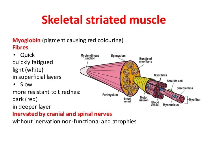
Skeletal striated muscle
Myoglobin (pigment causing red colouring)
Fibres
Quick
quickly fatigued
light (white)
in superficial layers
Slow
more
Skeletal striated muscle
Myoglobin (pigment causing red colouring)
Fibres
Quick
quickly fatigued
light (white)
in superficial layers
Slow
more
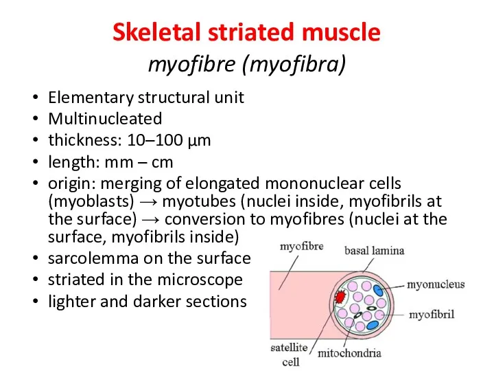
Skeletal striated muscle
myofibre (myofibra)
Elementary structural unit
Multinucleated
thickness: 10–100 µm
length: mm
Skeletal striated muscle
myofibre (myofibra)
Elementary structural unit
Multinucleated
thickness: 10–100 µm
length: mm
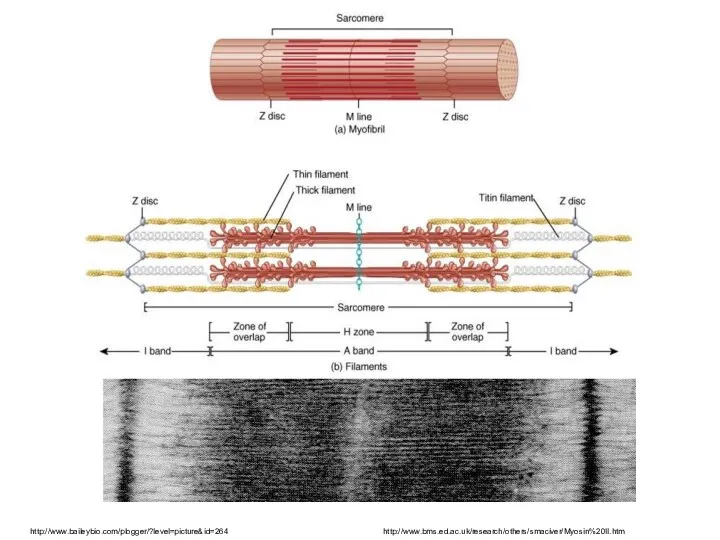
http://www.baileybio.com/plogger/?level=picture&id=264
http://www.bms.ed.ac.uk/research/others/smaciver/Myosin%20II.htm
http://www.baileybio.com/plogger/?level=picture&id=264
http://www.bms.ed.ac.uk/research/others/smaciver/Myosin%20II.htm
Functions of skeletal muscle
Movement of animal body
2. Control of body openings
Functions of skeletal muscle
Movement of animal body
2. Control of body openings
Basic muscle structure
striated muscle fibres
special muscle structures
primary muscle bundle
10-100 fibres
Basic muscle structure
striated muscle fibres
special muscle structures
primary muscle bundle
10-100 fibres
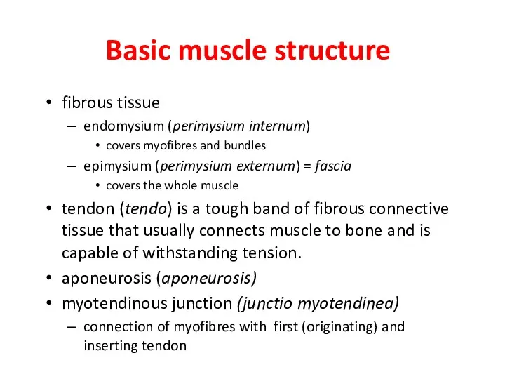
Basic muscle structure
fibrous tissue
endomysium (perimysium internum)
covers myofibres and bundles
epimysium (perimysium
Basic muscle structure
fibrous tissue
endomysium (perimysium internum)
covers myofibres and bundles
epimysium (perimysium
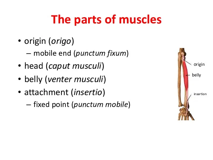
The parts of muscles
origin (origo)
mobile end (punctum fixum)
head (caput musculi)
belly (venter
The parts of muscles
origin (origo)
mobile end (punctum fixum)
head (caput musculi)
belly (venter
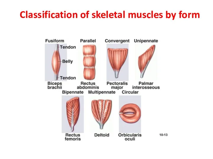
Classification of skeletal muscles by form
Classification of skeletal muscles by form
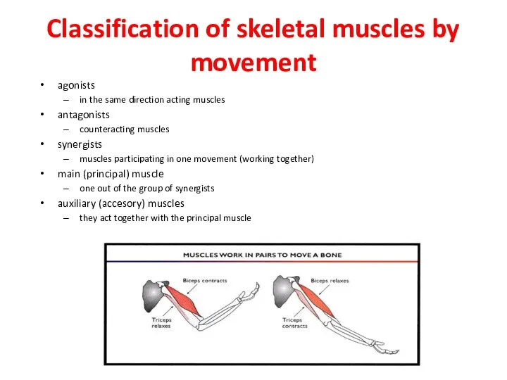
Classification of skeletal muscles by movement
agonists
in the same direction acting muscles
antagonists
counteracting
Classification of skeletal muscles by movement
agonists
in the same direction acting muscles
antagonists
counteracting
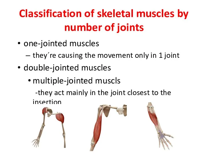
Classification of skeletal muscles by number of joints
one-jointed muscles
they´re causing the
Classification of skeletal muscles by number of joints
one-jointed muscles
they´re causing the
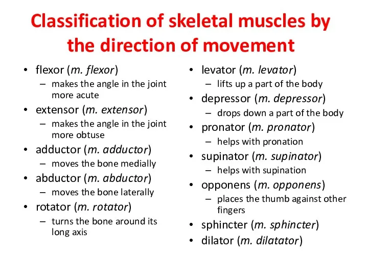
Classification of skeletal muscles by the direction of movement
flexor (m. flexor)
makes
Classification of skeletal muscles by the direction of movement
flexor (m. flexor)
makes
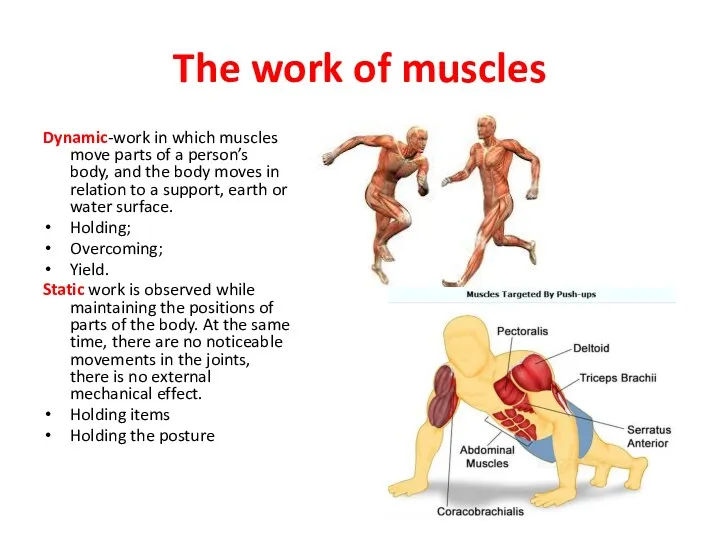
The work of muscles
Dynamic-work in which muscles move parts of a
The work of muscles
Dynamic-work in which muscles move parts of a
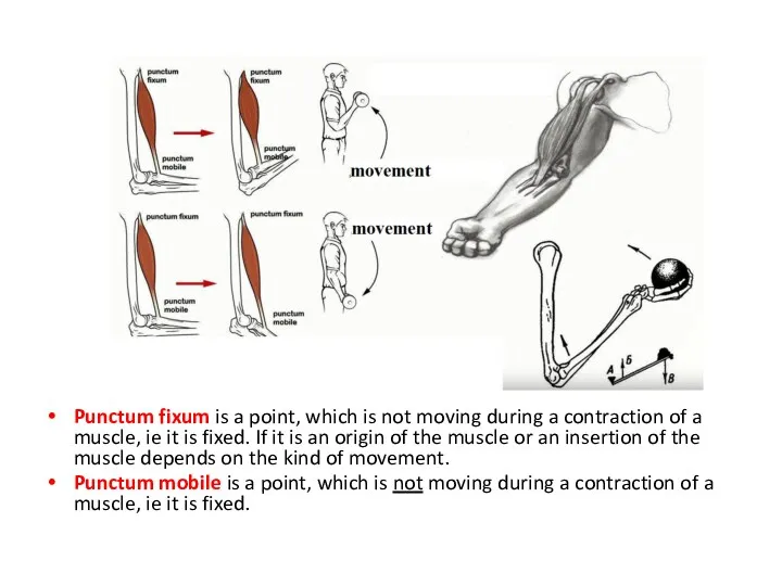
Punctum fixum is a point, which is not moving during a
Punctum fixum is a point, which is not moving during a
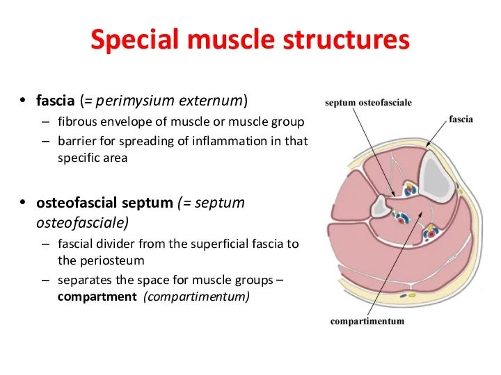
Special muscle structures
fascia (= perimysium externum)
fibrous envelope of muscle or muscle
Special muscle structures
fascia (= perimysium externum)
fibrous envelope of muscle or muscle
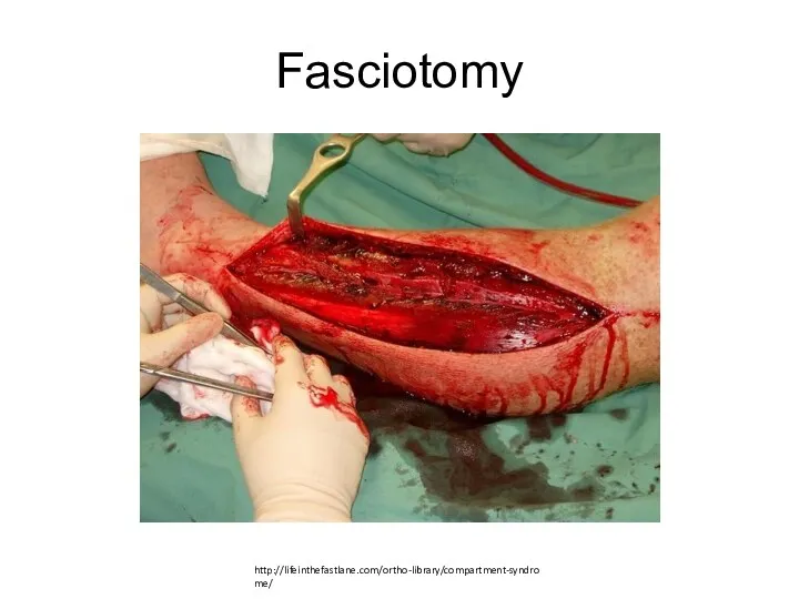
Fasciotomy
http://lifeinthefastlane.com/ortho-library/compartment-syndrome/
Fasciotomy
http://lifeinthefastlane.com/ortho-library/compartment-syndrome/
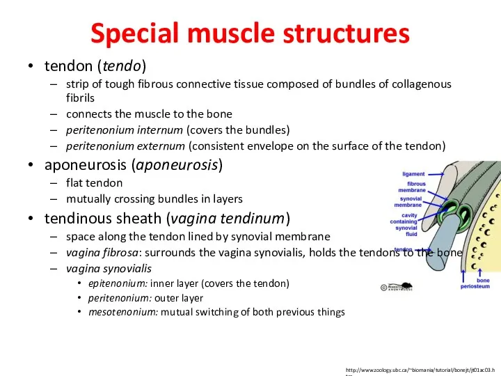
Special muscle structures
tendon (tendo)
strip of tough fibrous connective tissue composed of
Special muscle structures
tendon (tendo)
strip of tough fibrous connective tissue composed of
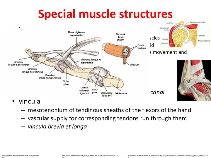
Special muscle structures
bursae mucosae
pouches in the vincinity of the joints, tendons
Special muscle structures
bursae mucosae
pouches in the vincinity of the joints, tendons
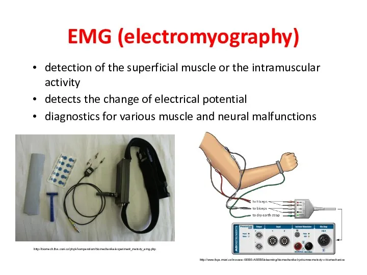
EMG (electromyography)
detection of the superficial muscle or the intramuscular activity
detects the
EMG (electromyography)
detection of the superficial muscle or the intramuscular activity
detects the
Functional muscle test
informs us about the muscle strength
helps to assess the
Functional muscle test
informs us about the muscle strength
helps to assess the
spasm – involuntary contraction of one muscle
cramp – painful spasm
tetanus –
spasm – involuntary contraction of one muscle
cramp – painful spasm
tetanus –
 Когда появилась одежда. (1 класс)
 Регистры. Счетчики
 Polymer Flooding for Enhanced Oil Recovery
 Автоматизация и механизация процессов листовой штамповки
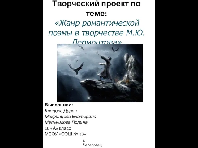 Творческий проект по теме: Жанр романтической поэмы в творчестве М.Ю. Лермонтова
 Аллергия. Аллергены
 Соединения на гвоздях
 Зима в картинах русских художников
 Автоматизация производства. ИП Акулов Николай Николаевич
 Атмосферные явления
 Религиозные праздники России в XVI веке
 Комплаентность пациента с артериальной гипертензией. Роль фельдшера
 Автоматизированные системы для мониторинга газовых выбросов из фиксированных источников
 Біблійна антропологія. Вчення про людину – розділ систематичної теології
 Залізничний транспорт
 Приоритеты в организации и содержании управления на основе выявленных проблем системы образования города Новосибирска
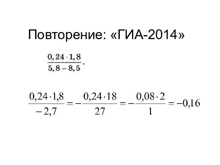 Повторение: подготовка к ГИА по математике: алгебра, геометрия, теория вероятностей (8, 9 класс) Диск Диск Диск Диск
 Принцесса Грёз или Женщина - Гражданка О чем писали женские журналы в 1917 году
 урок с презентацией химия 9класс Предмет органической химии
 Презентация Игрушки из скрученных полосок
 Возбуждение и рассмотрение дела об административном правонарушении
 Коммутация и программирование щитов
 Понятие о методах обучения и их классификация
 Сети. Интернет. Протоколы
 Шоколадное печенье в виде футбольного мяча
 Итоги обучения в 3 классе!
 Ішекті өңдеу. Ішек компектісі туралы түсінік. (Дәріс 11-12)
 Умножение многочленов. Формулы сокращенного умножения