Слайд 2
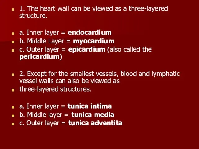
1. The heart wall can be viewed as a three-layered structure.
a.
Inner layer = endocardium
b. Middle Layer = myocardium
c. Outer layer = epicardium (also called the pericardium)
2. Except for the smallest vessels, blood and lymphatic vessel walls can also be viewed as
three-layered structures.
a. Inner layer = tunica intima
b. Middle layer = tunica media
c. Outer layer = tunica adventita
Слайд 3
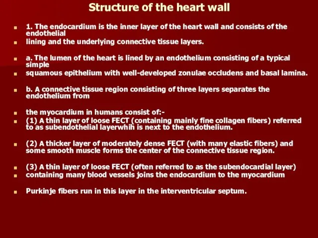
Structure of the heart wall
1. The endocardium is the inner layer
of the heart wall and consists of the endothelial
lining and the underlying connective tissue layers.
a. The lumen of the heart is lined by an endothelium consisting of a typical simple
squamous epithelium with well-developed zonulae occludens and basal lamina.
b. A connective tissue region consisting of three layers separates the endothelium from
the myocardium in humans consist of:-
(1) A thin layer of loose FECT (containing mainly fine collagen fibers) referred to as subendothelial layerwhih is next to the endothelium.
(2) A thicker layer of moderately dense FECT (with many elastic fibers) and some smooth muscle forms the center of the connective tissue region.
(3) A thin layer of loose FECT (often referred to as the subendocardial layer)
containing many blood vessels joins the endocardium to the myocardium
Purkinje fibers run in this layer in the interventricular septum.
Слайд 4
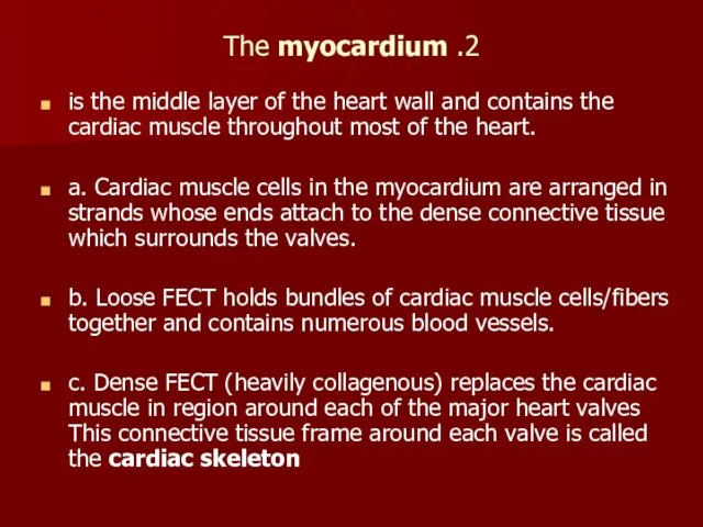
2. The myocardium
is the middle layer of the heart wall and
contains the cardiac muscle throughout most of the heart.
a. Cardiac muscle cells in the myocardium are arranged in strands whose ends attach to the dense connective tissue which surrounds the valves.
b. Loose FECT holds bundles of cardiac muscle cells/fibers together and contains numerous blood vessels.
c. Dense FECT (heavily collagenous) replaces the cardiac muscle in region around each of the major heart valves This connective tissue frame around each valve is called the cardiac skeleton
Слайд 5
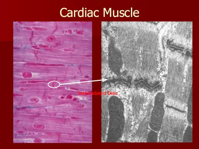
Cardiac Muscle
Intercalated Disc
Слайд 6
The epicardium
is the outer layer of the heart and consists of
a connective tissue region covered by a mesothelium on its outer surface.
a. The connective tissue region consists of three layers in humans.
(1) The inner two regions are referred to collectively as the subepicardial layer and contain large blood vessels (coronary vessels), nerves, and varying amounts of adipose tissue.
(a) A thin layer of loose FECT lies next to the myocardium.
(b) A thicker layer of slightly denser FECT lies outside the loose FECT layer.
(2) A thin layer of loose FECT with many elastic fibers connects the connective tissue layers of the epicardium to the mesothelial covering.
Слайд 7
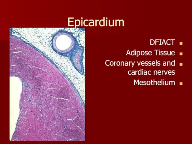
Epicardium
DFIACT
Adipose Tissue
Coronary vessels and cardiac nerves
Mesothelium
Слайд 8
b. A mesothelium (simple squamous epithelium) covers the outer surface of
the heart (except where the arteries leave and the great veins enter the heart). This covering epithelium closely resembles the mesothelial covering of the other thoracic and abdominal organs.
B. The thickness of the heart wall and the thickness of the layers within the heart wall varies with location.
1. The myocardium is thickest in the ventricular region, especially the left ventricle, and contains more cardiac muscle in the ventricles than in the atrium. The myocardium around the valves contains only dense collagenous CT which forms the cardiac skeleton.
2. The endocardium and epicardium are thinner in the ventricles than in the atria
In the atria, the cardiac muscle cells contain small granules (called atrial specific granules) in the perinuclear sarcoplasm which can be observed with TEM. These granules are the source of atrial natriuretic peptide (ANP), a hormone which influences blood pressure by affecting kidney function
Слайд 9
Special features of the heart
1. Valves are out growths from the
endocardium which prevent backflow
of blood. Valves contain three components.
.
2. The cardiac skeleton supports each of the heart valves. Cardiac
muscle in the myocardium is replaced by dense regular FECT (heavily collagenous)
3. Cardiac muscle fibers in the atria and ventricles are highly organized.
a. Cardiac muscle cells are attached end-to-end in branching strands.
b. The ends of most strands of cardiac muscle fibers are attached to the cardiac skeleton
Слайд 10
"Pacemakers"
in the heart are modified cardiac muscle cells.
a. Cardiac muscle
cells in the myocardium of the sinoatrial (SA) node are modified to serve as the pacemaker region. The plasma membrane of the cells has a high leakage rate, giving them the fastest intrinsic contraction rate among the populations
b. Cardiac muscle cells in the atrioventricular (AV) node have a similar histological appearance, but have a lower intrinsic rate of contraction, so these cells do not normally act as a pacemaker region. These cells receive the wave of excitation from the cardiac muscle of the atria and pass the excitation on to the bundle of His.
Слайд 11
The impulse-conducting system
which connects the atria with the ventricles serves
several functions.
a.
The impulse conducting system is made up of a series of Purkinje fibers which are specialized cardiac muscle cells.
(1) Purkinje fibers are organized into a branched bundle (Bundle of His) which
extends from the atrio-ventricular (AV) node, through the interventricular
septum down to the apex of the ventricles.
(2) Purkinje fibers are attached (by intercalated disks) to cardiac muscle cells in the
myocardium at the apex of the ventricles and along outer walls of both
ventricles
b. The impulse conducting system improves heart function in two ways
Слайд 12
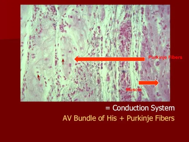
Conduction System =
AV Bundle of His + Purkinje Fibers
Purkinje Fibers
Muscle
Слайд 13
Microanatomy of Blood Vessels
Most larger blood vessel walls contain three major
layers with sublayering.
1. The tunica intima is the luminal layer.
a. The lumen is lined by an endothelium of simple squamous epithelium.
b. A subendothelial layer of loose FECT is present in most medium to large vessels
and may contain scattered smooth muscle in larger vessels.
2. An internal elastic lamina (elastica interna) marks the boundary between the tunica
intima and the tunica media.
3. The tunica media contains layers of either elastic laminae/lamellae (fenestrated sheets) or FECT alternating with layers of smooth muscle.
4. If present, the external elastic lamina (elastica externa) marks the boundary between
the tunica media and the tunica adventita.
5. The tunica adventita contains loose to moderately dense FECT, +/- scattered smooth
muscle cells. Small and medium arteries and veins are present in the tunica adventitia of large arteries and veins
Слайд 14
Large arteries (also called elastic arteries or conducting arteries)
include the aorta
and its largest main branches.
(a. Tunica intima - thin (relative to other layers in this type of vessel)
(1) Endothelium
(2) Subendothelial layer contains some smooth muscle, elastic fibers, collagen fibers
b. Internal elastic lamina - not as distinct as in other arteries
c. Tunica media - thick
(1) 40 - 60 distinct, concentrically arranged elastic laminae
(2) Between elastic laminae - fibroblasts, elastic fibers, collagen fibers, spiral (to circular) smooth muscle
d. Tunica adventita - thin; consists mainly of collagen fibers, blood vessels, nerves; some elastic fibers, fibroblasts, macrophages may also be present
2. Function = to conduct blood from the heart to smaller arteries and to even out blood pressure and flow. The presence of elastic laminae gives these vessels elastic properties. They expand as the heart contracts (to modulate blood pressure and store energy) and recoil during ventricular relaxation (to maintain more even pressure in large arteries).
Слайд 15
Medium to small arteries (also called muscular arteries)
Tunica intima - thin
(1)
Endothelium
(2) Thin subendothelial layer consisting of scattered fine collagen and elastic fibers and a few fibroblasts
b. Internal elastic lamina - very distinct, usually folded
c. Tunica media - thick
(1) Circular smooth muscle, 5 - 40 layers
(2) Small amount of CT with collagen fibers and elastic fibers (longitudinal orientation) between muscle
(3) Thickness decreases as diameter of vessel decreases
d. External elastic lamina (May be indistinct in smaller muscular arteries)
e. Tunica adventita - thick; loose FECT
2. Function - to distribute blood to smaller arterial vessels. The muscular wall resists damage due to relatively high blood pressure in these vessels
Слайд 16
Arterioles
1. Structure
a. Tunica intima - very thin consisting only of
endothelium
b. Internal elastic lamina - usually present except in smaller arterioles
c. Tunica media - 1 to 5 layers of smooth muscle, some elastic fibers
d. Tunica adventita - thin, consisting of longitudinally arranged collagen and elastic
fibers
2. Function - to redistribute blood flow to capillaries and to alter blood pressure by altering peripheral resistance to blood flow. Arterioles can change diameter very drastically therefore affecting blood pressure and flow patterns. Arterioles are referred to as peripheral resistance vessels.
Слайд 17
Capillaries
1. Structure - consist only of endothelium, but may be partially
surrounded by pericytes.
Three types of capillaries may be distinguished
.
a. Continuous (type I) capillaries have relatively thick cytoplasm and
the capillary wall is continuous. Lateral cell surfaces of cells are characterized by
zonula occludens (tight junctions), so materials move across cells via pinocytosis or
diffusion. These capillaries occur in most organs.
b. Fenestrated (type II) capillaries (Figure 13.18) have extremely thin cytoplasm and
the capillary wall is perforated at intervals by pores or fenestrations. Lateral cell
surfaces are characterized by zonula occludens (tight junctions). Materials
apparently cross the cells through the fenestrations. These capillaries are found in
the kidney and in endocrine glands.
c. Sinusoidal capillaries are larger in diameter than the other types and have wide
spaces between the lateral edges of the adjacent endothelial cells, so materials
(and some cells) can move freely in and out of the capillary. Sinusoidal capillaries
are found in the spleen, liver, and bone marrow.
2. Functions
a. Capillaries are the site of normal exchange of materials between blood and tissue
fluid.
b. Capillaries may be a site of exit of WBCs from blood into tissue under some conditions, although this is probably more frequent in venules.
Слайд 18
Venules
Size varies from 10 microns (post-capillary venules) to 1 mm (muscular
venules)
2. Post-capillary venules
a. Structure - larger diameter than capillaries; consist of endothelium surrounded by pericytes
b. Functions
(1) Collect blood from capillaries
(2) Respond to vasoactive agents (e.g., histamine, serotonin) by altering permeability
(3) Also a site of exchange of materials between tissue fluid and blood
(4) Site of exit of WBCs from blood into tissue
Слайд 19
Larger muscular venules
a. Structure
(1) Tunica intima - thin; endothelium surrounded by
outer sheath of collagen fibers
(2) Tunica media - thin; 1 - 3 layers of smooth muscle (circular) with collagen and elastic fibers between muscles
(3) Tunica adventita - thick; loose FECT containing longitudinal collagen fibers and scattered elastic fibers and fibroblasts
b. Function - to collect blood from post-capillary venules
Слайд 20
Small to medium veins
1. Structure
a. Tunica intima - thin
(1) Endothelium
(2) Thin
subendothelial layer
(3) May be folded to form valves
b. Tunica media - thin; circular smooth muscle, collagen fibers, some elastic fibers
c. Tunica adventita - well developed; loose FECT with longitudinally arranged collagen and elastic fibers, bundles of longitudinal smooth muscle
2. Function - to collect blood from smaller venous vessels
Слайд 21
Large veins - vena cavae and larger branches
1. Structure
a. Tunica intima
- thicker
(1) Endothelium
(2) Thin subendothelial layer
b. Internal elastic lamina - usually distinguishable
c. Tunica media - thin, poorly developed; mostly FECT; little smooth muscle
d. Tunica adventita - very thick; moderately dense FECT with spirally arranged collagen fibers, elastic laminae, longitudinal smooth muscle
2. Function - to collect blood from medium sized veins and return it to heart
Слайд 22
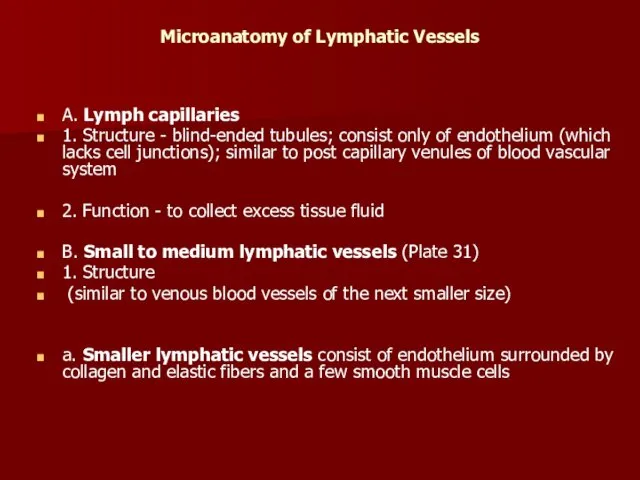
Microanatomy of Lymphatic Vessels
A. Lymph capillaries
1. Structure - blind-ended tubules; consist
only of endothelium (which lacks cell junctions); similar to post capillary venules of blood vascular system
2. Function - to collect excess tissue fluid
B. Small to medium lymphatic vessels (Plate 31)
1. Structure
(similar to venous blood vessels of the next smaller size)
a. Smaller lymphatic vessels consist of endothelium surrounded by collagen and elastic fibers and a few smooth muscle cells
Слайд 23
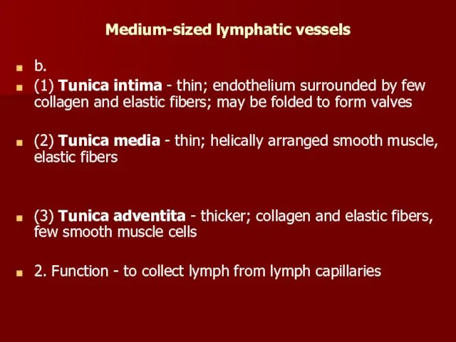
Medium-sized lymphatic vessels
b.
(1) Tunica intima - thin; endothelium surrounded by
few collagen and elastic fibers; may be folded to form valves
(2) Tunica media - thin; helically arranged smooth muscle, elastic fibers
(3) Tunica adventita - thicker; collagen and elastic fibers, few smooth muscle cells
2. Function - to collect lymph from lymph capillaries

 Презентация. Десять заповедей Януша Корчака для родителей
 Вербальные и невербальные коммуникации
 Каталитикалық риформинг қондырғысының реакторлар блогы
 Общий наркоз при лечении кариеса зубов у детей
 Urban style
 Разработка автоматизированной системы управления технологическим процессом получения горячей сетевой воды
 Презентация Кузьма Минин - великий гражданин России.
 Родовые понятия и методологические основания социологии Эмиля Дюркгейма в цитатах
 На звездной орбите. История праздника 12 апреля
 Цветная металлургия
 Внеклассное мероприятие в начальных классах к 8 марта Милые,добрые, нежные- ВАМ!
 Формы глагола Be в настоящем простом времени. GRAMMAR
 Палеозойская эра
 20230713_05._tehnicheskoe_konstruirovanie_i_modelirovanie
 Режимы работы редактора Word
 Общая характеристика спортивной подготовки
 Презентация: Столица моей родины - Москва
 Упражнения для развития памяти и внимания на уроках иностранного языка
 Российский университет дружбы народов Комиссия по связям с общественностью
 Презентация к проекту Что в имени моем...
 Внеклассные мероприятия
 Настоящее совершенное время Present Perfect Tense
 Творчество ненецкого художника К.Л.Панкова. Презентация
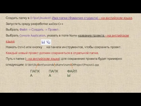 Создание программы
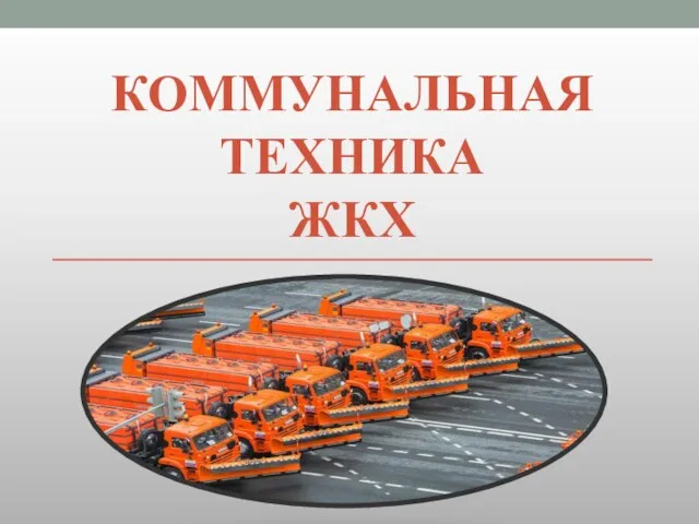 Коммунальная техника ЖКХ
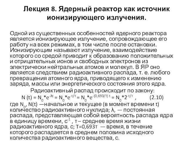 Ядерный реактор как источник ионизирующего излучения. Решение задач
 Магия намерения. Мужской и женский способ достижения желаемого
 Функциональные элементы САР ЭПС: исполнительные и управляющие элементы