- Optics of vision. Eye structure

Содержание
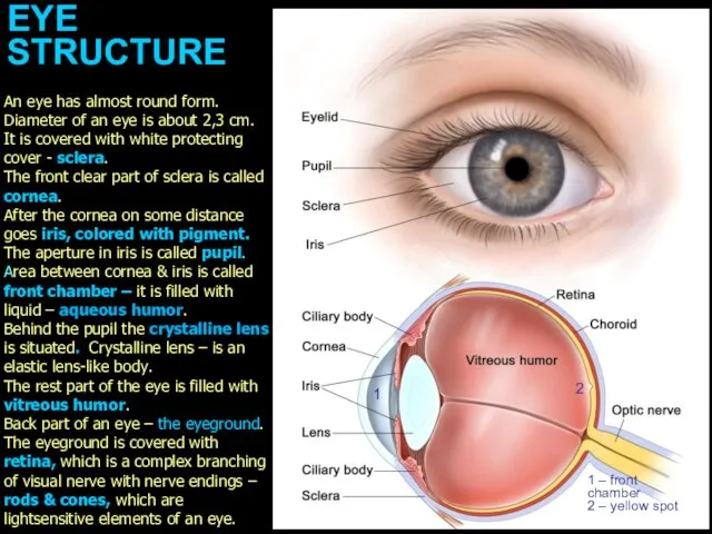
- 2. EYE STRUCTURE 1 1 – front chamber 2 – yellow spot An eye has almost round
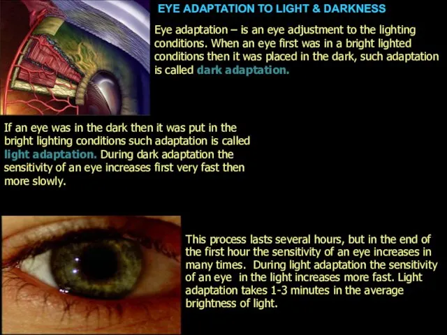
- 3. EYE ADAPTATION TO LIGHT & DARKNESS This process lasts several hours, but in the end of
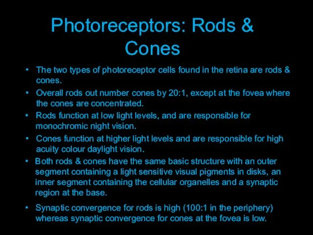
- 4. Photoreceptors: Rods & Cones Synaptic convergence for rods is high (100:1 in the periphery) whereas synaptic
- 5. Rods and cones.. ...are selective light transducers Dark pigment layer absorbs stray light & reduces reflection
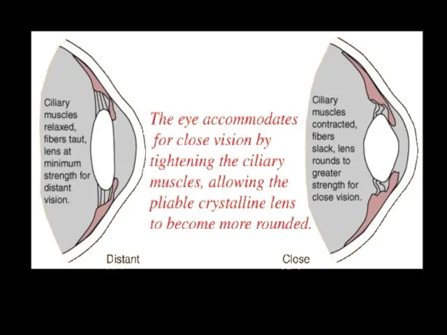
- 7. The cornea, clear liquid of front chamber, crystalline lens & vitreous humor are the optic system
- 9. Presbyopia: ciliary muscles can no longer contract as well; lens cannot be made round enough for
- 10. Length of Eyeball + Curvature of Cornea Emmetropia: objects focused on retina (normal)
- 11. Normal sight Myopia Myopia (nearsightedness): objects focused in front of retina
- 12. Length of Eyeball + Curvature of Cornea Myopia (nearsightedness): Axial myopia: eyeball too long (shown above)
- 13. concave lens: negative diopter; diffraction Myopia Correction laser surgery: remove corneal tissue in center to reduce
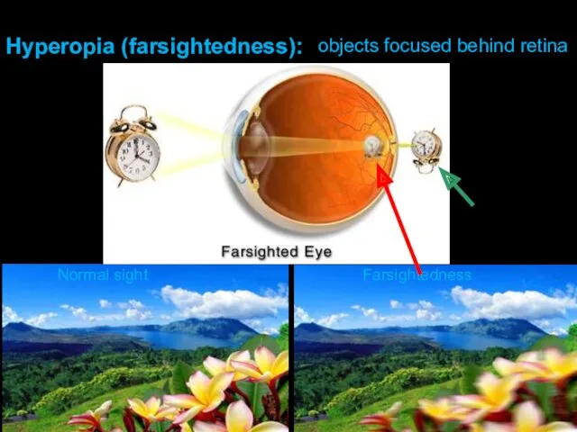
- 14. Normal sight Farsightedness Hyperopia (farsightedness): objects focused behind retina
- 15. convex lens: positive diopter; refraction Hyperopia Correction laser surgery: remove corneal tissue around sides to increase
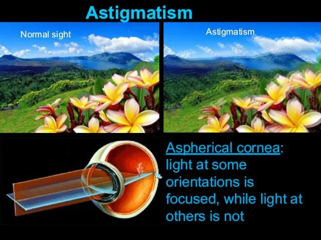
- 16. Normal sight Astigmatism Astigmatism Aspherical cornea: light at some orientations is focused, while light at others
- 18. Скачать презентацию
EYE
STRUCTURE
1
1 – front
chamber
2 – yellow spot
An eye has almost
EYE
STRUCTURE
1
1 – front
chamber
2 – yellow spot
An eye has almost
EYE ADAPTATION TO LIGHT & DARKNESS
This process lasts several hours,
EYE ADAPTATION TO LIGHT & DARKNESS
This process lasts several hours,
Photoreceptors: Rods & Cones
Synaptic convergence for rods is high (100:1 in
Photoreceptors: Rods & Cones
Synaptic convergence for rods is high (100:1 in
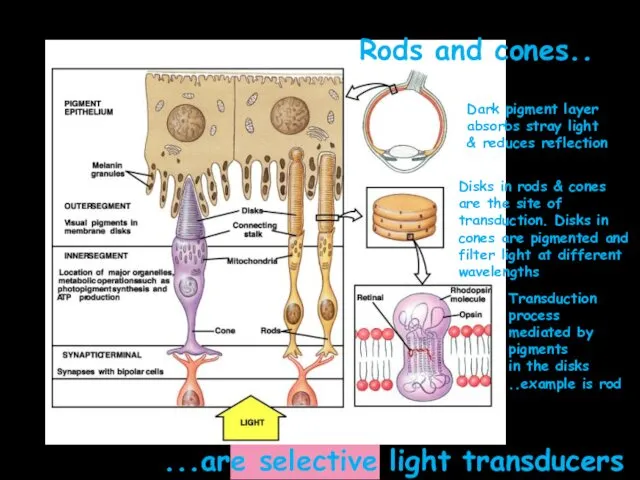
Rods and cones..
...are selective light transducers
Dark pigment layer
absorbs stray light
& reduces
Rods and cones..
...are selective light transducers
Dark pigment layer
absorbs stray light
& reduces
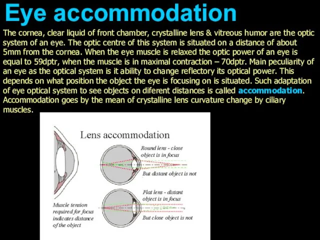
The cornea, clear liquid of front chamber, crystalline lens & vitreous
The cornea, clear liquid of front chamber, crystalline lens & vitreous
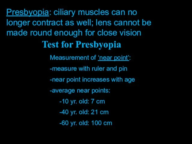
Presbyopia: ciliary muscles can no longer contract as well; lens cannot
Presbyopia: ciliary muscles can no longer contract as well; lens cannot
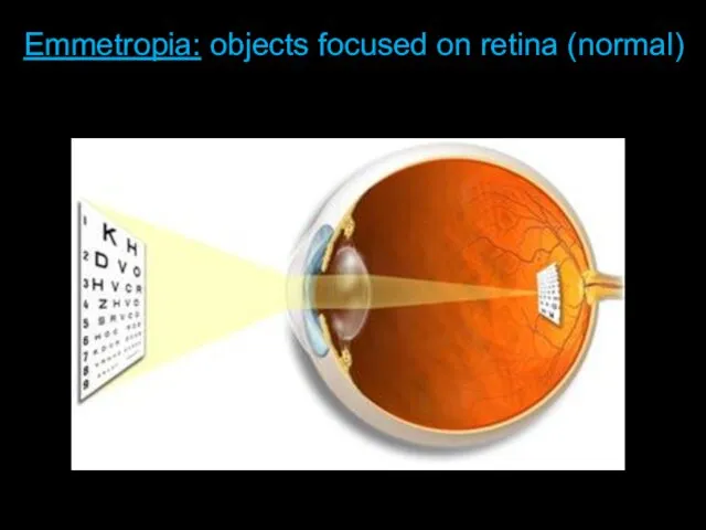
Length of Eyeball + Curvature of Cornea
Emmetropia: objects focused on retina
Length of Eyeball + Curvature of Cornea
Emmetropia: objects focused on retina
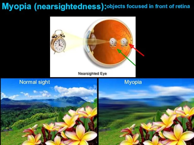
Normal sight
Myopia
Myopia (nearsightedness):
objects focused in front of retina
Normal sight
Myopia
Myopia (nearsightedness):
objects focused in front of retina
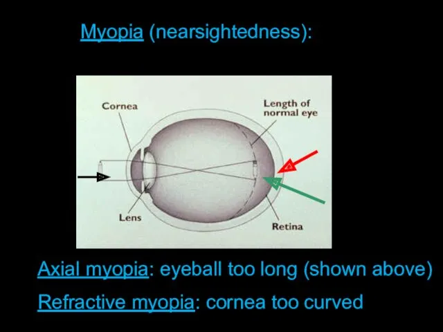
Length of Eyeball + Curvature of Cornea
Myopia (nearsightedness):
Axial myopia: eyeball too
Length of Eyeball + Curvature of Cornea
Myopia (nearsightedness):
Axial myopia: eyeball too
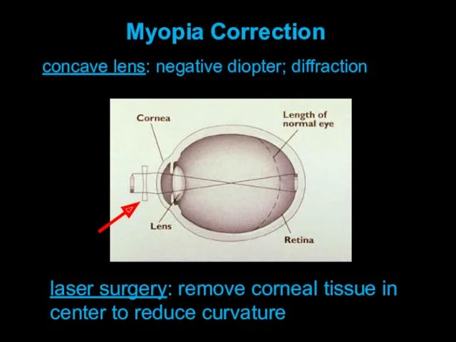
concave lens: negative diopter; diffraction
Myopia Correction
laser surgery: remove corneal tissue in
concave lens: negative diopter; diffraction
Myopia Correction
laser surgery: remove corneal tissue in
Normal sight
Farsightedness
Hyperopia (farsightedness):
objects focused behind retina
Normal sight
Farsightedness
Hyperopia (farsightedness):
objects focused behind retina
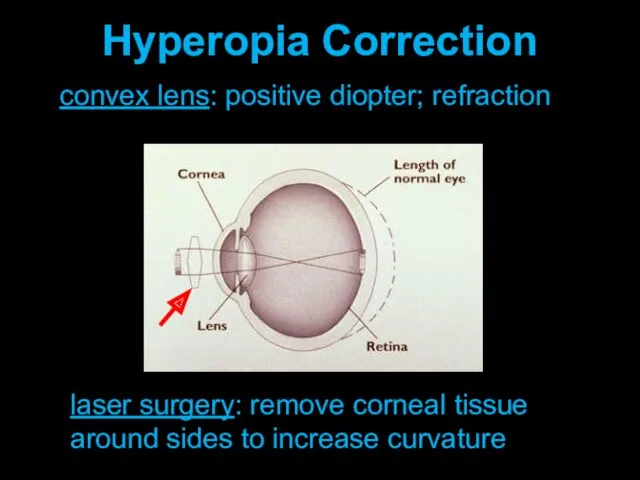
convex lens: positive diopter; refraction
Hyperopia Correction
laser surgery: remove corneal tissue around
convex lens: positive diopter; refraction
Hyperopia Correction
laser surgery: remove corneal tissue around
Normal sight
Astigmatism
Astigmatism
Aspherical cornea: light at some orientations is focused, while light
Normal sight
Astigmatism
Astigmatism
Aspherical cornea: light at some orientations is focused, while light
 Характеристика топливно-энергетической базы Крыма
 Вред курения
 Презентация. Летний оздоровительный лагерь.
 Анализ динамики экономических показателей России и США
 Биосфера. Среды жизни
 Аллергия. Аллергены
 Презентация Заповеди Блаженствпо предмету ОПК
 Облік, контроль і аналіз непрямих виробничих витрат
 Конспект внеклассного занятия на тему: Законы жизни класса.
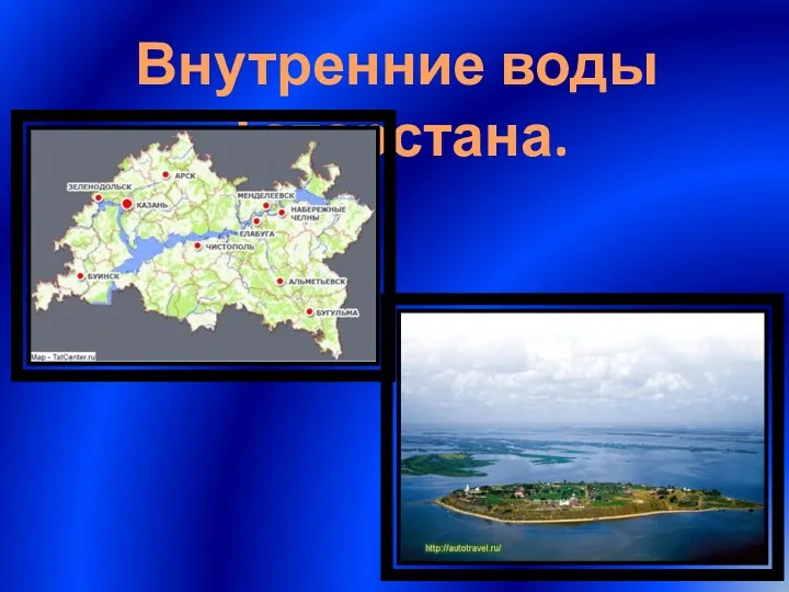 Внутренние воды РТ
 Формирование культурной среды небольшого города/села
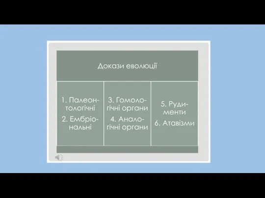 аналогічні-гомологічні органи
 Организаторская и воспитательная работа командира подразделения по укреплению воинской дисциплины. Тема № 5
 О мерах по поддержки генерирующих объектов на основе ВИЭ. Законодательная база поддержки генерации ВИЭ
 Интернет в жизни старшеклассника: за или против
 Тольятти. История любимого города
 Пейзаж — поэтичная и музыкальная живопись
 Особенности рельефа территории России
 Направления реализации Национальной стратегии по обращению с ТКО и ВМР
 ФЭМП 14.04.2020
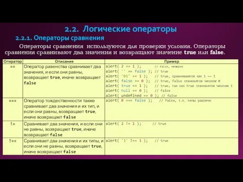 Логические операторы
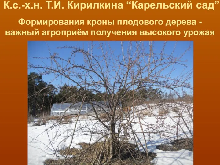 Обрезка яблони и груши
 Проектирование современного урока биологии, географии в соответствии с требованиями ФГОС
 Правовое регулирование предпринимательской деятельности
 Презентация для детей
 Миотоническая дистрофия Россолимо-Штейнерта-Куршманна-Баттена
 Плотность
 Праздники и календари. Основы мировых религиозных наук (4 класс)