- Anatomy of the human brain

Содержание
- 2. Anatomy of the human brain The Lateral Surface of the Brain The three major parts: the
- 3. Anatomy of the human brain Cerebral Lobes and the Insula The central sulcus divides the frontal
- 4. Anatomy of the human brain Selected Gyri, Sulci, and Fissures The surface of the human cerebrum
- 5. Anatomy of the human brain Selected Gyri, Sulci, and Fissures The postcentral gyrus lies immediately posterior
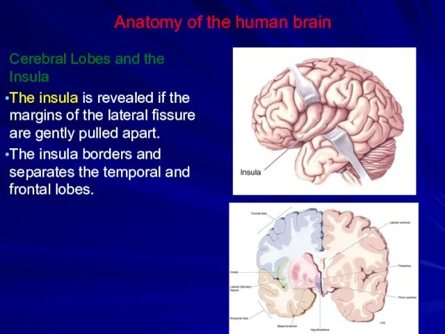
- 6. Anatomy of the human brain Cerebral Lobes and the Insula The insula is revealed if the
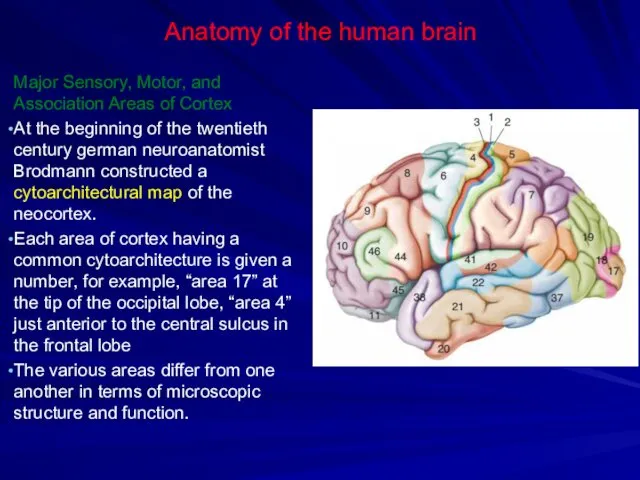
- 7. Anatomy of the human brain Major Sensory, Motor, and Association Areas of Cortex At the beginning
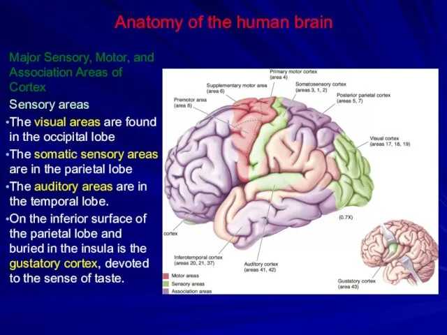
- 8. Anatomy of the human brain Major Sensory, Motor, and Association Areas of Cortex Sensory areas The
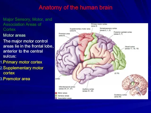
- 9. Anatomy of the human brain Major Sensory, Motor, and Association Areas of Cortex Motor areas The
- 10. Anatomy of the human brain Major Sensory, Motor, and Association Areas of Cortex The association areas
- 12. Anatomy of the human brain The Medial Surface of the Brain The brain stem consists of
- 13. Anatomy of the human brain Forebrain Structures Corpus callosum (connects the two sides of the cerebrum)
- 14. Anatomy of the human brain Forebrain Structures The amygdala is an important structure for regulating emotional
- 15. Anatomy of the human brain Ventricles the third ventricle the cerebral aqueduct the fourth ventricle the
- 16. Anatomy of the human brain Ventricles The lateral ventricles are paired structures that sprout like antlers
- 18. Anatomy of the human brain The Ventral Surface of the Brain the cranial nerves the optic
- 19. Anatomy of the human brain The cerebellum two hemispheres the vermis (midline region)
- 21. Anatomy of the human brain The brain stem the pineal body (involved in the regulation of
- 23. Cross-sectional anatomy of the brain Cross Section 1: Forebrain at Thalamus–Telencephalon Junction
- 24. Cross-sectional anatomy of the brain Cross Section 1: Forebrain at Thalamus–Telencephalon Junction (a) Gross Features the
- 25. Cross-sectional anatomy of the brain Cross Section 1: Forebrain at Thalamus–Telencephalon Junction (b) Selected Fiber Groups
- 26. Cross-sectional anatomy of the brain Cross Section 1: Forebrain at Thalamus–Telencephalon Junction (b) Selected Cell Groups
- 27. Cross-sectional anatomy of the brain Cross Section 2: Forebrain at Mid-Thalamus
- 28. Cross-sectional anatomy of the brain Cross Section 2: Forebrain at Mid-Thalamus (a) Gross Features As we
- 29. Cross-sectional anatomy of the brain Cross Section 2: Forebrain at Mid-Thalamus (b) Selected Cell Groups. the
- 30. Cross-sectional anatomy of the brain Cross Section 2: Forebrain at Mid-Thalamus (b) Selected Cell Groups. the
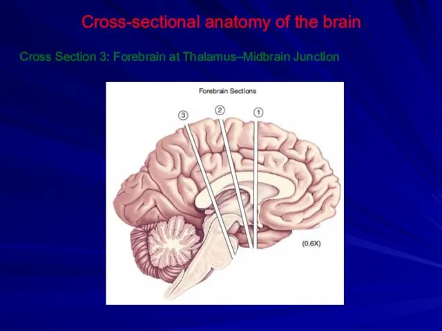
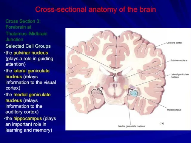
- 31. Cross-sectional anatomy of the brain Cross Section 3: Forebrain at Thalamus–Midbrain Junction
- 32. Cross-sectional anatomy of the brain Cross Section 3: Forebrain at Thalamus–Midbrain Junction Selected Cell Groups the
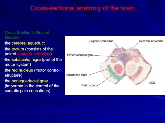
- 33. Cross-sectional anatomy of the brain Cross Section 4: Rostral Midbrain
- 34. Cross-sectional anatomy of the brain Cross Section 4: Rostral Midbrain the cerebral aqueduct the tectum (consists
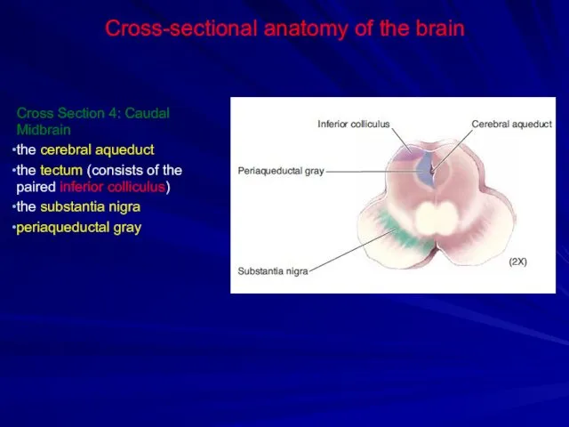
- 35. Cross-sectional anatomy of the brain Cross Section 5: Caudal Midbrain
- 36. Cross-sectional anatomy of the brain Cross Section 4: Caudal Midbrain the cerebral aqueduct the tectum (consists
- 37. Cross-sectional anatomy of the brain Cross Section 6: Pons and Cerebellum
- 38. Cross-sectional anatomy of the brain Cross Section 6: Pons and Cerebellum pontine nuclei (the input to
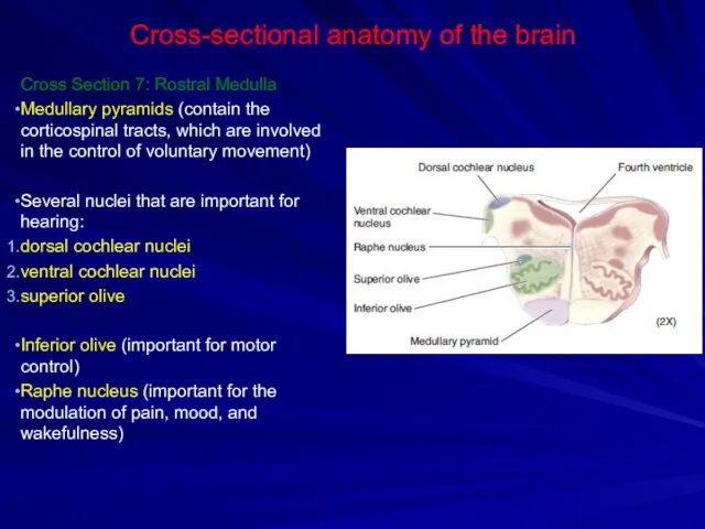
- 39. Cross-sectional anatomy of the brain Cross Section 7: Rostral Medulla
- 40. Cross-sectional anatomy of the brain Cross Section 7: Rostral Medulla Medullary pyramids (contain the corticospinal tracts,
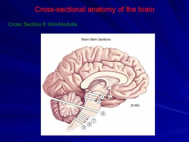
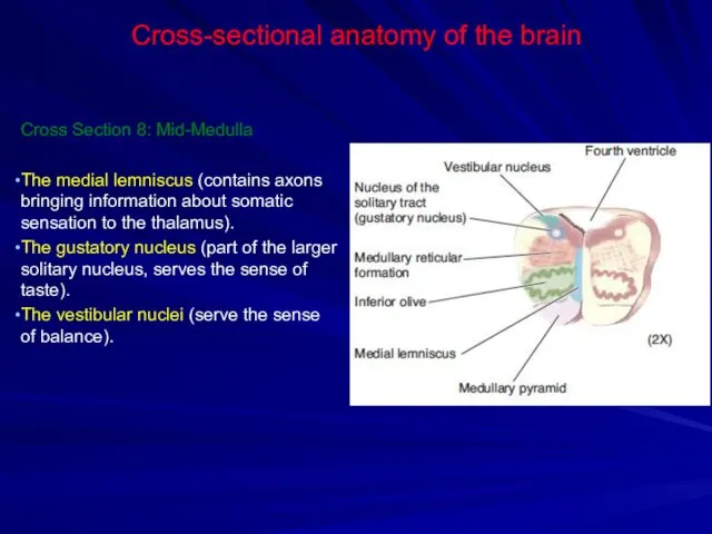
- 41. Cross-sectional anatomy of the brain Cross Section 8: Mid-Medulla
- 42. Cross-sectional anatomy of the brain Cross Section 8: Mid-Medulla The medial lemniscus (contains axons bringing information
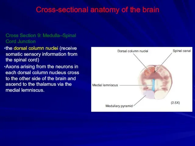
- 43. Cross-sectional anatomy of the brain Cross Section 9: Medulla–Spinal Cord Junction
- 44. Cross-sectional anatomy of the brain Cross Section 9: Medulla–Spinal Cord Junction the dorsal column nuclei (receive
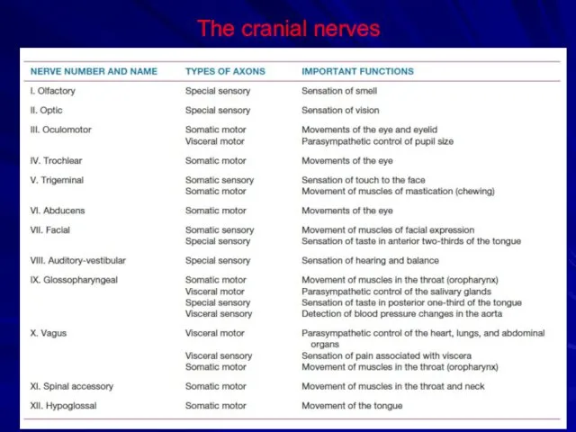
- 45. The cranial nerves Twelve pairs of cranial nerves emerge from the base of the brain. Single
- 47. The cranial nerves
- 49. The spinal cord Gross Anatomy The spinal cord is located within the vertebral canal The spinal
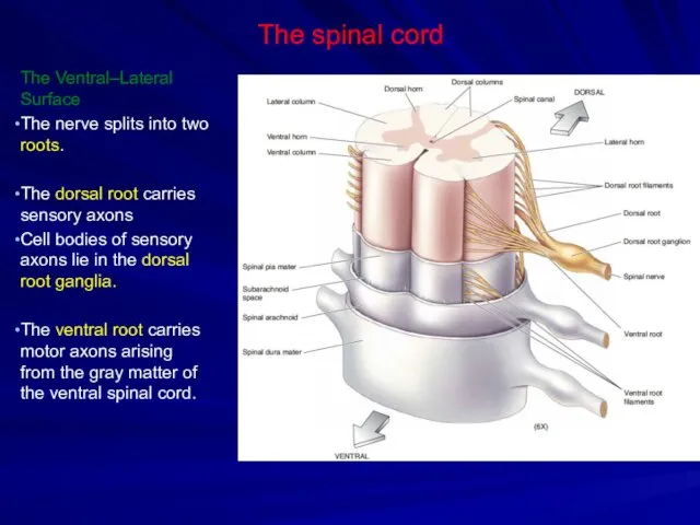
- 50. The spinal cord The Ventral–Lateral Surface The nerve splits into two roots. The dorsal root carries
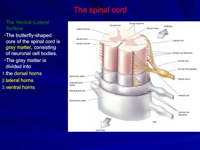
- 51. The spinal cord The Ventral–Lateral Surface The butterfly-shaped core of the spinal cord is gray matter,
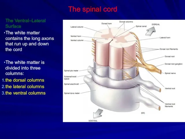
- 52. The spinal cord The Ventral–Lateral Surface The white matter contains the long axons that run up
- 53. The spinal cord Cross-Sectional Anatomy The white mutter consists of the ascending sensory pathways the descending
- 54. The spinal cord Cross-Sectional Anatomy Descending motor pathways The descending tracts contribute to two pathways: the
- 56. Скачать презентацию
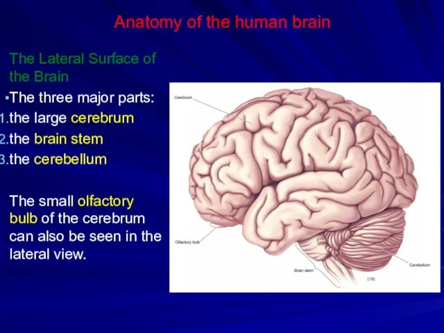
Anatomy of the human brain
The Lateral Surface of the Brain
The three
Anatomy of the human brain
The Lateral Surface of the Brain
The three
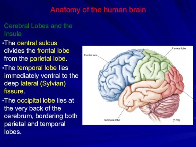
Anatomy of the human brain
Cerebral Lobes and the Insula
The central sulcus
Anatomy of the human brain
Cerebral Lobes and the Insula
The central sulcus
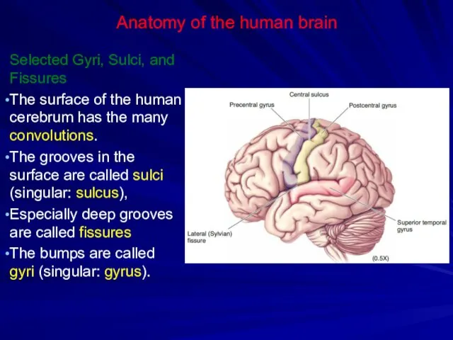
Anatomy of the human brain
Selected Gyri, Sulci, and Fissures
The surface of
Anatomy of the human brain
Selected Gyri, Sulci, and Fissures
The surface of
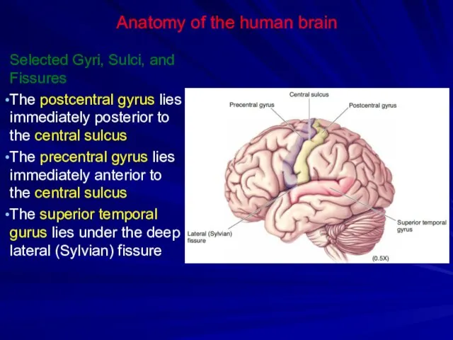
Anatomy of the human brain
Selected Gyri, Sulci, and Fissures
The postcentral gyrus
Anatomy of the human brain
Selected Gyri, Sulci, and Fissures
The postcentral gyrus
Anatomy of the human brain
Cerebral Lobes and the Insula
The insula is
Anatomy of the human brain
Cerebral Lobes and the Insula
The insula is
Anatomy of the human brain
Major Sensory, Motor, and Association Areas of
Anatomy of the human brain
Major Sensory, Motor, and Association Areas of
Anatomy of the human brain
Major Sensory, Motor, and Association Areas of
Anatomy of the human brain
Major Sensory, Motor, and Association Areas of
Anatomy of the human brain
Major Sensory, Motor, and Association Areas of
Anatomy of the human brain
Major Sensory, Motor, and Association Areas of
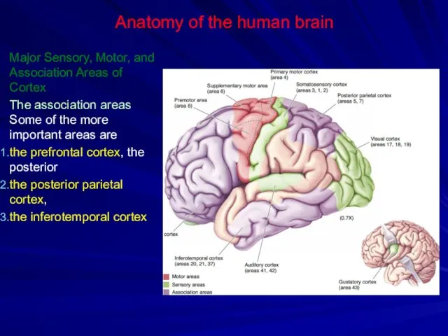
Anatomy of the human brain
Major Sensory, Motor, and Association Areas of
Anatomy of the human brain
Major Sensory, Motor, and Association Areas of
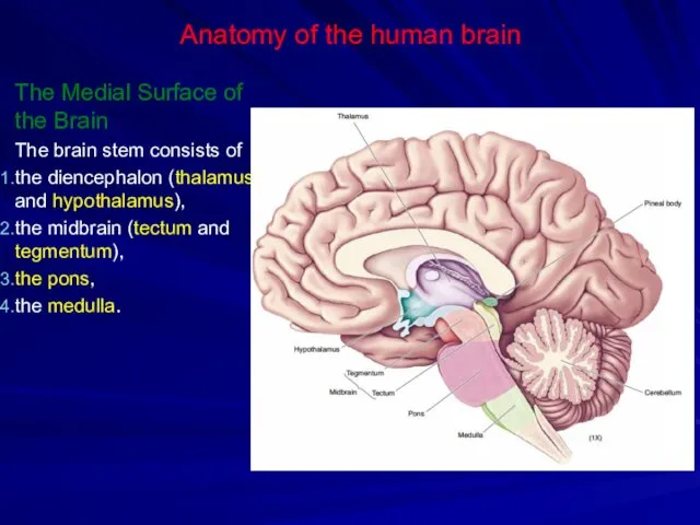
Anatomy of the human brain
The Medial Surface of the Brain
The brain
Anatomy of the human brain
The Medial Surface of the Brain
The brain

Anatomy of the human brain
Forebrain Structures
Corpus callosum (connects the two sides
Anatomy of the human brain
Forebrain Structures
Corpus callosum (connects the two sides
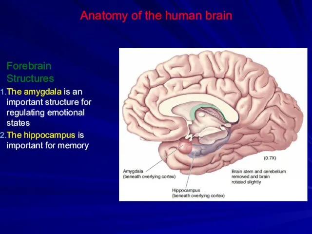
Anatomy of the human brain
Forebrain Structures
The amygdala is an important structure
Anatomy of the human brain
Forebrain Structures
The amygdala is an important structure
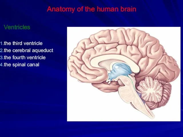
Anatomy of the human brain
Ventricles
the third ventricle
the cerebral aqueduct
the fourth ventricle
the
Anatomy of the human brain
Ventricles
the third ventricle
the cerebral aqueduct
the fourth ventricle
the
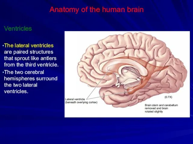
Anatomy of the human brain
Ventricles
The lateral ventricles are paired structures that
Anatomy of the human brain
Ventricles
The lateral ventricles are paired structures that

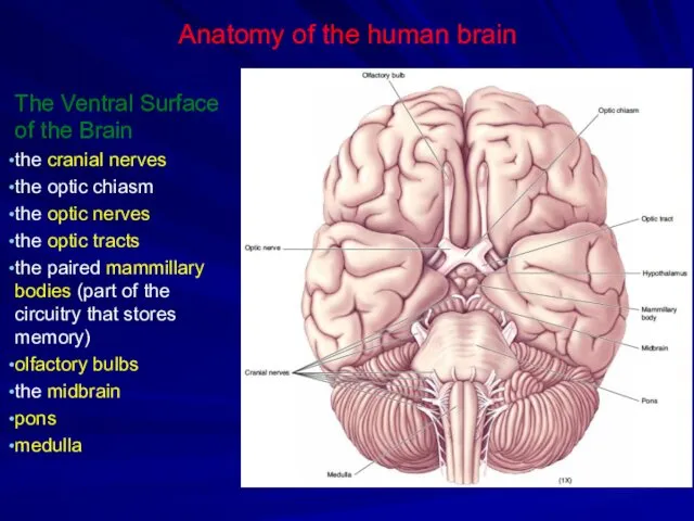
Anatomy of the human brain
The Ventral Surface of the Brain
the cranial
Anatomy of the human brain
The Ventral Surface of the Brain
the cranial
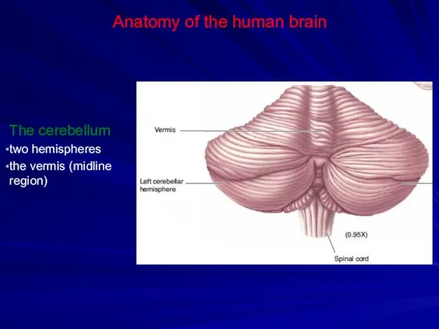
Anatomy of the human brain
The cerebellum
two hemispheres
the vermis (midline region)
Anatomy of the human brain
The cerebellum
two hemispheres
the vermis (midline region)

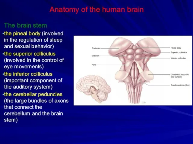
Anatomy of the human brain
The brain stem
the pineal body (involved in
Anatomy of the human brain
The brain stem
the pineal body (involved in

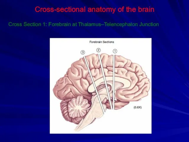
Cross-sectional anatomy of the brain
Cross Section 1: Forebrain at Thalamus–Telencephalon Junction
Cross-sectional anatomy of the brain
Cross Section 1: Forebrain at Thalamus–Telencephalon Junction
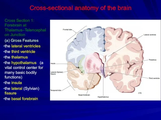
Cross-sectional anatomy of the brain
Cross Section 1: Forebrain at Thalamus–Telencephalon Junction
(a)
Cross-sectional anatomy of the brain
Cross Section 1: Forebrain at Thalamus–Telencephalon Junction
(a)
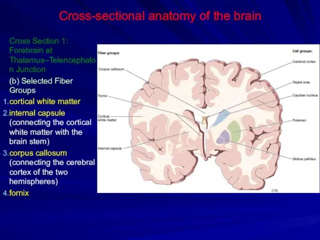
Cross-sectional anatomy of the brain
Cross Section 1: Forebrain at Thalamus–Telencephalon Junction
(b)
Cross-sectional anatomy of the brain
Cross Section 1: Forebrain at Thalamus–Telencephalon Junction
(b)
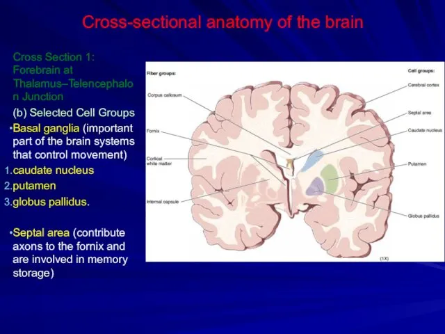
Cross-sectional anatomy of the brain
Cross Section 1: Forebrain at Thalamus–Telencephalon Junction
(b)
Cross-sectional anatomy of the brain
Cross Section 1: Forebrain at Thalamus–Telencephalon Junction
(b)

Cross-sectional anatomy of the brain
Cross Section 2: Forebrain at Mid-Thalamus
Cross-sectional anatomy of the brain
Cross Section 2: Forebrain at Mid-Thalamus
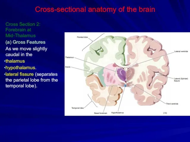
Cross-sectional anatomy of the brain
Cross Section 2: Forebrain at Mid-Thalamus
(a) Gross
Cross-sectional anatomy of the brain
Cross Section 2: Forebrain at Mid-Thalamus
(a) Gross
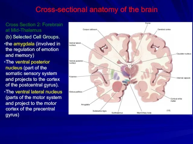
Cross-sectional anatomy of the brain
Cross Section 2: Forebrain at Mid-Thalamus
(b) Selected
Cross-sectional anatomy of the brain
Cross Section 2: Forebrain at Mid-Thalamus
(b) Selected
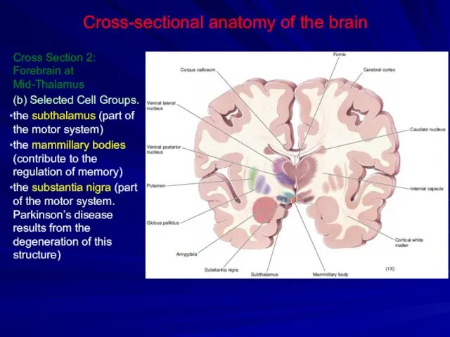
Cross-sectional anatomy of the brain
Cross Section 2: Forebrain at Mid-Thalamus
(b) Selected
Cross-sectional anatomy of the brain
Cross Section 2: Forebrain at Mid-Thalamus
(b) Selected
Cross-sectional anatomy of the brain
Cross Section 3: Forebrain at Thalamus–Midbrain Junction
Cross-sectional anatomy of the brain
Cross Section 3: Forebrain at Thalamus–Midbrain Junction
Cross-sectional anatomy of the brain
Cross Section 3: Forebrain at
Thalamus–Midbrain Junction
Selected Cell
Cross-sectional anatomy of the brain
Cross Section 3: Forebrain at
Thalamus–Midbrain Junction
Selected Cell

Cross-sectional anatomy of the brain
Cross Section 4: Rostral Midbrain
Cross-sectional anatomy of the brain
Cross Section 4: Rostral Midbrain
Cross-sectional anatomy of the brain
Cross Section 4: Rostral Midbrain
the cerebral aqueduct
the
Cross-sectional anatomy of the brain
Cross Section 4: Rostral Midbrain
the cerebral aqueduct
the

Cross-sectional anatomy of the brain
Cross Section 5: Caudal Midbrain
Cross-sectional anatomy of the brain
Cross Section 5: Caudal Midbrain
Cross-sectional anatomy of the brain
Cross Section 4: Caudal Midbrain
the cerebral aqueduct
the
Cross-sectional anatomy of the brain
Cross Section 4: Caudal Midbrain
the cerebral aqueduct
the

Cross-sectional anatomy of the brain
Cross Section 6: Pons and Cerebellum
Cross-sectional anatomy of the brain
Cross Section 6: Pons and Cerebellum

Cross-sectional anatomy of the brain
Cross Section 6: Pons and Cerebellum
pontine nuclei
Cross-sectional anatomy of the brain
Cross Section 6: Pons and Cerebellum
pontine nuclei

Cross-sectional anatomy of the brain
Cross Section 7: Rostral Medulla
Cross-sectional anatomy of the brain
Cross Section 7: Rostral Medulla
Cross-sectional anatomy of the brain
Cross Section 7: Rostral Medulla
Medullary pyramids (contain
Cross-sectional anatomy of the brain
Cross Section 7: Rostral Medulla
Medullary pyramids (contain
Cross-sectional anatomy of the brain
Cross Section 8: Mid-Medulla
Cross-sectional anatomy of the brain
Cross Section 8: Mid-Medulla
Cross-sectional anatomy of the brain
Cross Section 8: Mid-Medulla
The medial lemniscus (contains
Cross-sectional anatomy of the brain
Cross Section 8: Mid-Medulla
The medial lemniscus (contains

Cross-sectional anatomy of the brain
Cross Section 9: Medulla–Spinal Cord Junction
Cross-sectional anatomy of the brain
Cross Section 9: Medulla–Spinal Cord Junction
Cross-sectional anatomy of the brain
Cross Section 9: Medulla–Spinal Cord Junction
the dorsal
Cross-sectional anatomy of the brain
Cross Section 9: Medulla–Spinal Cord Junction
the dorsal
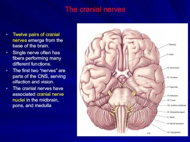
The cranial nerves
Twelve pairs of cranial nerves emerge from the base
The cranial nerves
Twelve pairs of cranial nerves emerge from the base

The cranial nerves
The cranial nerves

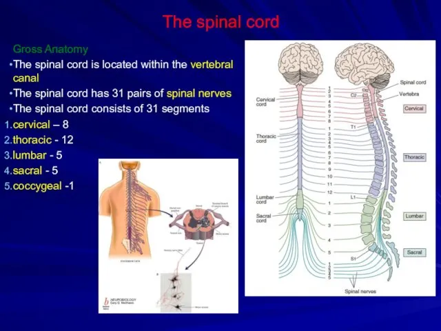
The spinal cord
Gross Anatomy
The spinal cord is located within the vertebral
The spinal cord
Gross Anatomy
The spinal cord is located within the vertebral
The spinal cord
The Ventral–Lateral Surface
The nerve splits into two roots.
The
The spinal cord
The Ventral–Lateral Surface
The nerve splits into two roots.
The
The spinal cord
The Ventral–Lateral Surface
The butterfly-shaped core of the spinal cord
The spinal cord
The Ventral–Lateral Surface
The butterfly-shaped core of the spinal cord
The spinal cord
The Ventral–Lateral Surface
The white matter contains the long axons
The spinal cord
The Ventral–Lateral Surface
The white matter contains the long axons
The spinal cord
Cross-Sectional Anatomy
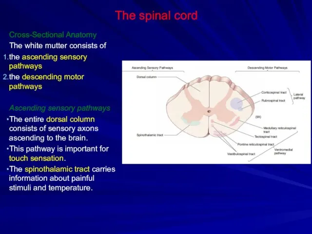
The white mutter consists of
the ascending sensory pathways
the
The spinal cord
Cross-Sectional Anatomy
The white mutter consists of
the ascending sensory pathways
the
The spinal cord
Cross-Sectional Anatomy
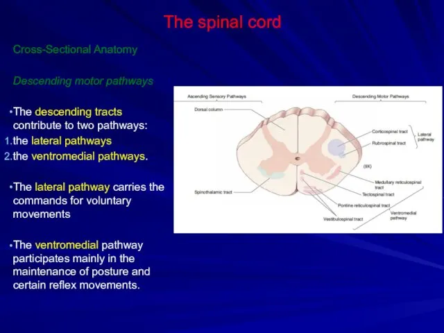
Descending motor pathways
The descending tracts contribute to two
The spinal cord
Cross-Sectional Anatomy
Descending motor pathways
The descending tracts contribute to two
 Класс земноводные или амфибии
 Методы биологических исследований
 Происхождение и начальные этапы развития жизни на Земле
 Сечоутворення і сечовиведення
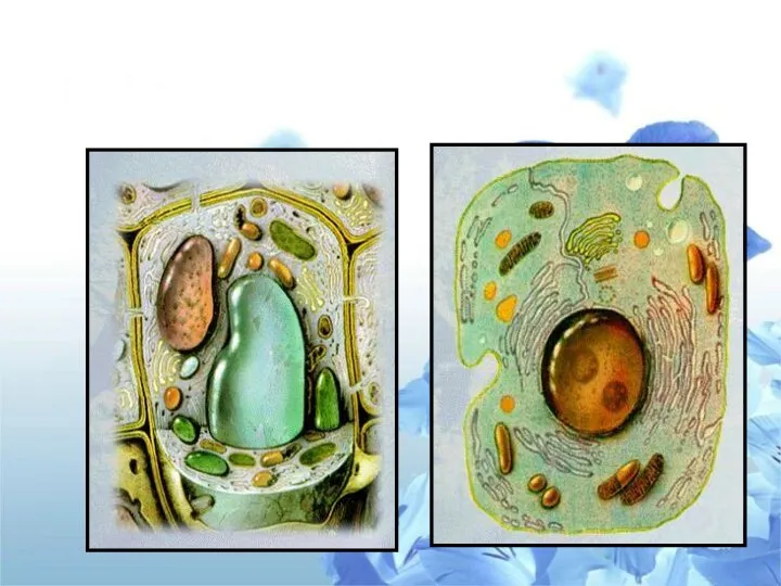 КЛЕТКА
 Гены и хромосомы
 Parts of human body
 Кишечнополостные
 Развитие ребёнка 7-9 месяцев
 Систематика
 Эволюциялық ұғымдардың қалыптасуы және дамуы. Ч. Дарвиннің эволюциялық ілімінің негізгі қағидалары
 Урок-практикум по решению задач по генетике
 Классификация возрастных периодов
 Первичноназемные позвоночные амниоты. Класс рептилии
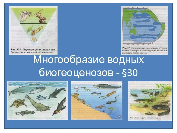 Многообразие водных биогеоценозов
 Екосистема- сад
 Консультация по разделу Насекомые Крыма
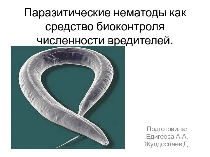 Паразитические нематоды как средство биоконтроля численности вредителей
 Ошущение. Возникновение ощущений
 презентация к уроку Многообразие покрытосеменных
 Тип Моллюски или Мягкотелые
 Одомашнивание животных
 Класс Двудольные
 Мать-и-мачеха. Рисуем по шагам
 Проблема безпритульних тварин
 Развитие представлений о возникновении жизни
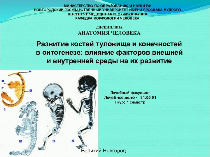 Развитие костей туловища и конечностей в онтогенезе: влияние факторов внешней и внутренней среды на их развитие
 Виды изменчивости. Модификационная изменчивость