- Circulation and Gas Exchange

Содержание
- 2. Overview: Trading Places Every organism must exchange materials with its environment. Exchanges ultimately occur at the
- 3. For most cells making up multicellular organisms, direct exchange with the environment is not possible. Gills
- 4. How does a feathery fringe help this animal survive?
- 5. Circulatory systems link exchange surfaces with cells throughout the body In small and/or thin animals, cells
- 6. Gastrovascular Cavities Simple animals, such as cnidarians, have a body wall that is only two cells
- 7. Internal transport in gastrovascular cavities Circular canal Radial canal Mouth (a) The moon jelly Aurelia, a
- 8. Open and Closed Circulatory Systems More complex animals have either open or closed circulatory systems. Both
- 9. In insects, other arthropods, and most molluscs, blood bathes the organs directly in an open circulatory
- 10. In a closed circulatory system, the blood is confined to vessels and is distinct from the
- 11. Open and closed circulatory systems Heart Hemolymph in sinuses surrounding organs Heart Interstitial fluid Small branch
- 12. Organization of Vertebrate Closed Circulatory Systems Humans and other vertebrates have a closed circulatory system, often
- 13. Arteries branch into arterioles and carry blood to capillaries. Networks of capillaries called capillary beds are
- 14. Vertebrate hearts contain two or more chambers. Blood enters through an atrium and is pumped out
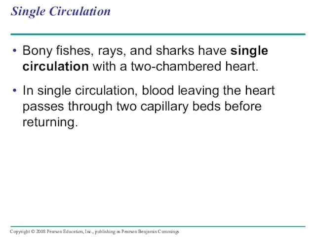
- 15. Single Circulation Bony fishes, rays, and sharks have single circulation with a two-chambered heart. In single
- 16. Single circulation in fishes Artery Ventricle Atrium Heart Vein Systemic capillaries Systemic circulation Gill circulation Gill
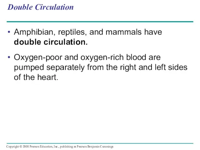
- 17. Double Circulation Amphibian, reptiles, and mammals have double circulation. Oxygen-poor and oxygen-rich blood are pumped separately
- 18. Double circulation in vertebrates Amphibians Lung and skin capillaries Pulmocutaneous circuit Atrium (A) Ventricle (V) Atrium
- 19. In reptiles and mammals, oxygen-poor blood flows through the pulmonary circuit to pick up oxygen through
- 20. Adaptations of Double Circulatory Systems Amphibians: Frogs / amphibians have a three-chambered heart: 2 atria and
- 21. Reptiles (Except Birds) Turtles, snakes, and lizards have a three-chambered heart: two atria and one ventricle.
- 22. Mammals Mammals and birds have a four-chambered heart with two atria and two ventricles. The left
- 23. Coordinated cycles of heart contraction drive double circulation in mammals Blood begins its flow with the
- 24. Blood returns to the heart through the superior vena cava (deoxygenated blood from head, neck, and
- 25. mammalian cardiovascular system Superior vena cava Returns deoxygenated blood from body to heart RA Pulmonary artery
- 26. The Mammalian Heart: A Closer Look A closer look at the mammalian heart provides a better
- 27. Mammalian Heart Pulmonary artery - to lungs Right Atrium RA Receives Deoxygented Blood from body Semilunar
- 28. The heart contracts and relaxes in a rhythmic cycle called the cardiac cycle. The contraction, or
- 29. Cardiac cycle Semilunar valves closed 0.4 sec AV valves open Atrial and ventricular diastole 1 2
- 30. The heart rate, also called the pulse, is the number of beats per minute. The stroke
- 31. Four valves prevent backflow of blood in the heart: The atrioventricular (AV) valves separate each atrium
- 32. Maintaining the Heart’s Rhythmic Beat Some cardiac muscle cells are self-excitable = they contract without any
- 33. Control of heart rhythm Signals spread throughout ventricles. 4 Purkinje Fibers: ventricles contract Pacemaker generates wave
- 34. Patterns of blood pressure and flow reflect the structure and arrangement of blood vessels The physical
- 35. Structure of blood vessels Artery Vein SEM 100 µm Endothelium Artery Smooth muscle Connective tissue Capillary
- 36. Capillaries have thin walls, the endothelium plus its basement membrane, to facilitate the exchange of materials.
- 37. Blood Flow Velocity Physical laws governing movement of fluids through pipes affect blood flow and blood
- 38. The interrelationship of cross-sectional area of blood vessels, blood flow velocity, and blood pressure. 5,000 4,000
- 39. Blood Pressure Blood pressure is the hydrostatic pressure that blood exerts against the wall of a
- 40. Changes in Blood Pressure During the Cardiac Cycle Systolic pressure is the pressure in the arteries
- 41. Regulation of Blood Pressure Blood pressure is determined by cardiac output and peripheral resistance due to
- 42. Vasoconstriction and vasodilation help maintain adequate blood flow as the body’s demands change. The peptide endothelin
- 43. Question: How do endothelial cells control vasoconstriction? Ser RESULTS Ser Ser Cys Cys —NH3+ Leu Met
- 44. Measurement of blood pressure: sphygmomanometer Pressure in cuff greater than 120 mm Hg Rubber cuff inflated
- 45. Fainting is caused by inadequate blood flow to the head. Animals with longer necks require a
- 46. Blood flow in veins Blood flow in veins Direction of blood flow in vein (toward heart)
- 47. Capillary Function Capillaries in major organs are usually filled to capacity. Blood supply varies in many
- 48. Blood flow in capillary beds Precapillary sphincters Thoroughfare channel Arteriole Capillaries Venule (a) Sphincters relaxed (b)
- 49. The critical exchange of substances between the blood and interstitial fluid takes place across the thin
- 50. Fluid exchange between capillaries and the interstitial fluid Body tissue Capillary INTERSTITIAL FLUID Net fluid movement
- 51. Fluid Return by the Lymphatic System The lymphatic system - returns fluid that leaks out in
- 52. Lymph nodes are organs that produce phagocytic white blood cells and filter lymph - an important
- 53. Blood Composition and Function Blood consists of several kinds of blood cells suspended in a liquid
- 54. Composition of mammalian blood Plasma 55% Constituent Major functions Water Solvent for carrying other substances Ions
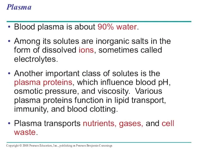
- 55. Plasma Blood plasma is about 90% water. Among its solutes are inorganic salts in the form
- 56. Cellular Elements Suspended in blood plasma are two types of cells: Red blood cells rbc =
- 57. Red blood cells, or erythrocytes, are by far the most numerous blood cells. They transport oxygen
- 58. Leukocytes - Defense There are five major types of white blood cells, or leukocytes: monocytes, neutrophils,
- 59. Platelets - Blood Clotting Platelets are fragments of cells and function in blood clotting. When the
- 60. Collagen fibers Platelet plug Platelet releases chemicals that make nearby platelets sticky Clotting factors from: Platelets
- 61. Stem Cells and the Replacement of Cellular Elements The cellular elements of blood wear out and
- 62. Differentiation of Blood Cells Stem cells in bone marrow Myeloid stem cells Lymphoid stem cells Lymphocytes
- 63. Cardiovascular Disease = Disorders of the Heart and the Blood Vessels One type of cardiovascular disease,
- 64. Atherosclerosis Connective tissue Smooth muscle Endothelium Plaque (a) Normal artery (b) Partly clogged artery 50 µm
- 65. Treatment and Diagnosis of Cardiovascular Disease Cholesterol is a major contributor to atherosclerosis. Low-density lipoproteins (LDLs)
- 66. Gas exchange occurs across specialized respiratory surfaces Gas exchange supplies oxygen for cellular respiration and disposes
- 67. Respiratory Media Animals can use air or water as a source of O2, or respiratory medium.
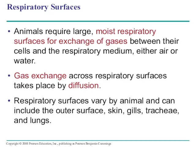
- 68. Respiratory Surfaces Animals require large, moist respiratory surfaces for exchange of gases between their cells and
- 69. Gills are outfoldings of the body that create a large surface area for gas exchange Parapodium
- 70. Ventilation moves the respiratory medium over the respiratory surface. Aquatic animals move through water or move
- 71. Structure and function of fish gills Anatomy of gills Gill arch Water flow Operculum Gill arch
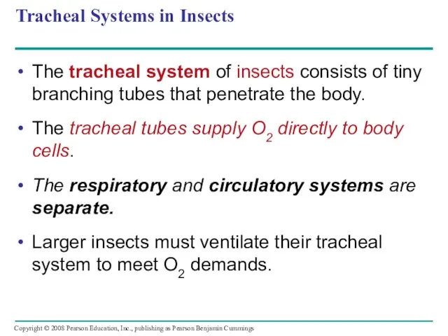
- 72. Tracheal Systems in Insects The tracheal system of insects consists of tiny branching tubes that penetrate
- 73. Tracheal systems Air sacs Tracheae = air tubes External opening: spiracles Body cell Air sac Tracheole
- 74. Lungs = Infoldings of the body surface The circulatory system (open or closed) transports gases between
- 75. Mammalian Respiratory Systems: A Closer Look A system of branching ducts / air tubes conveys air
- 76. Mammalian Respiratory System Pharynx Larynx (Esophagus) Trachea Right lung Bronchus Bronchiole Diaphragm Heart SEM Left lung
- 77. Breathing Ventilates the Lungs by Inhalation and Exhalation of Air Amphibians, such as a frog, ventilates
- 78. Negative pressure breathing: H --> L Lung Diaphragm Air inhaled Rib cage expands as rib muscles
- 79. How a Bird Breathes Birds have eight or nine air sacs that function as bellows that
- 80. The Avian Respiratory System Anterior air sacs Posterior air sacs Lungs Air Lungs Air 1 mm
- 81. Control of Breathing in Humans In humans, the main breathing control centers are in two regions
- 82. Sensors in the aorta and carotid arteries monitor O2 and CO2 concentrations in the blood. These
- 83. Automatic control of breathing Breathing control centers Cerebrospinal fluid Pons Medulla oblongata Carotid arteries Aorta Diaphragm
- 84. Adaptations for gas exchange include pigments that bind and transport gases The metabolic demands of many
- 85. Loading and unloading of respiratory gases Alveolus PO2 = 100 mm Hg PO2 = 40 PO2
- 86. Respiratory Pigments Respiratory pigments = proteins that transport oxygen, greatly increase the amount of oxygen that
- 87. Hemoglobin A single hemoglobin molecule can carry four molecules of O2 The hemoglobin dissociation curve shows
- 88. β Chains Iron Heme α Chains Hemoglobin
- 89. Dissociation curves for hemoglobin at 37ºC O2 unloaded to tissues at rest O2 unloaded to tissues
- 90. Carbon Dioxide Transport Hemoglobin also helps transport CO2 and assists in buffering. CO2 from respiring cells
- 91. Carbon dioxide transport in the blood Body tissue CO2 produced CO2 transport from tissues Capillary wall
- 92. Elite Animal Athletes Migratory and diving mammals have evolutionary adaptations that allow them to perform extraordinary
- 93. Review Inhaled air Exhaled air Alveolar epithelial cells Lungs - Alveolar Air Spaces GAS EXCHANGE CO2
- 94. You should now be able to: Compare and contrast open and closed circulatory systems. Compare and
- 95. Define cardiac cycle and explain the role of the sinoatrial node. Relate the structures of capillaries,
- 96. Describe the role played by the lymphatic system in relation to the circulatory system. Describe the
- 98. Скачать презентацию
Overview: Trading Places
Every organism must exchange materials with its environment.
Exchanges ultimately
Overview: Trading Places
Every organism must exchange materials with its environment.
Exchanges ultimately
For most cells making up multicellular organisms, direct exchange with the
For most cells making up multicellular organisms, direct exchange with the
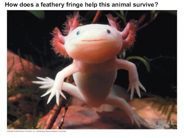
How does a feathery fringe help this animal survive?
How does a feathery fringe help this animal survive?
Circulatory systems link exchange surfaces with cells throughout the body
In small
Circulatory systems link exchange surfaces with cells throughout the body
In small
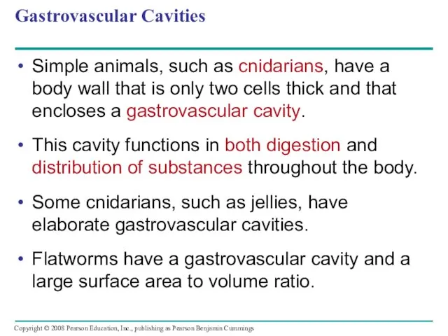
Gastrovascular Cavities
Simple animals, such as cnidarians, have a body wall that
Gastrovascular Cavities
Simple animals, such as cnidarians, have a body wall that
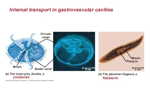
Internal transport in gastrovascular cavities
Circular
canal
Radial canal
Mouth
(a) The moon jelly Aurelia,
Internal transport in gastrovascular cavities
Circular
canal
Radial canal
Mouth
(a) The moon jelly Aurelia,
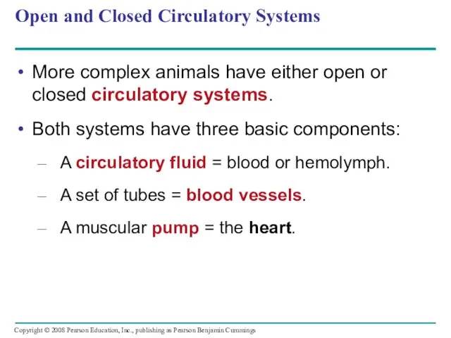
Open and Closed Circulatory Systems
More complex animals have either open or
Open and Closed Circulatory Systems
More complex animals have either open or
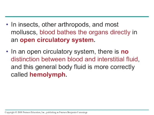
In insects, other arthropods, and most molluscs, blood bathes the organs
In insects, other arthropods, and most molluscs, blood bathes the organs
In a closed circulatory system, the blood is confined to vessels
In a closed circulatory system, the blood is confined to vessels

Open and closed circulatory systems
Heart
Hemolymph in
sinuses
surrounding organs
Heart
Interstitial
fluid
Small branch vessels
In
Open and closed circulatory systems
Heart
Hemolymph in
sinuses
surrounding organs
Heart
Interstitial
fluid
Small branch vessels
In
Organization of Vertebrate Closed Circulatory Systems
Humans and other vertebrates have a
Organization of Vertebrate Closed Circulatory Systems
Humans and other vertebrates have a
Arteries branch into arterioles and carry blood to capillaries.
Networks of
Arteries branch into arterioles and carry blood to capillaries.
Networks of
Vertebrate hearts contain two or more chambers.
Blood enters through an atrium
Vertebrate hearts contain two or more chambers.
Blood enters through an atrium
Single Circulation
Bony fishes, rays, and sharks have single circulation with a
Single Circulation
Bony fishes, rays, and sharks have single circulation with a
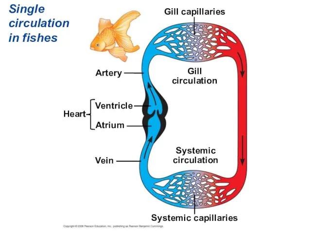
Single circulation in fishes
Artery
Ventricle
Atrium
Heart
Vein
Systemic capillaries
Systemic
circulation
Gill
circulation
Gill capillaries
Single circulation in fishes
Artery
Ventricle
Atrium
Heart
Vein
Systemic capillaries
Systemic
circulation
Gill
circulation
Gill capillaries
Double Circulation
Amphibian, reptiles, and mammals have double circulation.
Oxygen-poor and oxygen-rich blood
Double Circulation
Amphibian, reptiles, and mammals have double circulation.
Oxygen-poor and oxygen-rich blood
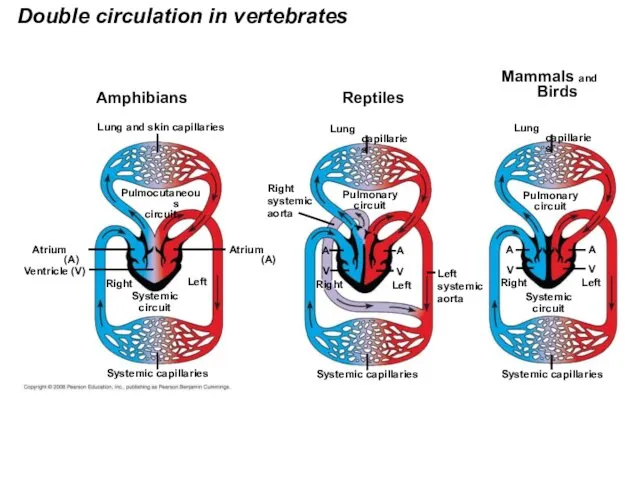
Double circulation in vertebrates
Amphibians
Lung and skin capillaries
Pulmocutaneous
circuit
Atrium (A)
Ventricle (V)
Atrium (A)
Systemic
circuit
Right
Left
Systemic capillaries
Double circulation in vertebrates
Amphibians
Lung and skin capillaries
Pulmocutaneous
circuit
Atrium (A)
Ventricle (V)
Atrium (A)
Systemic
circuit
Right
Left
Systemic capillaries
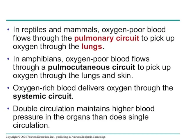
In reptiles and mammals, oxygen-poor blood flows through the pulmonary circuit
In reptiles and mammals, oxygen-poor blood flows through the pulmonary circuit
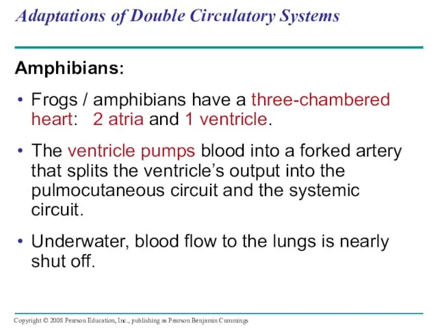
Adaptations of Double Circulatory Systems
Amphibians:
Frogs / amphibians have a three-chambered heart:
Adaptations of Double Circulatory Systems
Amphibians:
Frogs / amphibians have a three-chambered heart:

Reptiles (Except Birds)
Turtles, snakes, and lizards have a three-chambered heart: two
Reptiles (Except Birds)
Turtles, snakes, and lizards have a three-chambered heart: two
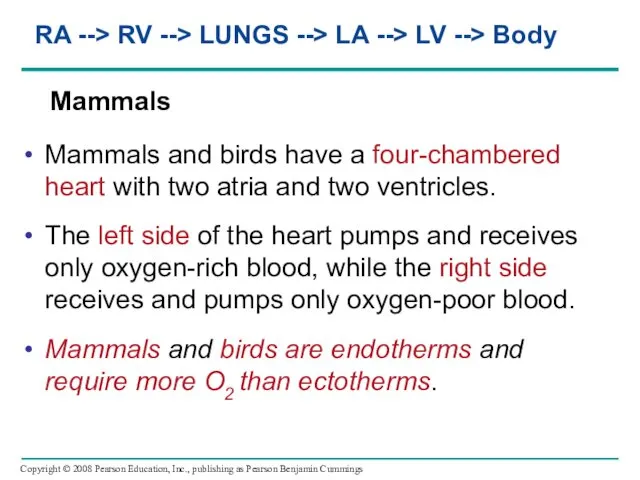
Mammals
Mammals and birds have a four-chambered heart with two atria
Mammals
Mammals and birds have a four-chambered heart with two atria

Coordinated cycles of heart contraction drive double circulation in mammals
Blood begins
Coordinated cycles of heart contraction drive double circulation in mammals
Blood begins
Blood returns to the heart through the superior vena cava (deoxygenated
Blood returns to the heart through the superior vena cava (deoxygenated
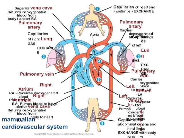
mammalian cardiovascular system
Superior vena cava
Returns deoxygenated blood from
body to heart
mammalian cardiovascular system
Superior vena cava
Returns deoxygenated blood from
body to heart
The Mammalian Heart: A Closer Look
A closer look at the mammalian
The Mammalian Heart: A Closer Look
A closer look at the mammalian
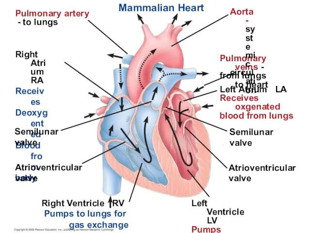
Mammalian Heart
Pulmonary artery
- to lungs
Right Atrium RA
Receives
Deoxygented
Blood from
body
Semilunar
valve
Atrioventricular
valve
Right
Mammalian Heart
Pulmonary artery
- to lungs
Right Atrium RA
Receives
Deoxygented
Blood from
body
Semilunar
valve
Atrioventricular
valve
Right
The heart contracts and relaxes in a rhythmic cycle called the
The heart contracts and relaxes in a rhythmic cycle called the
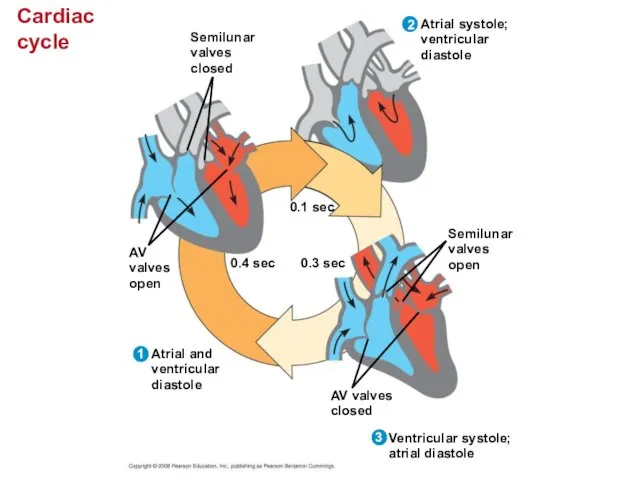
Cardiac cycle
Semilunar
valves
closed
0.4 sec
AV
valves
open
Atrial and
ventricular
diastole
1
2
0.1 sec
Atrial systole;
ventricular
diastole
3
0.3 sec
Semilunar
valves
open
AV valves
closed
Ventricular systole;
atrial diastole
Cardiac cycle
Semilunar
valves
closed
0.4 sec
AV
valves
open
Atrial and
ventricular
diastole
1
2
0.1 sec
Atrial systole;
ventricular
diastole
3
0.3 sec
Semilunar
valves
open
AV valves
closed
Ventricular systole;
atrial diastole
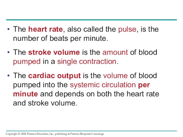
The heart rate, also called the pulse, is the number of
The heart rate, also called the pulse, is the number of
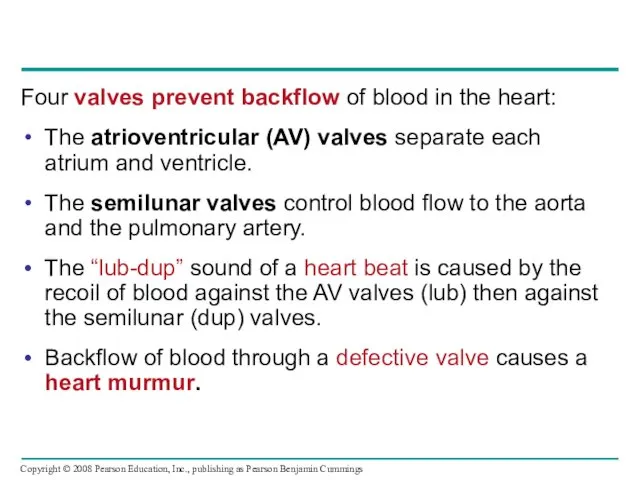
Four valves prevent backflow of blood in the heart:
The atrioventricular (AV)
Four valves prevent backflow of blood in the heart:
The atrioventricular (AV)
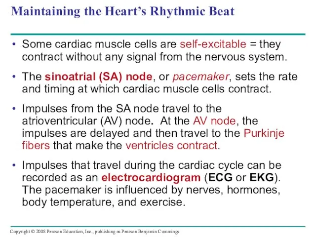
Maintaining the Heart’s Rhythmic Beat
Some cardiac muscle cells are self-excitable =
Maintaining the Heart’s Rhythmic Beat
Some cardiac muscle cells are self-excitable =
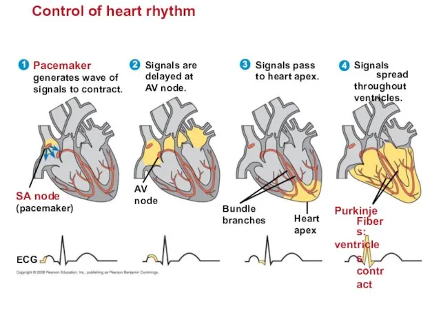
Control of heart rhythm
Signals spread
throughout
ventricles.
4
Purkinje Fibers:
ventricles contract
Pacemaker
generates wave of
signals
Control of heart rhythm
Signals spread
throughout
ventricles.
4
Purkinje Fibers:
ventricles contract
Pacemaker
generates wave of
signals
Patterns of blood pressure and flow reflect the structure and arrangement
Patterns of blood pressure and flow reflect the structure and arrangement
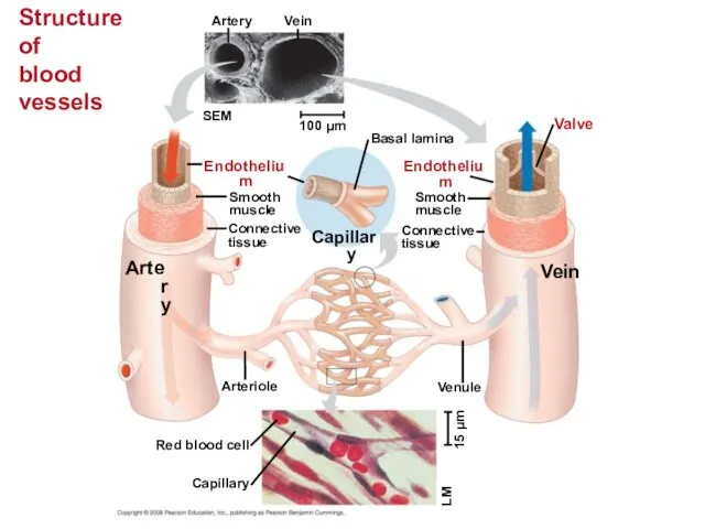
Structure
of
blood vessels
Artery
Vein
SEM
100 µm
Endothelium
Artery
Smooth
muscle
Connective
tissue
Capillary
Basal lamina
Endothelium
Smooth
muscle
Connective
tissue
Valve
Vein
Arteriole
Venule
Red blood cell
Capillary
15 µm
LM
Structure
of
blood vessels
Artery
Vein
SEM
100 µm
Endothelium
Artery
Smooth
muscle
Connective
tissue
Capillary
Basal lamina
Endothelium
Smooth
muscle
Connective
tissue
Valve
Vein
Arteriole
Venule
Red blood cell
Capillary
15 µm
LM

Capillaries have thin walls, the endothelium plus its basement membrane, to
Capillaries have thin walls, the endothelium plus its basement membrane, to
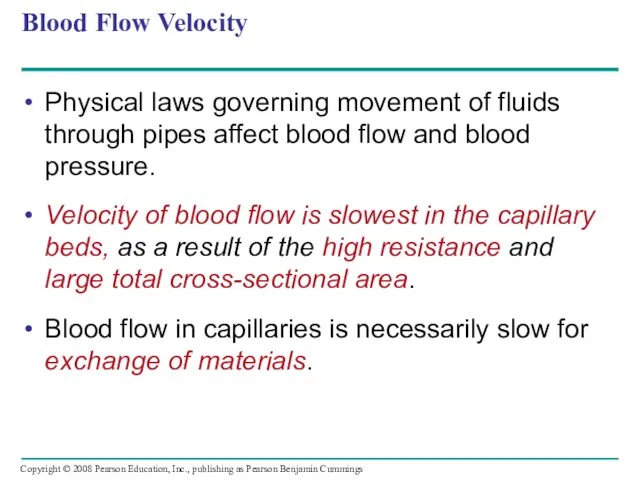
Blood Flow Velocity
Physical laws governing movement of fluids through pipes affect
Blood Flow Velocity
Physical laws governing movement of fluids through pipes affect

The interrelationship of cross-sectional area of blood vessels,
blood flow velocity,
The interrelationship of cross-sectional area of blood vessels, blood flow velocity,
Blood Pressure
Blood pressure is the hydrostatic pressure that blood exerts against
Blood Pressure
Blood pressure is the hydrostatic pressure that blood exerts against
Changes in Blood Pressure During the Cardiac Cycle
Systolic pressure is the
Changes in Blood Pressure During the Cardiac Cycle
Systolic pressure is the
Regulation of Blood Pressure
Blood pressure is determined by cardiac output and
Regulation of Blood Pressure
Blood pressure is determined by cardiac output and
Vasoconstriction and vasodilation help maintain adequate blood flow as the body’s
Vasoconstriction and vasodilation help maintain adequate blood flow as the body’s
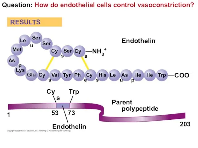
Question: How do endothelial cells control vasoconstriction?
Ser
RESULTS
Ser
Ser
Cys
Cys
—NH3+
Leu
Met
Asp
Lys
Glu
Cys
Val
Tyr
Phe
Cys
His
Leu
Asp
Ile
Ile
Trp
—COO–
Endothelin
Parent polypeptide
Trp
Cys
Endothelin
53
73
1
203
Question: How do endothelial cells control vasoconstriction?
Ser
RESULTS
Ser
Ser
Cys
Cys
—NH3+
Leu
Met
Asp
Lys
Glu
Cys
Val
Tyr
Phe
Cys
His
Leu
Asp
Ile
Ile
Trp
—COO–
Endothelin
Parent polypeptide
Trp
Cys
Endothelin
53
73
1
203
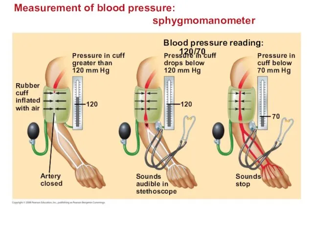
Measurement of blood pressure:
sphygmomanometer
Pressure in cuff
greater than
120 mm Hg
Rubber
cuff
inflated
with
Measurement of blood pressure:
sphygmomanometer
Pressure in cuff
greater than
120 mm Hg
Rubber
cuff
inflated
with
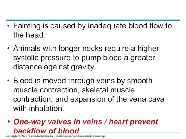
Fainting is caused by inadequate blood flow to the head.
Animals with
Fainting is caused by inadequate blood flow to the head.
Animals with
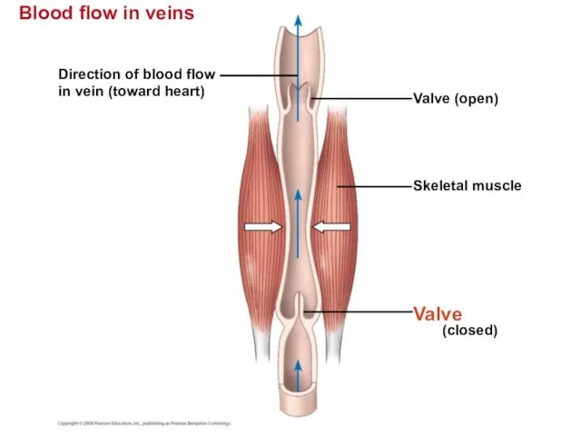
Blood flow in veins
Blood flow in veins
Direction of blood flow
in vein
Blood flow in veins
Blood flow in veins
Direction of blood flow
in vein
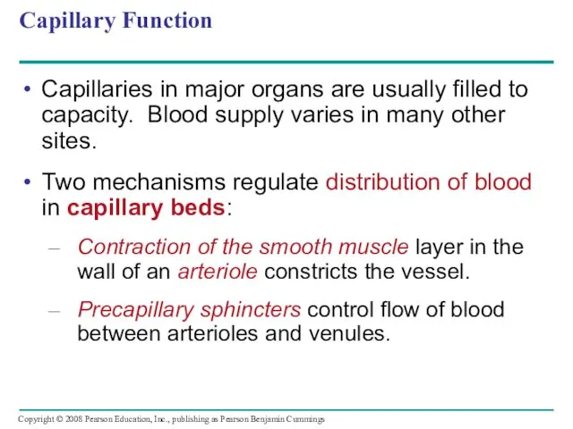
Capillary Function
Capillaries in major organs are usually filled to capacity. Blood
Capillary Function
Capillaries in major organs are usually filled to capacity. Blood
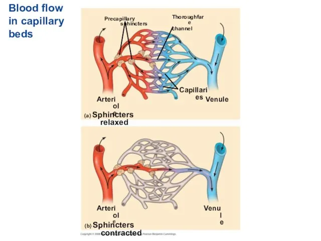
Blood flow in capillary beds
Precapillary sphincters
Thoroughfare
channel
Arteriole
Capillaries
Venule
(a) Sphincters relaxed
(b) Sphincters contracted
Arteriole
Venule
Blood flow in capillary beds
Precapillary sphincters
Thoroughfare
channel
Arteriole
Capillaries
Venule
(a) Sphincters relaxed
(b) Sphincters contracted
Arteriole
Venule
The critical exchange of substances between the blood and interstitial fluid
The critical exchange of substances between the blood and interstitial fluid

Fluid exchange between capillaries and the interstitial fluid
Body tissue
Capillary
INTERSTITIAL FLUID
Net fluid
movement
Fluid exchange between capillaries and the interstitial fluid
Body tissue
Capillary
INTERSTITIAL FLUID
Net fluid
movement
Fluid Return by the Lymphatic System
The lymphatic system - returns fluid
Fluid Return by the Lymphatic System
The lymphatic system - returns fluid
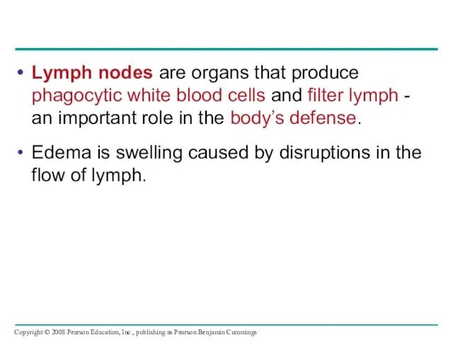
Lymph nodes are organs that produce phagocytic white blood cells and
Lymph nodes are organs that produce phagocytic white blood cells and
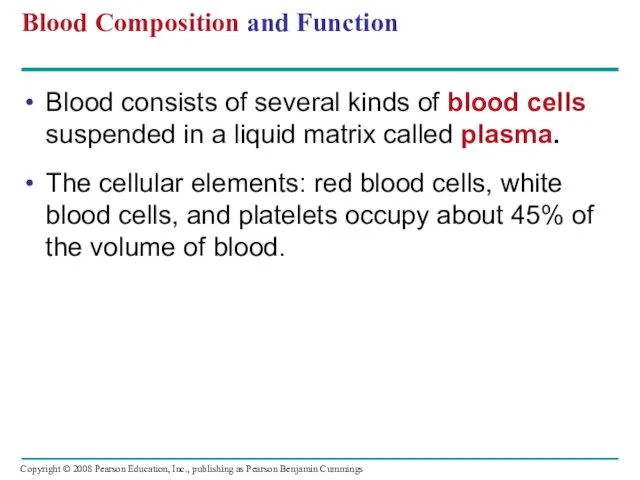
Blood Composition and Function
Blood consists of several kinds of blood cells
Blood Composition and Function
Blood consists of several kinds of blood cells
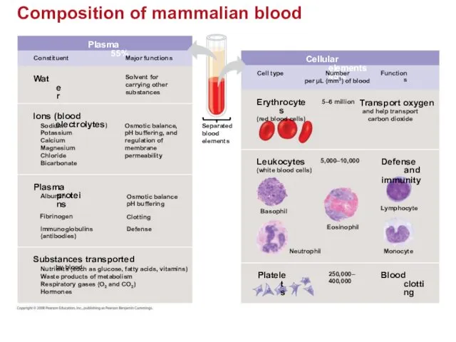
Composition of mammalian blood
Plasma 55%
Constituent
Major functions
Water
Solvent for
carrying other
substances
Ions (blood electrolytes)
Osmotic
Composition of mammalian blood
Plasma 55%
Constituent
Major functions
Water
Solvent for
carrying other
substances
Ions (blood electrolytes)
Osmotic
Plasma
Blood plasma is about 90% water.
Among its solutes are inorganic salts
Plasma
Blood plasma is about 90% water.
Among its solutes are inorganic salts
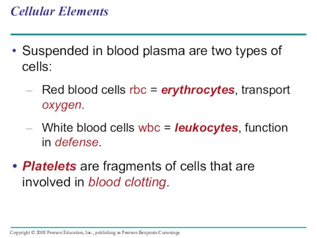
Cellular Elements
Suspended in blood plasma are two types of cells:
Red blood
Cellular Elements
Suspended in blood plasma are two types of cells:
Red blood
Red blood cells, or erythrocytes, are by far the most numerous
Red blood cells, or erythrocytes, are by far the most numerous
Leukocytes - Defense
There are five major types of white blood cells,
Leukocytes - Defense
There are five major types of white blood cells,
Platelets - Blood Clotting
Platelets are fragments of cells and function in
Platelets - Blood Clotting
Platelets are fragments of cells and function in
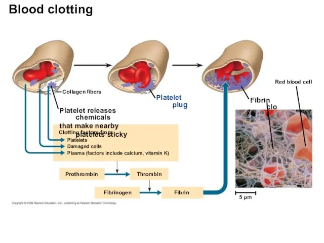
Collagen fibers
Platelet plug
Platelet releases chemicals
that make nearby platelets sticky
Clotting factors from:
Platelets
Damaged
Collagen fibers
Platelet plug
Platelet releases chemicals
that make nearby platelets sticky
Clotting factors from:
Platelets
Damaged
Stem Cells and the Replacement of Cellular Elements
The cellular elements of
Stem Cells and the Replacement of Cellular Elements
The cellular elements of
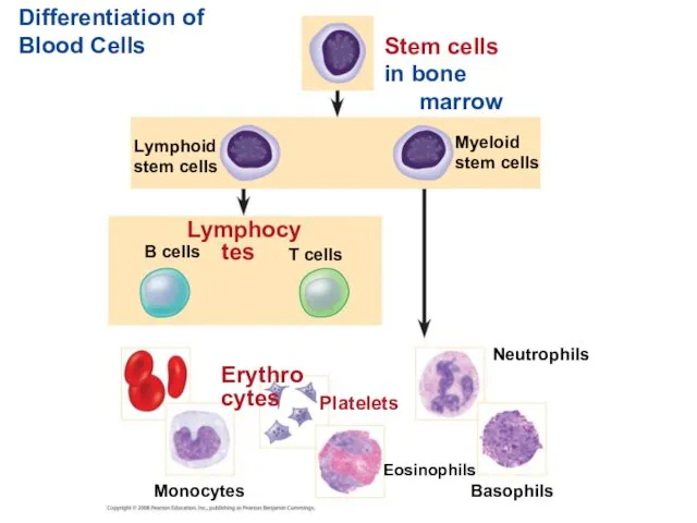
Differentiation of Blood Cells
Stem cells
in bone marrow
Myeloid
stem cells
Lymphoid
stem cells
Lymphocytes
B cells
T cells
Differentiation of Blood Cells
Stem cells
in bone marrow
Myeloid
stem cells
Lymphoid
stem cells
Lymphocytes
B cells
T cells
Cardiovascular Disease = Disorders of the Heart and the Blood Vessels
One
Cardiovascular Disease = Disorders of the Heart and the Blood Vessels
One
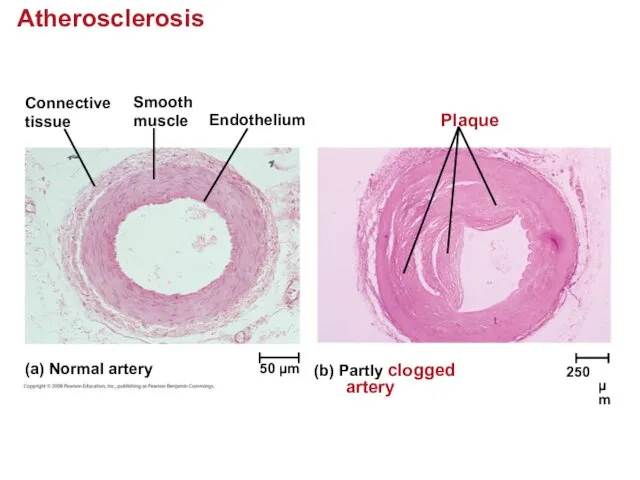
Atherosclerosis
Connective
tissue
Smooth
muscle
Endothelium
Plaque
(a) Normal artery
(b) Partly clogged artery
50 µm
250 µm
Atherosclerosis
Connective
tissue
Smooth
muscle
Endothelium
Plaque
(a) Normal artery
(b) Partly clogged artery
50 µm
250 µm
Treatment and Diagnosis of Cardiovascular Disease
Cholesterol is a major contributor to
Treatment and Diagnosis of Cardiovascular Disease
Cholesterol is a major contributor to
Gas exchange occurs across specialized respiratory surfaces
Gas exchange supplies oxygen for
Gas exchange occurs across specialized respiratory surfaces
Gas exchange supplies oxygen for
Respiratory Media
Animals can use air or water as a source of
Respiratory Media
Animals can use air or water as a source of
Respiratory Surfaces
Animals require large, moist respiratory surfaces for exchange of gases
Respiratory Surfaces
Animals require large, moist respiratory surfaces for exchange of gases
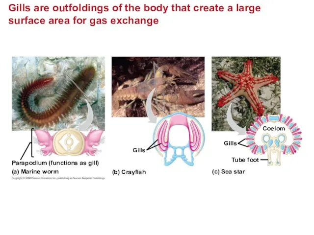
Gills are outfoldings of the body that create a large surface
Gills are outfoldings of the body that create a large surface
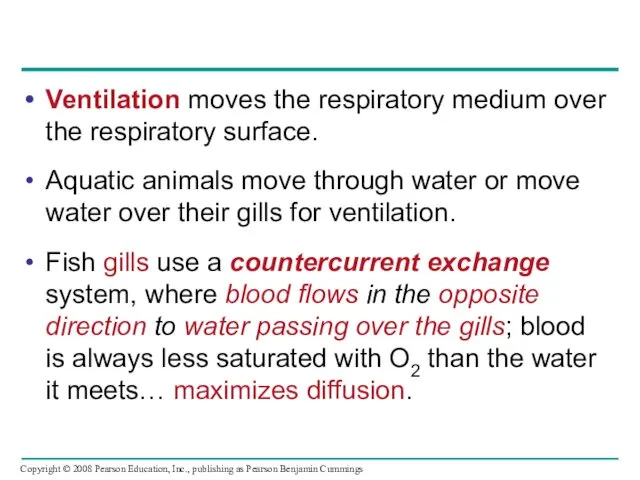
Ventilation moves the respiratory medium over the respiratory surface.
Aquatic animals move
Ventilation moves the respiratory medium over the respiratory surface.
Aquatic animals move
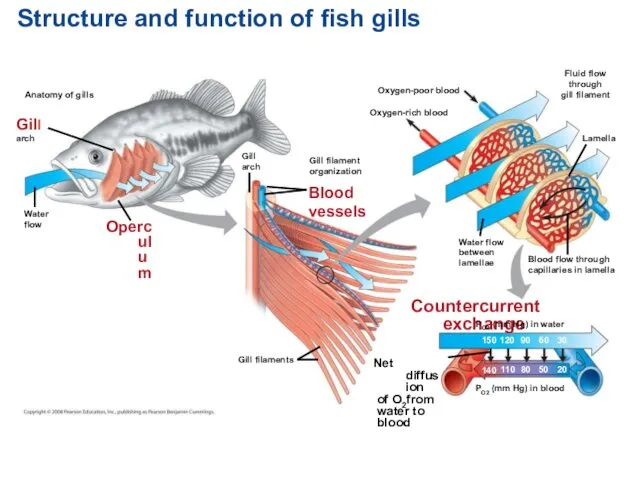
Structure and function of fish gills
Anatomy of gills
Gill
arch
Water
flow
Operculum
Gill
arch
Gill filament
organization
Blood
vessels
Oxygen-poor blood
Oxygen-rich blood
Fluid
Structure and function of fish gills
Anatomy of gills
Gill
arch
Water
flow
Operculum
Gill
arch
Gill filament
organization
Blood
vessels
Oxygen-poor blood
Oxygen-rich blood
Fluid
Tracheal Systems in Insects
The tracheal system of insects consists of tiny
Tracheal Systems in Insects
The tracheal system of insects consists of tiny
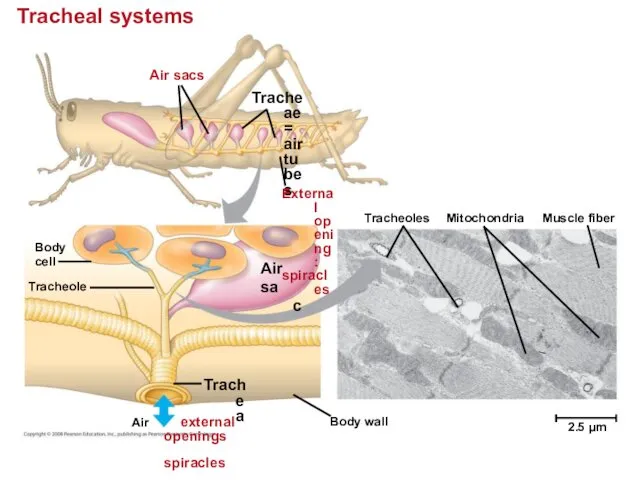
Tracheal systems
Air sacs
Tracheae = air tubes
External opening:
spiracles
Body
cell
Air
sac
Tracheole
Tracheoles
Mitochondria
Muscle fiber
2.5 µm
Body wall
Trachea
Air
Tracheal systems
Air sacs
Tracheae = air tubes
External opening:
spiracles
Body
cell
Air
sac
Tracheole
Tracheoles
Mitochondria
Muscle fiber
2.5 µm
Body wall
Trachea
Air
Lungs = Infoldings of the body surface
The circulatory system
Lungs = Infoldings of the body surface
The circulatory system
Mammalian Respiratory Systems: A Closer Look
A system of branching ducts /
Mammalian Respiratory Systems: A Closer Look
A system of branching ducts /
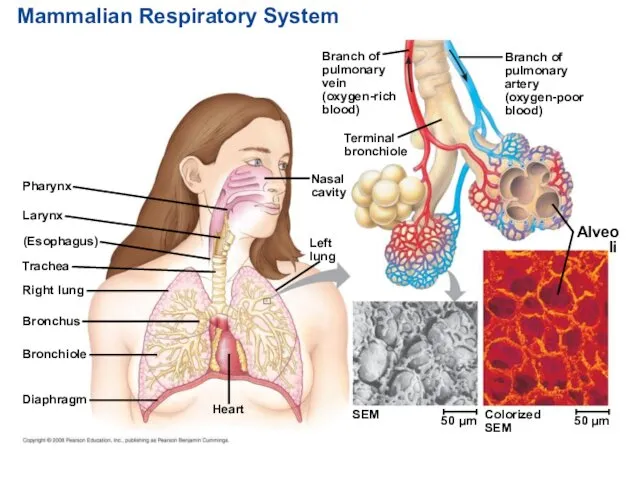
Mammalian Respiratory System
Pharynx
Larynx
(Esophagus)
Trachea
Right lung
Bronchus
Bronchiole
Diaphragm
Heart
SEM
Left
lung
Nasal
cavity
Terminal
bronchiole
Branch of
pulmonary
vein
(oxygen-rich
blood)
Branch of
pulmonary
artery
(oxygen-poor
blood)
Alveoli
Colorized
SEM
50 µm
50 µm
Mammalian Respiratory System
Pharynx
Larynx
(Esophagus)
Trachea
Right lung
Bronchus
Bronchiole
Diaphragm
Heart
SEM
Left
lung
Nasal
cavity
Terminal
bronchiole
Branch of
pulmonary
vein
(oxygen-rich
blood)
Branch of
pulmonary
artery
(oxygen-poor
blood)
Alveoli
Colorized
SEM
50 µm
50 µm
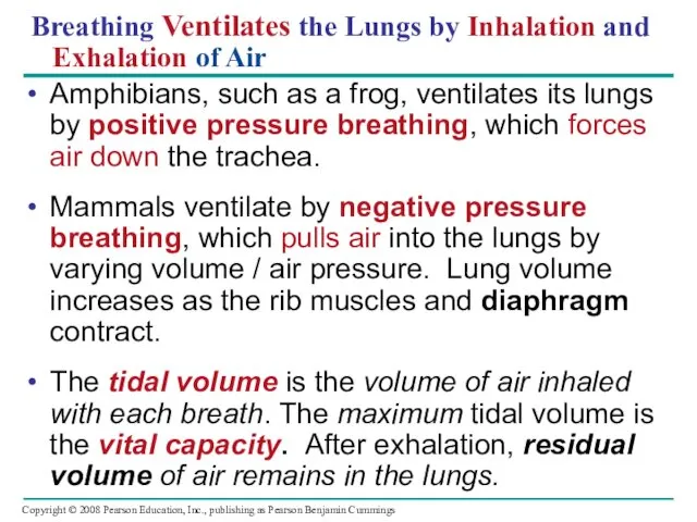
Breathing Ventilates the Lungs by Inhalation and
Exhalation of Air
Amphibians,
Breathing Ventilates the Lungs by Inhalation and
Exhalation of Air
Amphibians,
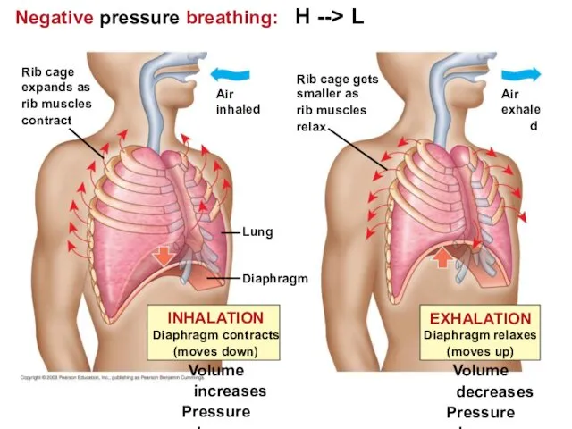
Negative pressure breathing: H --> L
Lung
Diaphragm
Air
inhaled
Rib cage
expands as
rib muscles
contract
Rib cage gets
smaller
Negative pressure breathing: H --> L
Lung
Diaphragm
Air
inhaled
Rib cage
expands as
rib muscles
contract
Rib cage gets
smaller
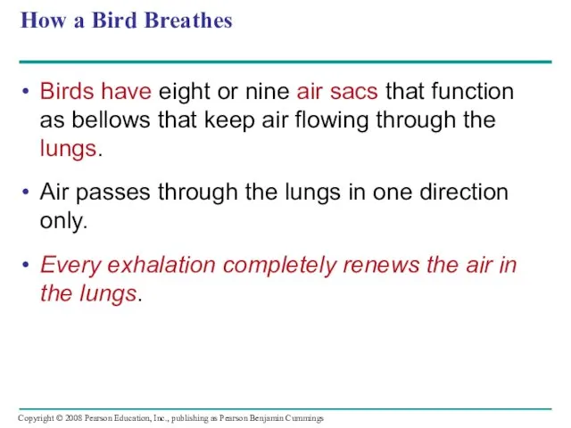
How a Bird Breathes
Birds have eight or nine air sacs that
How a Bird Breathes
Birds have eight or nine air sacs that
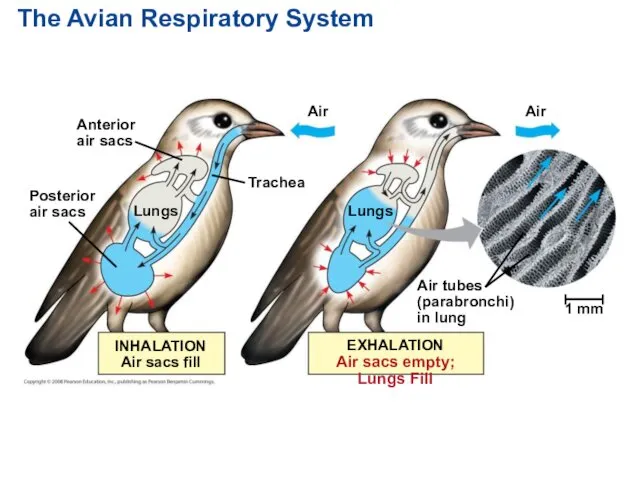
The Avian Respiratory System
Anterior
air sacs
Posterior
air sacs
Lungs
Air
Lungs
Air
1 mm
Trachea
Air tubes
(parabronchi)
in lung
EXHALATION
Air sacs empty;
The Avian Respiratory System
Anterior
air sacs
Posterior
air sacs
Lungs
Air
Lungs
Air
1 mm
Trachea
Air tubes
(parabronchi)
in lung
EXHALATION
Air sacs empty;
Control of Breathing in Humans
In humans, the main breathing control centers
Control of Breathing in Humans
In humans, the main breathing control centers
Sensors in the aorta and carotid arteries monitor O2 and CO2
Sensors in the aorta and carotid arteries monitor O2 and CO2
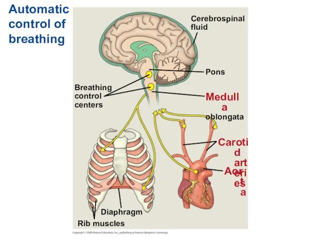
Automatic control of breathing
Breathing
control
centers
Cerebrospinal
fluid
Pons
Medulla
oblongata
Carotid arteries
Aorta
Diaphragm
Rib muscles
Automatic control of breathing
Breathing
control
centers
Cerebrospinal
fluid
Pons
Medulla
oblongata
Carotid arteries
Aorta
Diaphragm
Rib muscles
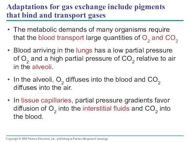
Adaptations for gas exchange include pigments that bind and transport gases
The
Adaptations for gas exchange include pigments that bind and transport gases
The
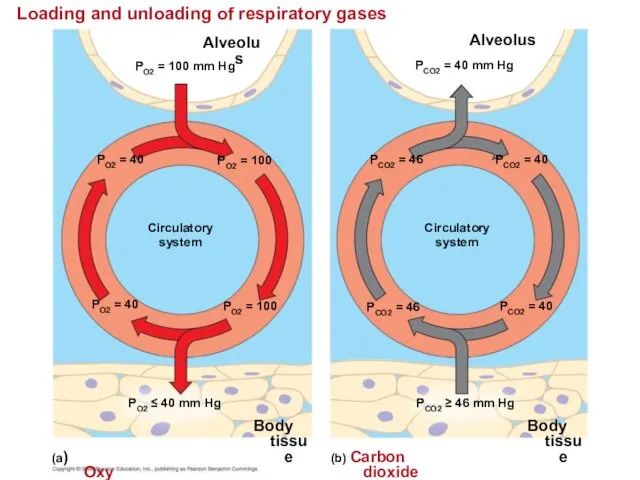
Loading and unloading of respiratory gases
Alveolus
PO2 = 100 mm Hg
PO2 =
Loading and unloading of respiratory gases
Alveolus
PO2 = 100 mm Hg
PO2 =
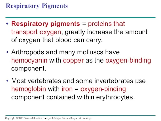
Respiratory Pigments
Respiratory pigments = proteins that transport oxygen, greatly increase the
Respiratory Pigments
Respiratory pigments = proteins that transport oxygen, greatly increase the
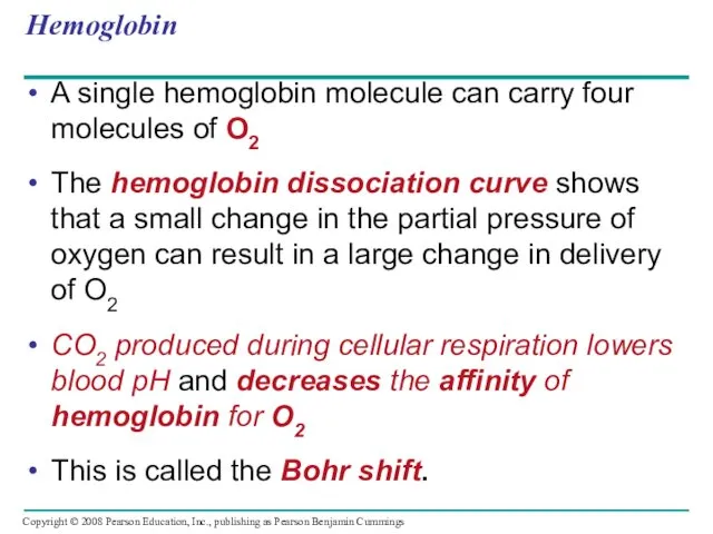
Hemoglobin
A single hemoglobin molecule can carry four molecules of O2
The hemoglobin
Hemoglobin
A single hemoglobin molecule can carry four molecules of O2
The hemoglobin
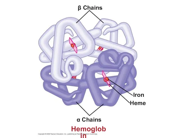
β Chains
Iron
Heme
α Chains
Hemoglobin
β Chains
Iron
Heme
α Chains
Hemoglobin
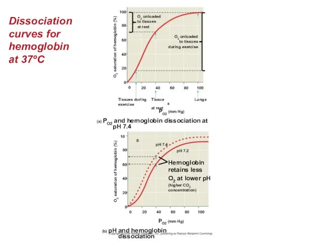
Dissociation curves for hemoglobin at 37ºC
O2 unloaded
to tissues
at rest
O2 unloaded
to tissues
during
Dissociation curves for hemoglobin at 37ºC
O2 unloaded
to tissues
at rest
O2 unloaded
to tissues
during
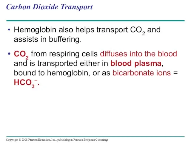
Carbon Dioxide Transport
Hemoglobin also helps transport CO2 and assists in buffering.
CO2
Carbon Dioxide Transport
Hemoglobin also helps transport CO2 and assists in buffering.
CO2
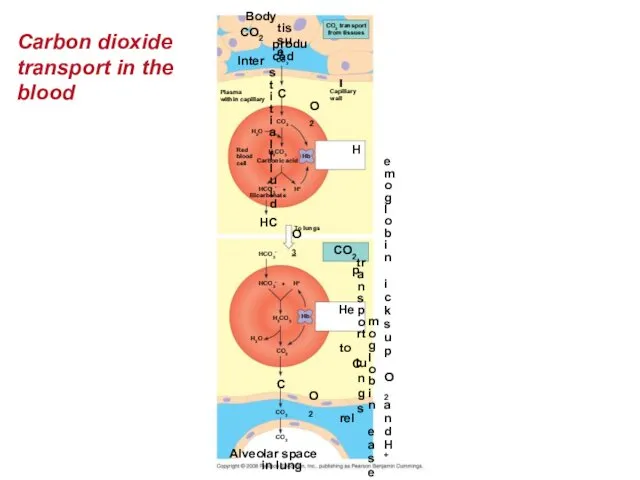
Carbon dioxide transport in the blood
Body tissue
CO2 produced
CO2 transport
from tissues
Capillary
wall
Interstitial fluid
Plasma
within
Carbon dioxide transport in the blood
Body tissue
CO2 produced
CO2 transport
from tissues
Capillary
wall
Interstitial fluid
Plasma
within
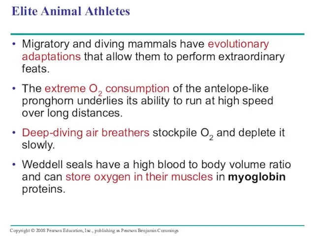
Elite Animal Athletes
Migratory and diving mammals have evolutionary adaptations that allow
Elite Animal Athletes
Migratory and diving mammals have evolutionary adaptations that allow
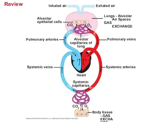
Review
Inhaled air
Exhaled air
Alveolar
epithelial cells
Lungs - Alveolar Air Spaces
GAS EXCHANGE
CO2
O2
CO2
O2
Alveolar
capillaries of
lung
Pulmonary veins
Pulmonary
Review
Inhaled air
Exhaled air
Alveolar
epithelial cells
Lungs - Alveolar Air Spaces
GAS EXCHANGE
CO2
O2
CO2
O2
Alveolar
capillaries of
lung
Pulmonary veins
Pulmonary
You should now be able to:
Compare and contrast open and closed
You should now be able to:
Compare and contrast open and closed
Define cardiac cycle and explain the role of the sinoatrial node.
Relate
Define cardiac cycle and explain the role of the sinoatrial node.
Relate
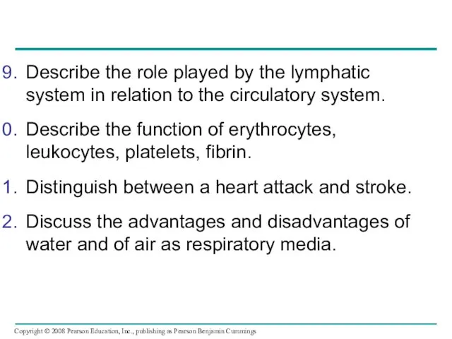
Describe the role played by the lymphatic system in relation to
Describe the role played by the lymphatic system in relation to
 Тип Моллюски. Класс Брюхоногие. Класс Двустворчатые. Класс Головоногие
 Углерод
 Первичные продуценты, в основе новообразования органического вещества которых лежит хемосинтез
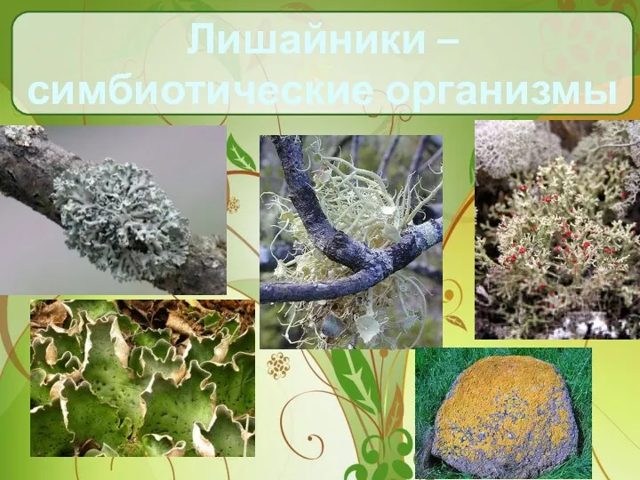 Лишайники – симбиотические организмы
 Ферменттің қасиеті мен құрылысы
 Покормите птиц зимой. (5 класс)
 Популяция. Типы экологических взаимодействий
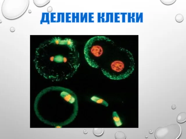 Митоз и мейоз
 Как стать городским фермером. Вертикальные фермы
 Хрящевые рыбы
 Запилення квіткових рослин
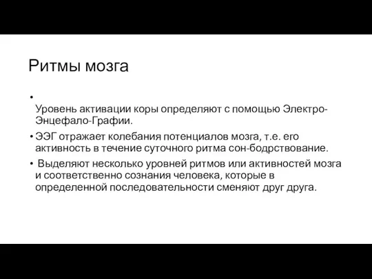 Ритмы мозга
 Характеристика костной системы
 Эндокринная система человека
 Энзимопатия. Энзимотерапия. Энзимодиагностика
 Почему дети похожи на родителей
 Анатомо-физиологические особенности человека в подростковом возрасте
 Жизнедеятельность клетки
 Овощи
Овощи Экология микроорганизмов
 Пищеварение в желудке и кишечнике
 Деревья и кустарники
 Строение и функции кожи
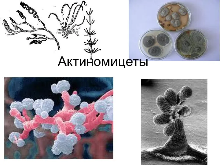 Актиномицеты (actinomycetes)
 Кожные покровы хордовых
 Презентация к уроку биологии в 7 классе Особенности организации одноклеточных, их классификация
 растения
 Интересные факты о коже. Презентация.