- Membrane Structure and Function

Содержание
- 2. Overview: Life at the Edge The plasma membrane is the boundary that separates the living cell
- 3. Fig. 7-1
- 4. Concept 7.1: Cellular membranes are fluid mosaics of lipids and proteins Phospholipids are the most abundant
- 5. Membrane Models: Scientific Inquiry Membranes have been chemically analyzed and found to be made of proteins
- 6. Fig. 7-2 Hydrophilic head WATER Hydrophobic tail WATER
- 7. In 1935, Hugh Davson and James Danielli proposed a sandwich model in which the phospholipid bilayer
- 8. Fig. 7-3 Phospholipid bilayer Hydrophobic regions of protein Hydrophilic regions of protein
- 9. Freeze-fracture studies of the plasma membrane supported the fluid mosaic model Freeze-fracture is a specialized preparation
- 10. Fig. 7-4 TECHNIQUE Extracellular layer Knife Proteins Inside of extracellular layer RESULTS Inside of cytoplasmic layer
- 11. The Fluidity of Membranes Phospholipids in the plasma membrane can move within the bilayer Most of
- 12. Fig. 7-5 Lateral movement (~107 times per second) Flip-flop (~ once per month) (a) Movement of
- 13. Fig. 7-5a (a) Movement of phospholipids Lateral movement (~107 times per second) Flip-flop (~ once per
- 14. Fig. 7-6 RESULTS Membrane proteins Mouse cell Human cell Hybrid cell Mixed proteins after 1 hour
- 15. As temperatures cool, membranes switch from a fluid state to a solid state The temperature at
- 16. Fig. 7-5b (b) Membrane fluidity Fluid Unsaturated hydrocarbon tails with kinks Viscous Saturated hydro- carbon tails
- 17. The steroid cholesterol has different effects on membrane fluidity at different temperatures At warm temperatures (such
- 18. Fig. 7-5c Cholesterol (c) Cholesterol within the animal cell membrane
- 19. Membrane Proteins and Their Functions A membrane is a collage of different proteins embedded in the
- 20. Fig. 7-7 Fibers of extracellular matrix (ECM) Glyco- protein Microfilaments of cytoskeleton Cholesterol Peripheral proteins Integral
- 21. Peripheral proteins are bound to the surface of the membrane Integral proteins penetrate the hydrophobic core
- 22. Fig. 7-8 N-terminus C-terminus α Helix CYTOPLASMIC SIDE EXTRACELLULAR SIDE
- 23. Six major functions of membrane proteins: Transport Enzymatic activity Signal transduction Cell-cell recognition Intercellular joining Attachment
- 24. Fig. 7-9 (a) Transport ATP (b) Enzymatic activity Enzymes (c) Signal transduction Signal transduction Signaling molecule
- 25. Fig. 7-9ac (a) Transport (b) Enzymatic activity (c) Signal transduction ATP Enzymes Signal transduction Signaling molecule
- 26. Fig. 7-9df (d) Cell-cell recognition Glyco- protein (e) Intercellular joining (f) Attachment to the cytoskeleton and
- 27. The Role of Membrane Carbohydrates in Cell-Cell Recognition Cells recognize each other by binding to surface
- 28. Synthesis and Sidedness of Membranes Membranes have distinct inside and outside faces The asymmetrical distribution of
- 29. Fig. 7-10 ER 1 Transmembrane glycoproteins Secretory protein Glycolipid 2 Golgi apparatus Vesicle 3 4 Secreted
- 30. Concept 7.2: Membrane structure results in selective permeability A cell must exchange materials with its surroundings,
- 31. The Permeability of the Lipid Bilayer Hydrophobic (nonpolar) molecules, such as hydrocarbons, can dissolve in the
- 32. Transport Proteins Transport proteins allow passage of hydrophilic substances across the membrane Some transport proteins, called
- 33. Other transport proteins, called carrier proteins, bind to molecules and change shape to shuttle them across
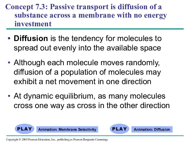
- 34. Concept 7.3: Passive transport is diffusion of a substance across a membrane with no energy investment
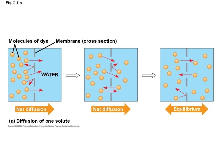
- 35. Fig. 7-11 Molecules of dye Membrane (cross section) WATER Net diffusion Net diffusion Equilibrium (a) Diffusion
- 36. Molecules of dye Fig. 7-11a Membrane (cross section) WATER Net diffusion Net diffusion (a) Diffusion of
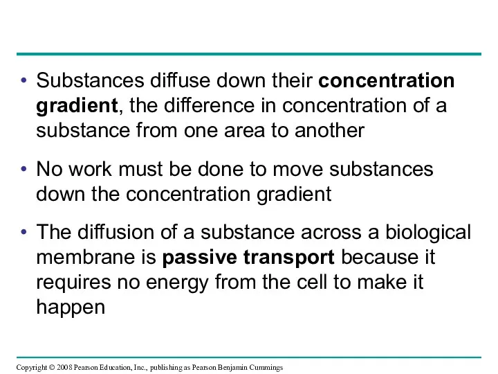
- 37. Substances diffuse down their concentration gradient, the difference in concentration of a substance from one area
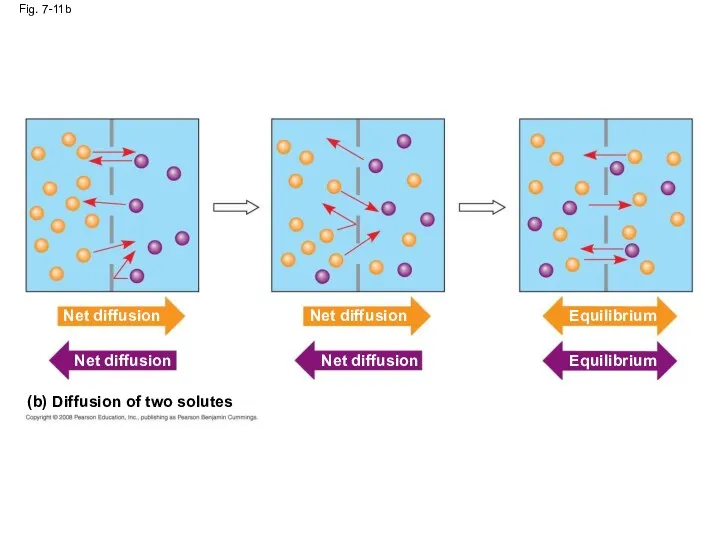
- 38. (b) Diffusion of two solutes Fig. 7-11b Net diffusion Net diffusion Net diffusion Net diffusion Equilibrium
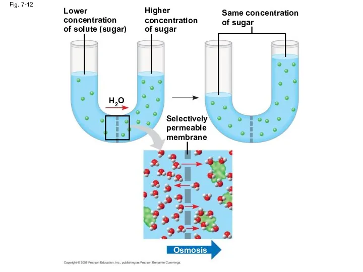
- 39. Effects of Osmosis on Water Balance Osmosis is the diffusion of water across a selectively permeable
- 40. Lower concentration of solute (sugar) Fig. 7-12 H2O Higher concentration of sugar Selectively permeable membrane Same
- 41. Water Balance of Cells Without Walls Tonicity is the ability of a solution to cause a
- 42. Fig. 7-13 Hypotonic solution (a) Animal cell (b) Plant cell H2O Lysed H2O Turgid (normal) H2O
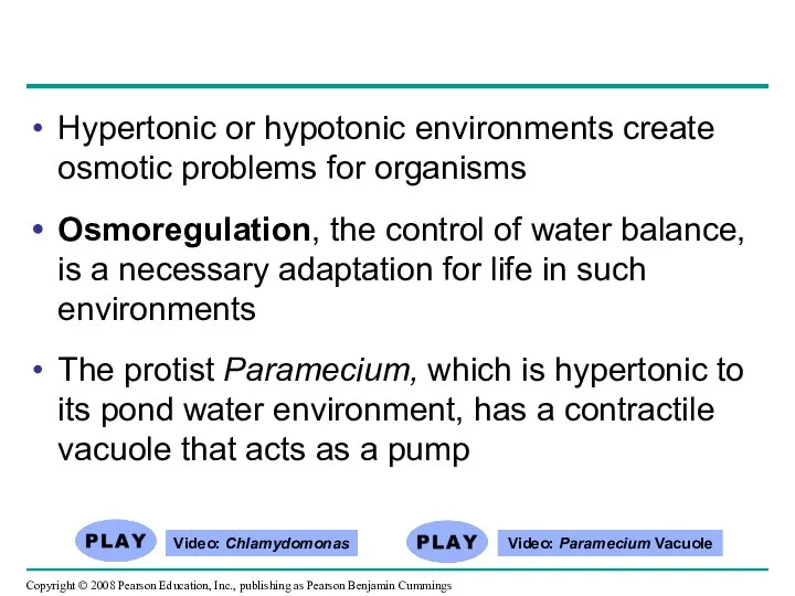
- 43. Hypertonic or hypotonic environments create osmotic problems for organisms Osmoregulation, the control of water balance, is
- 44. Fig. 7-14 Filling vacuole 50 µm (a) A contractile vacuole fills with fluid that enters from
- 45. Water Balance of Cells with Walls Cell walls help maintain water balance A plant cell in
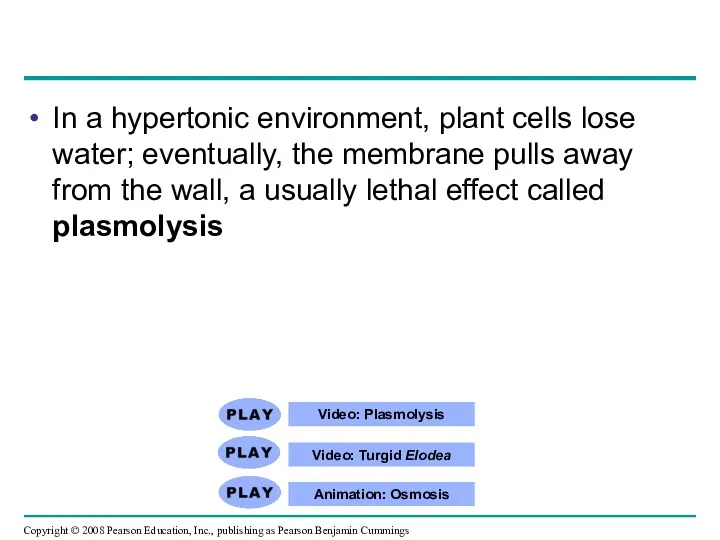
- 46. Video: Plasmolysis Video: Turgid Elodea Animation: Osmosis In a hypertonic environment, plant cells lose water; eventually,
- 47. Facilitated Diffusion: Passive Transport Aided by Proteins In facilitated diffusion, transport proteins speed the passive movement
- 48. Fig. 7-15 EXTRACELLULAR FLUID Channel protein (a) A channel protein Solute CYTOPLASM Solute Carrier protein (b)
- 49. Carrier proteins undergo a subtle change in shape that translocates the solute-binding site across the membrane
- 50. Some diseases are caused by malfunctions in specific transport systems, for example the kidney disease cystinuria
- 51. Concept 7.4: Active transport uses energy to move solutes against their gradients Facilitated diffusion is still
- 52. The Need for Energy in Active Transport Active transport moves substances against their concentration gradient Active
- 53. Active transport allows cells to maintain concentration gradients that differ from their surroundings The sodium-potassium pump
- 54. Fig. 7-16-1 EXTRACELLULAR FLUID [Na+] high [K+] low Na+ Na+ Na+ [Na+] low [K+] high CYTOPLASM
- 55. Na+ binding stimulates phosphorylation by ATP. Fig. 7-16-2 Na+ Na+ Na+ ATP P ADP 2
- 56. Fig. 7-16-3 Phosphorylation causes the protein to change its shape. Na+ is expelled to the outside.
- 57. Fig. 7-16-4 K+ binds on the extracellular side and triggers release of the phosphate group. P
- 58. Fig. 7-16-5 Loss of the phosphate restores the protein’s original shape. K+ K+ 5
- 59. Fig. 7-16-6 K+ is released, and the cycle repeats. K+ K+ 6
- 60. 2 EXTRACELLULAR FLUID [Na+] high [K+] low [Na+] low [K+] high Na+ Na+ Na+ Na+ Na+
- 61. Fig. 7-17 Passive transport Diffusion Facilitated diffusion Active transport ATP
- 62. How Ion Pumps Maintain Membrane Potential Membrane potential is the voltage difference across a membrane Voltage
- 63. Two combined forces, collectively called the electrochemical gradient, drive the diffusion of ions across a membrane:
- 64. An electrogenic pump is a transport protein that generates voltage across a membrane The sodium-potassium pump
- 65. Fig. 7-18 EXTRACELLULAR FLUID H+ H+ H+ H+ Proton pump + + + H+ H+ +
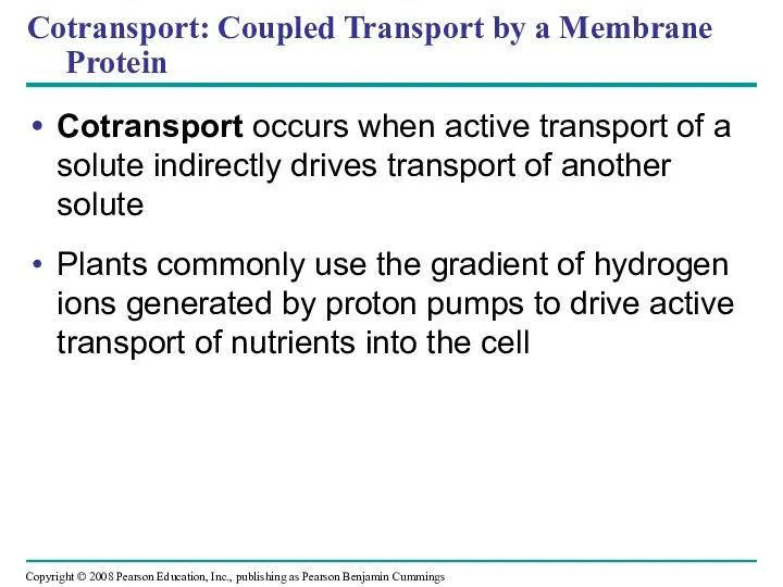
- 66. Cotransport: Coupled Transport by a Membrane Protein Cotransport occurs when active transport of a solute indirectly
- 67. Fig. 7-19 Proton pump – – – – – – + + + + + +
- 68. Concept 7.5: Bulk transport across the plasma membrane occurs by exocytosis and endocytosis Small molecules and
- 69. Exocytosis In exocytosis, transport vesicles migrate to the membrane, fuse with it, and release their contents
- 70. Endocytosis In endocytosis, the cell takes in macromolecules by forming vesicles from the plasma membrane Endocytosis
- 71. In phagocytosis a cell engulfs a particle in a vacuole The vacuole fuses with a lysosome
- 72. Fig. 7-20 PHAGOCYTOSIS EXTRACELLULAR FLUID CYTOPLASM Pseudopodium “Food”or other particle Food vacuole PINOCYTOSIS 1 µm Pseudopodium
- 73. Fig. 7-20a PHAGOCYTOSIS CYTOPLASM EXTRACELLULAR FLUID Pseudopodium “Food” or other particle Food vacuole Food vacuole Bacterium
- 74. In pinocytosis, molecules are taken up when extracellular fluid is “gulped” into tiny vesicles Animation: Pinocytosis
- 75. Fig. 7-20b PINOCYTOSIS Plasma membrane Vesicle 0.5 µm Pinocytosis vesicles forming (arrows) in a cell lining
- 76. In receptor-mediated endocytosis, binding of ligands to receptors triggers vesicle formation A ligand is any molecule
- 77. Fig. 7-20c RECEPTOR-MEDIATED ENDOCYTOSIS Receptor Coat protein Coated pit Ligand Coat protein Plasma membrane 0.25 µm
- 78. Fig. 7-UN1 Passive transport: Facilitated diffusion Channel protein Carrier protein
- 79. Fig. 7-UN2 Active transport: ATP
- 80. Fig. 7-UN3 Environment: 0.01 M sucrose 0.01 M glucose 0.01 M fructose “Cell” 0.03 M sucrose
- 81. Fig. 7-UN4
- 82. You should now be able to: Define the following terms: amphipathic molecules, aquaporins, diffusion Explain how
- 84. Скачать презентацию
Overview: Life at the Edge
The plasma membrane is the boundary that
Overview: Life at the Edge
The plasma membrane is the boundary that
Fig. 7-1
Fig. 7-1
Concept 7.1: Cellular membranes are fluid mosaics of lipids and proteins
Phospholipids
Concept 7.1: Cellular membranes are fluid mosaics of lipids and proteins
Phospholipids
Membrane Models: Scientific Inquiry
Membranes have been chemically analyzed and found to
Membrane Models: Scientific Inquiry
Membranes have been chemically analyzed and found to
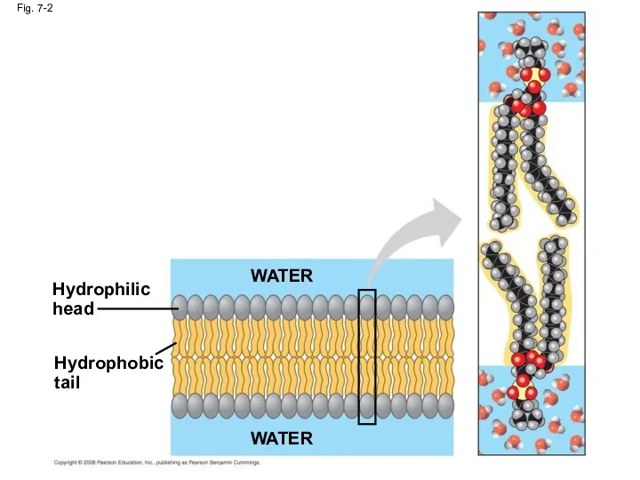
Fig. 7-2
Hydrophilic
head
WATER
Hydrophobic
tail
WATER
Fig. 7-2
Hydrophilic
head
WATER
Hydrophobic
tail
WATER
In 1935, Hugh Davson and James Danielli proposed a sandwich model
In 1935, Hugh Davson and James Danielli proposed a sandwich model
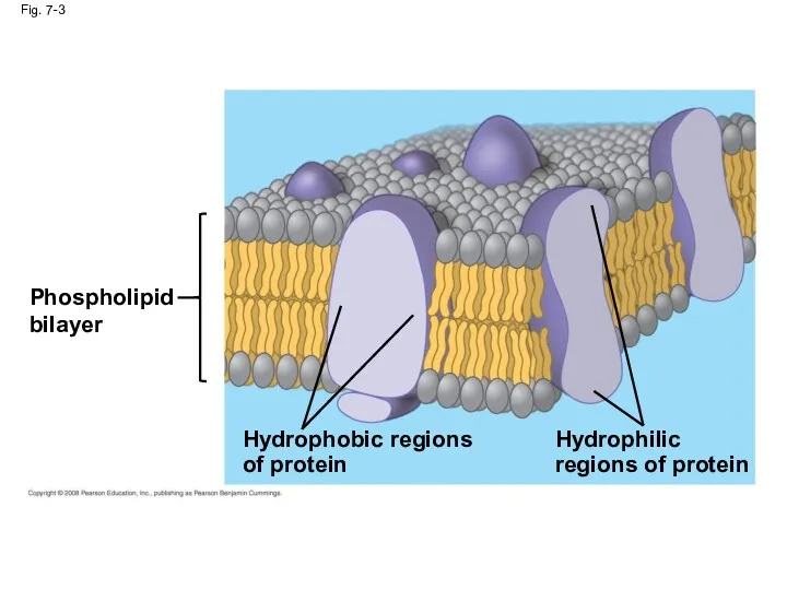
Fig. 7-3
Phospholipid
bilayer
Hydrophobic regions
of protein
Hydrophilic
regions of protein
Fig. 7-3
Phospholipid
bilayer
Hydrophobic regions
of protein
Hydrophilic
regions of protein
Freeze-fracture studies of the plasma membrane supported the fluid mosaic model
Freeze-fracture studies of the plasma membrane supported the fluid mosaic model
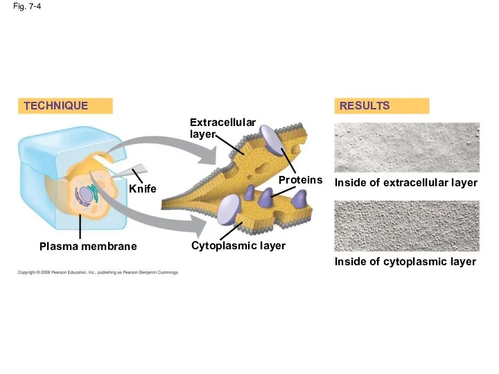
Fig. 7-4
TECHNIQUE
Extracellular
layer
Knife
Proteins
Inside of extracellular layer
RESULTS
Inside of cytoplasmic layer
Cytoplasmic layer
Plasma membrane
Fig. 7-4
TECHNIQUE
Extracellular
layer
Knife
Proteins
Inside of extracellular layer
RESULTS
Inside of cytoplasmic layer
Cytoplasmic layer
Plasma membrane
The Fluidity of Membranes
Phospholipids in the plasma membrane can move within
The Fluidity of Membranes
Phospholipids in the plasma membrane can move within
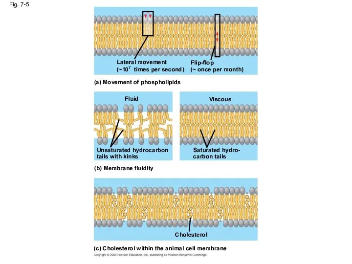
Fig. 7-5
Lateral movement
(~107 times per second)
Flip-flop
(~ once per month)
(a) Movement of
Fig. 7-5
Lateral movement
(~107 times per second)
Flip-flop
(~ once per month)
(a) Movement of
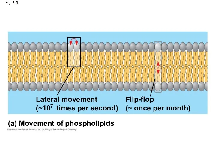
Fig. 7-5a
(a) Movement of phospholipids
Lateral movement
(~107 times per second)
Flip-flop
(~ once per
Fig. 7-5a
(a) Movement of phospholipids
Lateral movement
(~107 times per second)
Flip-flop
(~ once per
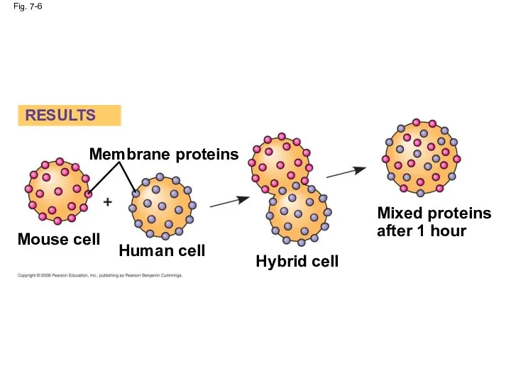
Fig. 7-6
RESULTS
Membrane proteins
Mouse cell
Human cell
Hybrid cell
Mixed proteins
after 1 hour
Fig. 7-6
RESULTS
Membrane proteins
Mouse cell
Human cell
Hybrid cell
Mixed proteins
after 1 hour
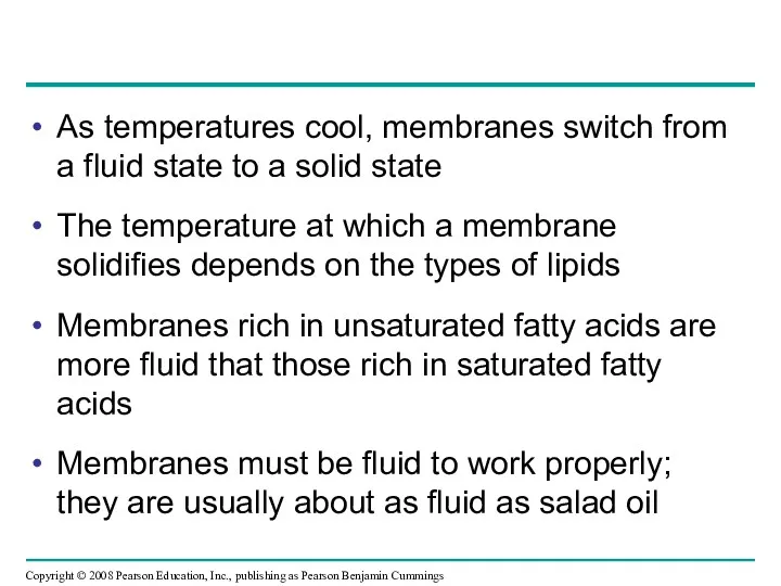
As temperatures cool, membranes switch from a fluid state to a
As temperatures cool, membranes switch from a fluid state to a
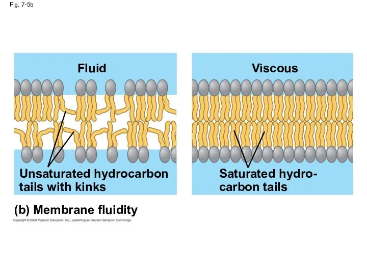
Fig. 7-5b
(b) Membrane fluidity
Fluid
Unsaturated hydrocarbon
tails with kinks
Viscous
Saturated hydro-
carbon tails
Fig. 7-5b
(b) Membrane fluidity
Fluid
Unsaturated hydrocarbon
tails with kinks
Viscous
Saturated hydro-
carbon tails
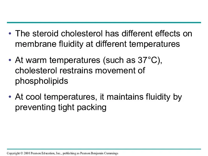
The steroid cholesterol has different effects on membrane fluidity at different
The steroid cholesterol has different effects on membrane fluidity at different
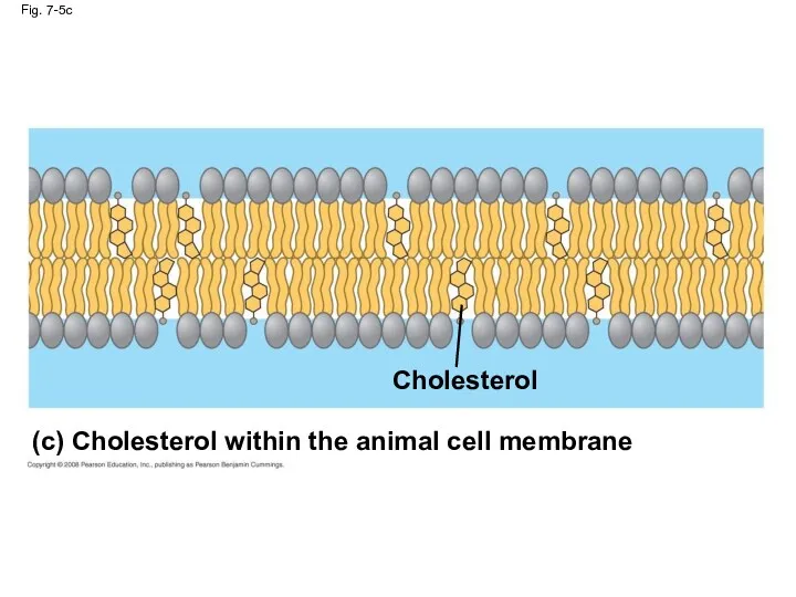
Fig. 7-5c
Cholesterol
(c) Cholesterol within the animal cell membrane
Fig. 7-5c
Cholesterol
(c) Cholesterol within the animal cell membrane
Membrane Proteins and Their Functions
A membrane is a collage of different
Membrane Proteins and Their Functions
A membrane is a collage of different
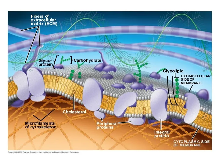
Fig. 7-7
Fibers of
extracellular
matrix (ECM)
Glyco-
protein
Microfilaments
of cytoskeleton
Cholesterol
Peripheral
proteins
Integral
protein
CYTOPLASMIC SIDE
OF MEMBRANE
Glycolipid
EXTRACELLULAR
SIDE OF
MEMBRANE
Carbohydrate
Fig. 7-7
Fibers of
extracellular
matrix (ECM)
Glyco-
protein
Microfilaments
of cytoskeleton
Cholesterol
Peripheral
proteins
Integral
protein
CYTOPLASMIC SIDE
OF MEMBRANE
Glycolipid
EXTRACELLULAR
SIDE OF
MEMBRANE
Carbohydrate
Peripheral proteins are bound to the surface of the membrane
Integral proteins
Peripheral proteins are bound to the surface of the membrane
Integral proteins
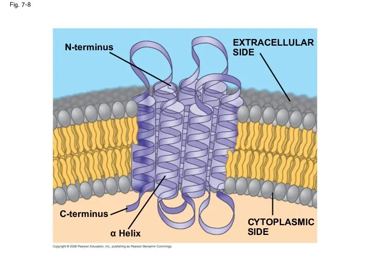
Fig. 7-8
N-terminus
C-terminus
α Helix
CYTOPLASMIC
SIDE
EXTRACELLULAR
SIDE
Fig. 7-8
N-terminus
C-terminus
α Helix
CYTOPLASMIC
SIDE
EXTRACELLULAR
SIDE
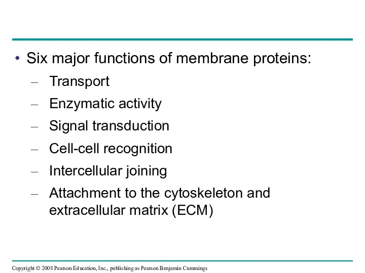
Six major functions of membrane proteins:
Transport
Enzymatic activity
Signal transduction
Cell-cell recognition
Intercellular joining
Attachment to
Six major functions of membrane proteins:
Transport
Enzymatic activity
Signal transduction
Cell-cell recognition
Intercellular joining
Attachment to
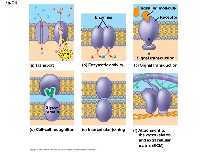
Fig. 7-9
(a) Transport
ATP
(b) Enzymatic activity
Enzymes
(c) Signal transduction
Signal transduction
Signaling molecule
Receptor
(d) Cell-cell recognition
Glyco-
protein
(e)
Fig. 7-9
(a) Transport
ATP
(b) Enzymatic activity
Enzymes
(c) Signal transduction
Signal transduction
Signaling molecule
Receptor
(d) Cell-cell recognition
Glyco-
protein
(e)
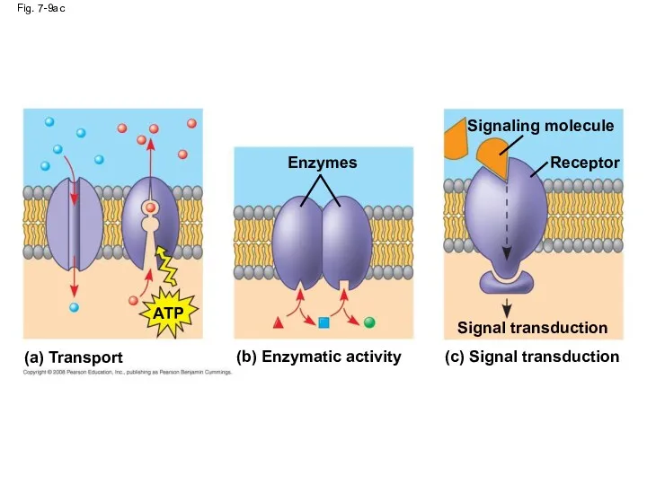
Fig. 7-9ac
(a) Transport
(b) Enzymatic activity
(c) Signal transduction
ATP
Enzymes
Signal transduction
Signaling molecule
Receptor
Fig. 7-9ac
(a) Transport
(b) Enzymatic activity
(c) Signal transduction
ATP
Enzymes
Signal transduction
Signaling molecule
Receptor
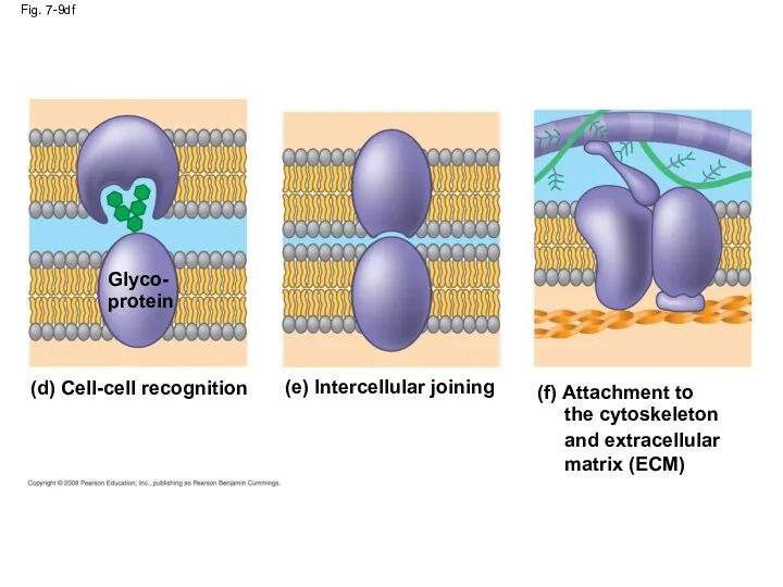
Fig. 7-9df
(d) Cell-cell recognition
Glyco-
protein
(e) Intercellular joining
(f) Attachment to
the cytoskeleton
and
Fig. 7-9df
(d) Cell-cell recognition
Glyco-
protein
(e) Intercellular joining
(f) Attachment to
the cytoskeleton
and
The Role of Membrane Carbohydrates in Cell-Cell Recognition
Cells recognize each other
The Role of Membrane Carbohydrates in Cell-Cell Recognition
Cells recognize each other
Synthesis and Sidedness of Membranes
Membranes have distinct inside and outside faces
The
Synthesis and Sidedness of Membranes
Membranes have distinct inside and outside faces
The
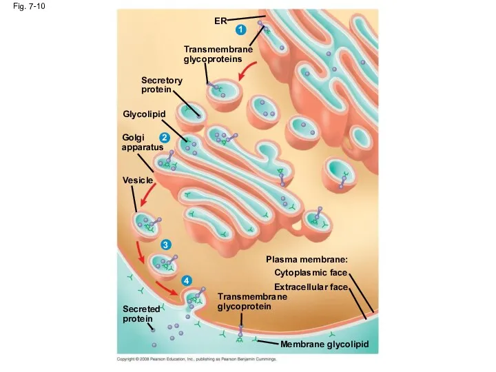
Fig. 7-10
ER
1
Transmembrane
glycoproteins
Secretory
protein
Glycolipid
2
Golgi
apparatus
Vesicle
3
4
Secreted
protein
Transmembrane
glycoprotein
Plasma membrane:
Cytoplasmic face
Extracellular face
Membrane glycolipid
Fig. 7-10
ER
1
Transmembrane
glycoproteins
Secretory
protein
Glycolipid
2
Golgi
apparatus
Vesicle
3
4
Secreted
protein
Transmembrane
glycoprotein
Plasma membrane:
Cytoplasmic face
Extracellular face
Membrane glycolipid
Concept 7.2: Membrane structure results in selective permeability
A cell must exchange
Concept 7.2: Membrane structure results in selective permeability
A cell must exchange
The Permeability of the Lipid Bilayer
Hydrophobic (nonpolar) molecules, such as hydrocarbons,
The Permeability of the Lipid Bilayer
Hydrophobic (nonpolar) molecules, such as hydrocarbons,
Transport Proteins
Transport proteins allow passage of hydrophilic substances across the membrane
Some
Transport Proteins
Transport proteins allow passage of hydrophilic substances across the membrane
Some
Other transport proteins, called carrier proteins, bind to molecules and change
Other transport proteins, called carrier proteins, bind to molecules and change
Concept 7.3: Passive transport is diffusion of a substance across a
Concept 7.3: Passive transport is diffusion of a substance across a
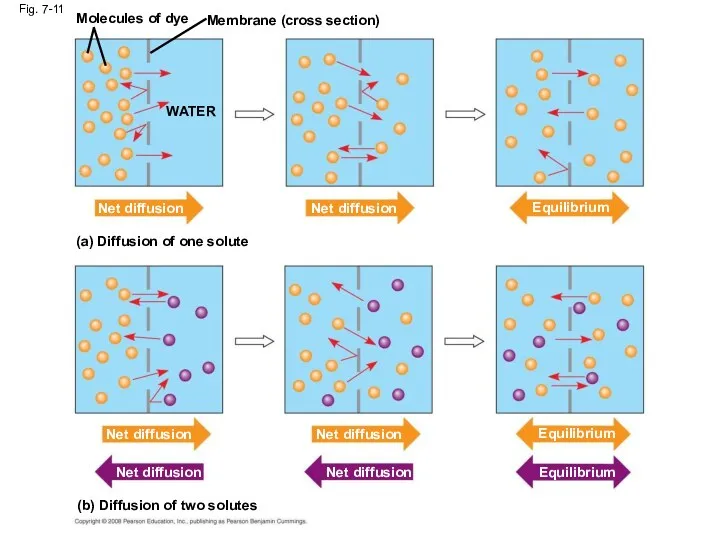
Fig. 7-11
Molecules of dye
Membrane (cross section)
WATER
Net diffusion
Net diffusion
Equilibrium
(a) Diffusion of one
Fig. 7-11
Molecules of dye
Membrane (cross section)
WATER
Net diffusion
Net diffusion
Equilibrium
(a) Diffusion of one
Molecules of dye
Fig. 7-11a
Membrane (cross section)
WATER
Net diffusion
Net diffusion
(a) Diffusion of one
Molecules of dye
Fig. 7-11a
Membrane (cross section)
WATER
Net diffusion
Net diffusion
(a) Diffusion of one
Substances diffuse down their concentration gradient, the difference in concentration of
Substances diffuse down their concentration gradient, the difference in concentration of
(b) Diffusion of two solutes
Fig. 7-11b
Net diffusion
Net diffusion
Net diffusion
Net diffusion
Equilibrium
Equilibrium
(b) Diffusion of two solutes
Fig. 7-11b
Net diffusion
Net diffusion
Net diffusion
Net diffusion
Equilibrium
Equilibrium
Effects of Osmosis on Water Balance
Osmosis is the diffusion of water
Effects of Osmosis on Water Balance
Osmosis is the diffusion of water
Lower
concentration
of solute (sugar)
Fig. 7-12
H2O
Higher concentration
of sugar
Selectively
permeable
membrane
Same concentration
of sugar
Osmosis
Lower
concentration
of solute (sugar)
Fig. 7-12
H2O
Higher concentration
of sugar
Selectively
permeable
membrane
Same concentration
of sugar
Osmosis
Water Balance of Cells Without Walls
Tonicity is the ability of a
Water Balance of Cells Without Walls
Tonicity is the ability of a
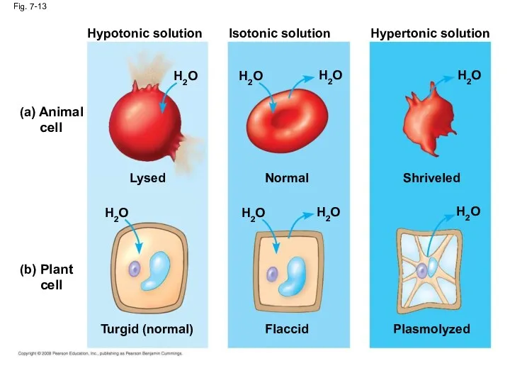
Fig. 7-13
Hypotonic solution
(a) Animal
cell
(b) Plant
cell
H2O
Lysed
H2O
Turgid (normal)
H2O
H2O
H2O
H2O
Normal
Isotonic solution
Flaccid
H2O
H2O
Shriveled
Plasmolyzed
Hypertonic solution
Fig. 7-13
Hypotonic solution
(a) Animal
cell
(b) Plant
cell
H2O
Lysed
H2O
Turgid (normal)
H2O
H2O
H2O
H2O
Normal
Isotonic solution
Flaccid
H2O
H2O
Shriveled
Plasmolyzed
Hypertonic solution
Hypertonic or hypotonic environments create osmotic problems for organisms
Osmoregulation, the control
Hypertonic or hypotonic environments create osmotic problems for organisms
Osmoregulation, the control
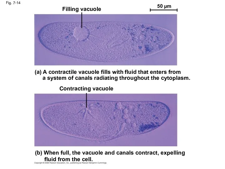
Fig. 7-14
Filling vacuole
50 µm
(a) A contractile vacuole fills with fluid
Fig. 7-14
Filling vacuole
50 µm
(a) A contractile vacuole fills with fluid
Water Balance of Cells with Walls
Cell walls help maintain water balance
A
Water Balance of Cells with Walls
Cell walls help maintain water balance
A
Video: Plasmolysis
Video: Turgid Elodea
Animation: Osmosis
In a hypertonic environment, plant cells lose
Video: Plasmolysis
Video: Turgid Elodea
Animation: Osmosis
In a hypertonic environment, plant cells lose
Facilitated Diffusion: Passive Transport Aided by Proteins
In facilitated diffusion, transport proteins
Facilitated Diffusion: Passive Transport Aided by Proteins
In facilitated diffusion, transport proteins
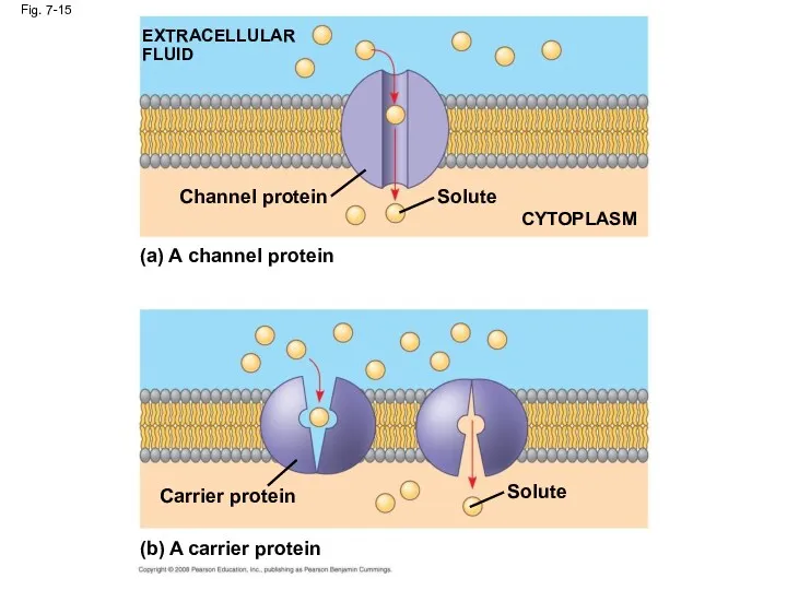
Fig. 7-15
EXTRACELLULAR FLUID
Channel protein
(a) A channel protein
Solute
CYTOPLASM
Fig. 7-15
EXTRACELLULAR FLUID
Channel protein
(a) A channel protein
Solute
CYTOPLASM
Carrier proteins undergo a subtle change in shape that translocates the
Carrier proteins undergo a subtle change in shape that translocates the
Some diseases are caused by malfunctions in specific transport systems, for
Some diseases are caused by malfunctions in specific transport systems, for
Concept 7.4: Active transport uses energy to move solutes against their
Concept 7.4: Active transport uses energy to move solutes against their
The Need for Energy in Active Transport
Active transport moves substances against
The Need for Energy in Active Transport
Active transport moves substances against
Active transport allows cells to maintain concentration gradients that differ from
Active transport allows cells to maintain concentration gradients that differ from
![Fig. 7-16-1 EXTRACELLULAR FLUID [Na+] high [K+] low Na+ Na+](/_ipx/f_webp&q_80&fit_contain&s_1440x1080/imagesDir/jpg/355141/slide-53.jpg)
Fig. 7-16-1
EXTRACELLULAR
FLUID
[Na+] high
[K+] low
Na+
Na+
Na+
[Na+] low
[K+]
Fig. 7-16-1
EXTRACELLULAR
FLUID
[Na+] high
[K+] low
Na+
Na+
Na+
[Na+] low
[K+]
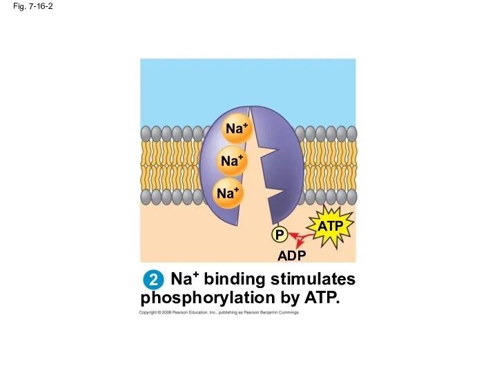
Na+ binding stimulates
phosphorylation by ATP.
Fig. 7-16-2
Na+
Na+
Na+
ATP
Na+ binding stimulates
phosphorylation by ATP.
Fig. 7-16-2
Na+
Na+
Na+
ATP
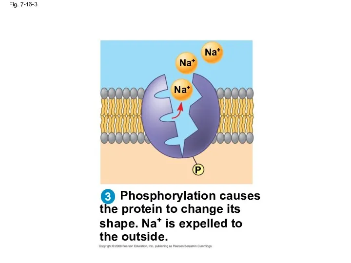
Fig. 7-16-3
Phosphorylation causes
the protein to change its
shape. Na+ is expelled
Fig. 7-16-3
Phosphorylation causes
the protein to change its
shape. Na+ is expelled
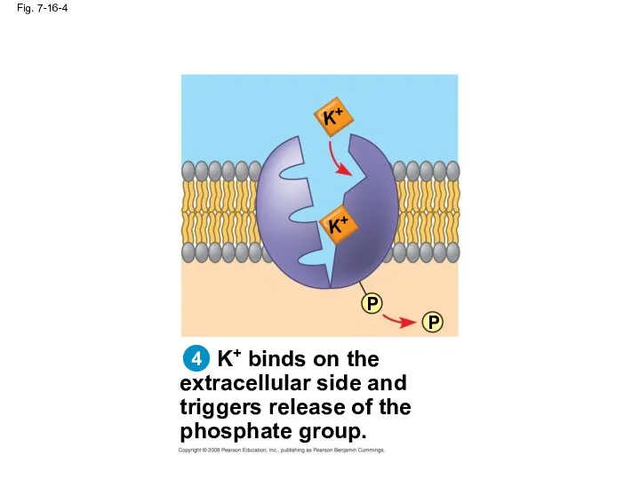
Fig. 7-16-4
K+ binds on the
extracellular side and
triggers release of the
phosphate
Fig. 7-16-4
K+ binds on the
extracellular side and
triggers release of the
phosphate
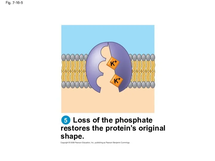
Fig. 7-16-5
Loss of the phosphate
restores the protein’s original
shape.
K+
K+
Fig. 7-16-5
Loss of the phosphate
restores the protein’s original
shape.
K+
K+
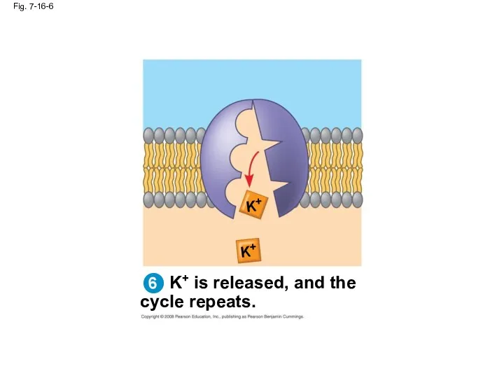
Fig. 7-16-6
K+ is released, and the
cycle repeats.
K+
K+
6
Fig. 7-16-6
K+ is released, and the
cycle repeats.
K+
K+
6
![2 EXTRACELLULAR FLUID [Na+] high [K+] low [Na+] low [K+]](/_ipx/f_webp&q_80&fit_contain&s_1440x1080/imagesDir/jpg/355141/slide-59.jpg)
2
EXTRACELLULAR
FLUID
[Na+] high
[K+] low
[Na+] low
[K+] high
Na+
Na+
Na+
2
EXTRACELLULAR
FLUID
[Na+] high
[K+] low
[Na+] low
[K+] high
Na+
Na+
Na+
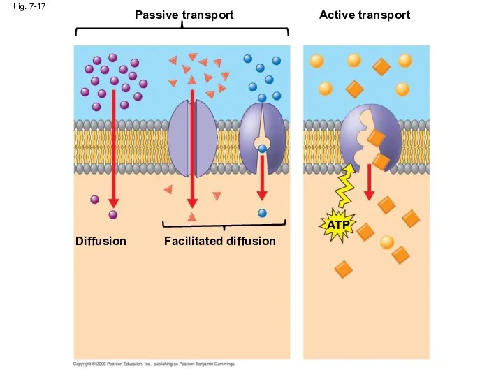
Fig. 7-17
Passive transport
Diffusion
Facilitated diffusion
Active transport
ATP
Fig. 7-17
Passive transport
Diffusion
Facilitated diffusion
Active transport
ATP
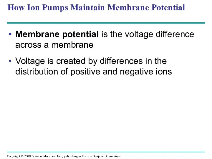
How Ion Pumps Maintain Membrane Potential
Membrane potential is the voltage difference
How Ion Pumps Maintain Membrane Potential
Membrane potential is the voltage difference
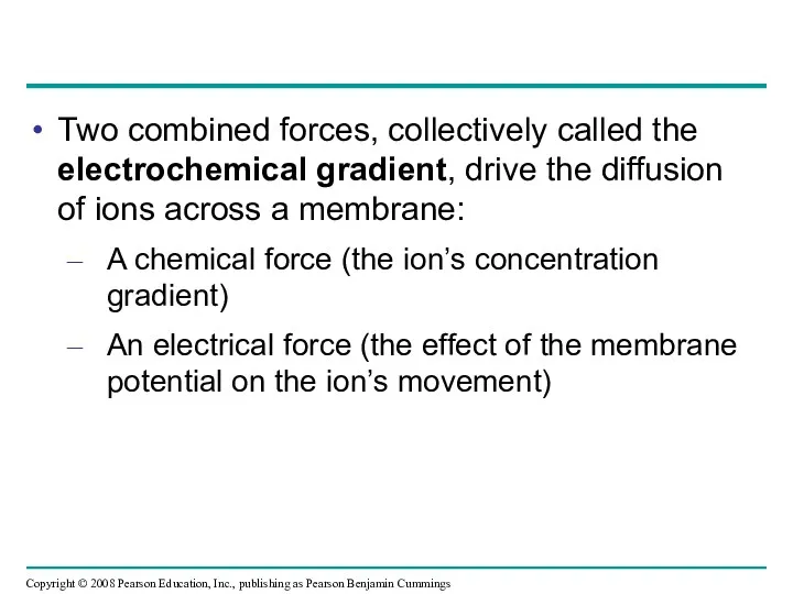
Two combined forces, collectively called the electrochemical gradient, drive the diffusion
Two combined forces, collectively called the electrochemical gradient, drive the diffusion
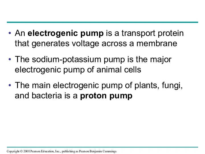
An electrogenic pump is a transport protein that generates voltage across
An electrogenic pump is a transport protein that generates voltage across
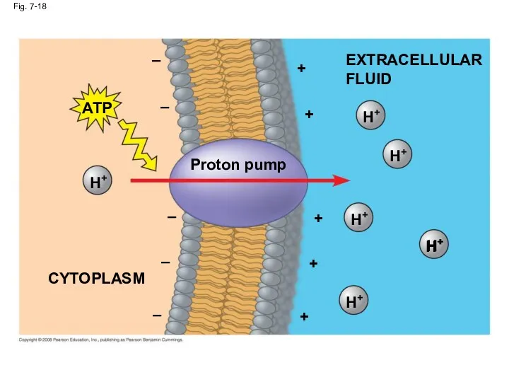
Fig. 7-18
EXTRACELLULAR
FLUID
H+
H+
H+
H+
Proton pump
+
+
+
Fig. 7-18
EXTRACELLULAR
FLUID
H+
H+
H+
H+
Proton pump
+
+
+
Cotransport: Coupled Transport by a Membrane Protein
Cotransport occurs when active transport
Cotransport: Coupled Transport by a Membrane Protein
Cotransport occurs when active transport
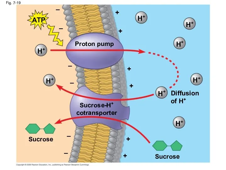
Fig. 7-19
Proton pump
–
–
–
–
–
–
+
+
+
+
+
+
ATP
H+
H+
H+
H+
H+
H+
H+
H+
Diffusion
of H+
Sucrose-H+
cotransporter
Fig. 7-19
Proton pump
–
–
–
–
–
–
+
+
+
+
+
+
ATP
H+
H+
H+
H+
H+
H+
H+
H+
Diffusion
of H+
Sucrose-H+
cotransporter
Concept 7.5: Bulk transport across the plasma membrane occurs by exocytosis
Concept 7.5: Bulk transport across the plasma membrane occurs by exocytosis
Exocytosis
In exocytosis, transport vesicles migrate to the membrane, fuse with it,
Exocytosis
In exocytosis, transport vesicles migrate to the membrane, fuse with it,
Endocytosis
In endocytosis, the cell takes in macromolecules by forming vesicles from
Endocytosis
In endocytosis, the cell takes in macromolecules by forming vesicles from
In phagocytosis a cell engulfs a particle in a vacuole
The vacuole
In phagocytosis a cell engulfs a particle in a vacuole
The vacuole
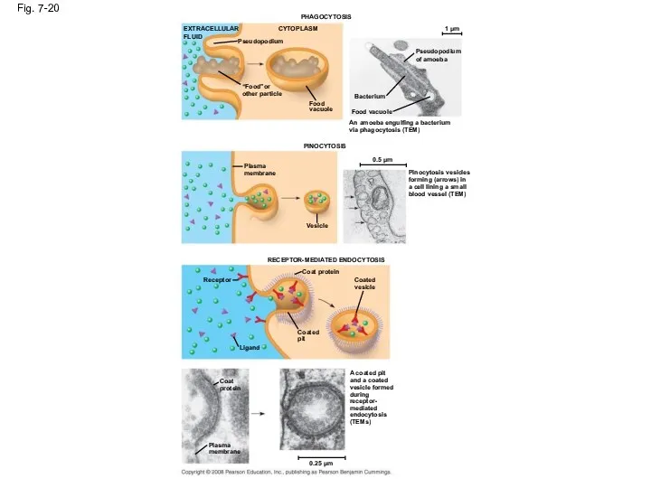
Fig. 7-20
PHAGOCYTOSIS
EXTRACELLULAR
FLUID
CYTOPLASM
Pseudopodium
“Food”or
other particle
Food
vacuole
PINOCYTOSIS
1 µm
Pseudopodium
of
Fig. 7-20
PHAGOCYTOSIS
EXTRACELLULAR
FLUID
CYTOPLASM
Pseudopodium
“Food”or
other particle
Food
vacuole
PINOCYTOSIS
1 µm
Pseudopodium
of
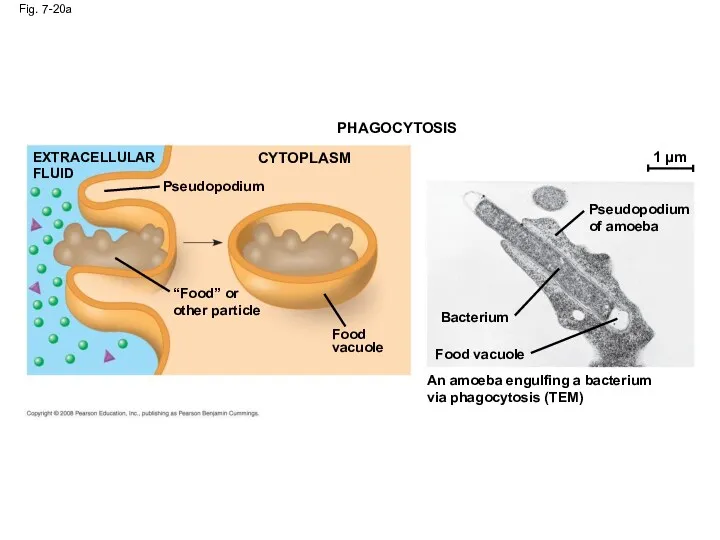
Fig. 7-20a
PHAGOCYTOSIS
CYTOPLASM
EXTRACELLULAR
FLUID
Pseudopodium
“Food” or
other particle
Food
vacuole
Food vacuole
Fig. 7-20a
PHAGOCYTOSIS
CYTOPLASM
EXTRACELLULAR
FLUID
Pseudopodium
“Food” or
other particle
Food
vacuole
Food vacuole
In pinocytosis, molecules are taken up when extracellular fluid is “gulped”
In pinocytosis, molecules are taken up when extracellular fluid is “gulped”
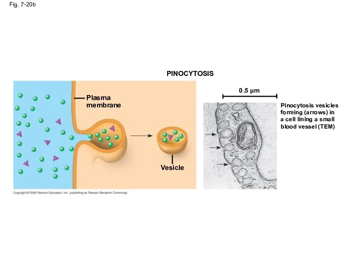
Fig. 7-20b
PINOCYTOSIS
Plasma
membrane
Vesicle
0.5 µm
Pinocytosis vesicles
forming (arrows) in
a cell
Fig. 7-20b
PINOCYTOSIS
Plasma
membrane
Vesicle
0.5 µm
Pinocytosis vesicles
forming (arrows) in
a cell
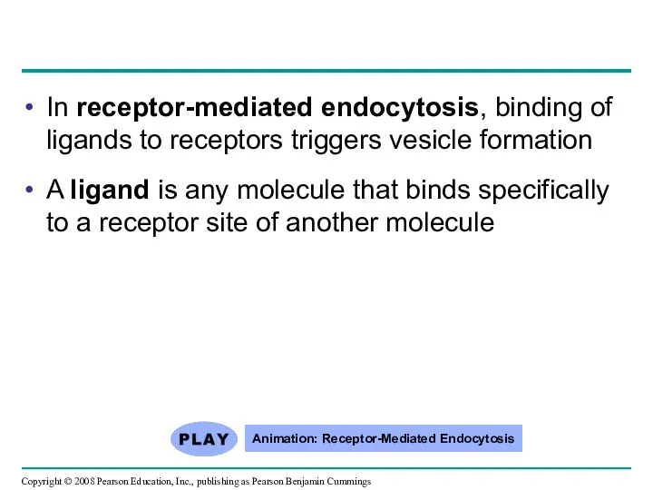
In receptor-mediated endocytosis, binding of ligands to receptors triggers vesicle
In receptor-mediated endocytosis, binding of ligands to receptors triggers vesicle
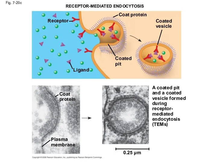
Fig. 7-20c
RECEPTOR-MEDIATED ENDOCYTOSIS
Receptor
Coat protein
Coated
pit
Ligand
Coat
protein
Plasma
membrane
0.25 µm
Coated
vesicle
A coated pit
and
Fig. 7-20c
RECEPTOR-MEDIATED ENDOCYTOSIS
Receptor
Coat protein
Coated
pit
Ligand
Coat
protein
Plasma
membrane
0.25 µm
Coated
vesicle
A coated pit
and
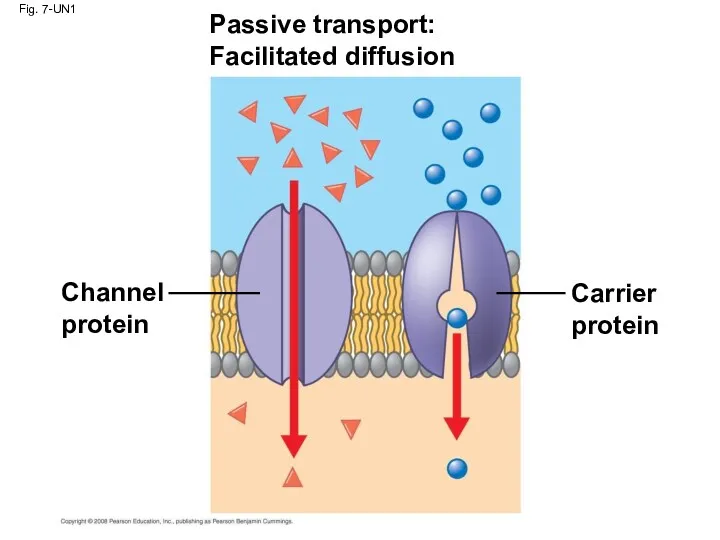
Fig. 7-UN1
Passive transport:
Facilitated diffusion
Channel
protein
Carrier
protein
Fig. 7-UN1
Passive transport:
Facilitated diffusion
Channel
protein
Carrier
protein
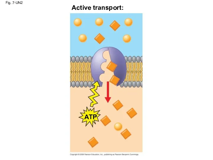
Fig. 7-UN2
Active transport:
ATP
Fig. 7-UN2
Active transport:
ATP
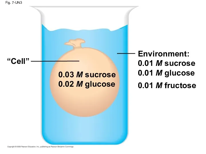
Fig. 7-UN3
Environment:
0.01 M sucrose
0.01 M glucose
0.01 M fructose
“Cell”
0.03 M
Fig. 7-UN3
Environment:
0.01 M sucrose
0.01 M glucose
0.01 M fructose
“Cell”
0.03 M
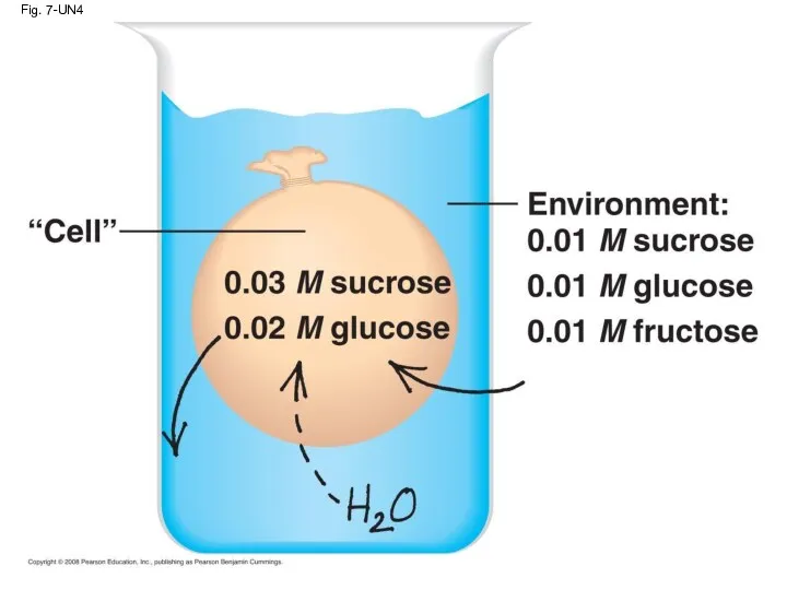
Fig. 7-UN4
Fig. 7-UN4
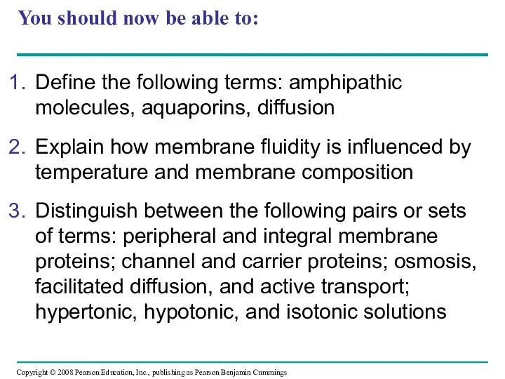
You should now be able to:
Define the following terms: amphipathic molecules,
You should now be able to:
Define the following terms: amphipathic molecules,
 Биологическое и свободное окисление
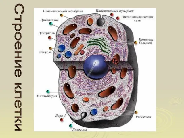 Строение клетки. Этапы формирования и развития представлений о клетке
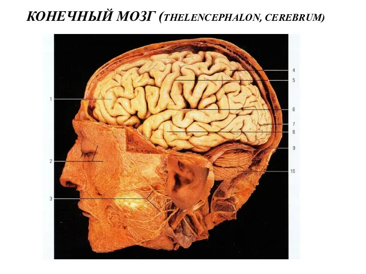 Конечный мозг (thelencephalon, cerebrum)
 Чувствительность. Рецепторы
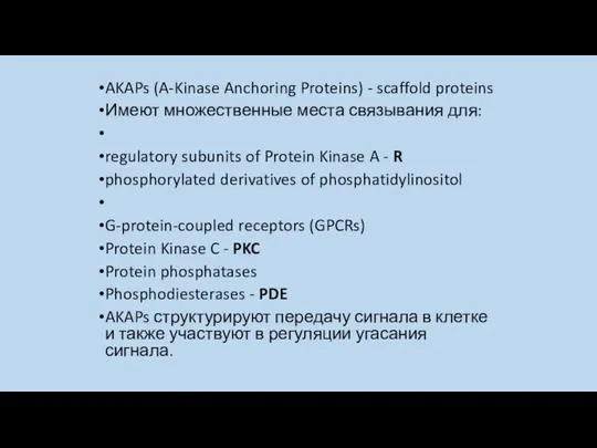 AKAPs. Общий признак всех AKAPs
 В гости к осени. 2 класс
 Ангиология. Введение в изучение сердечно-сосудистой системы
 Будова тварин: клітини
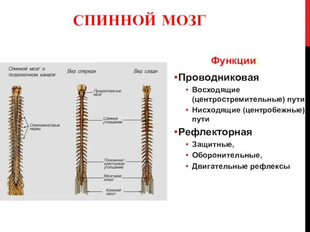 Спинной мозг (medulla spinalis). Лекция №3
 Функциональная анатомия мышц туловища и конечностей
 Skeleton Quiz
 Среда обитания
 Жири. Склад жирів, їх утворення. Жири у природі. Біологічна роль жирів
 Интересные факты о змеях
 Особенности адаптации респираторной системы к длительным физическим нагрузкам
 Маргаритки.Двухлетние цветочные растения.
 Строение волоса
 Биосинтез холестерина, жирных кислот. Липопротеины. Регуляция и патология липидного обмена
 Мутации, виды мутаций и их последствия
 Тип Круглые черви. Тип Кольчатые черви
 Энергетический обмен, катаболизм
 Введение в анатомию. Общая анатомия скелета
 Редуцирленетін және редуцирленбейтін қанттар
 Селекция ягодных культур
 Открытый урок по темеЛист. Внешнее и внутреннее строение
 Хим.состав клетки
 Радиационный контроль яиц
 Защита овощных и цветочных культур в защищенном грунте препаратами ООО Сингента