- Pathologic Protozoa (Lesson 1)

Содержание
- 2. CHARACTERISTICS OF PROTOZOA 1. Unicellular 2. Chemoheterotrophs (get their energy by breaking down organic matter). 3.
- 3. CHARACTERISTICS OF PROTOZOA 4. The vegetative form is the TROPHOZOA (tropho = movement; zoite = animal;
- 4. CHARACTERISTICS OF PROTOZOA 6. Some produce cysts. These are not tissue cysts like a human gets
- 5. TERMS: Host Types The definitive host is the one in which the parasite completes its sexual
- 6. TERMS: Host Types Its sexual life cycle also starts in the human, so that can be
- 7. TERMS Trophozoite: any stage in a protozoa’s life cycle which can ingest food. In practice it
- 9. Phylum Euglenozoa
- 10. MASTIGOPHORA DISEASES Trypanosomiasis Leishmaniasis
- 11. TERMS Mastigote = flagella Promastigote: has single flagella Amastigote: has no flagella Kinetoplast: round mass of
- 12. Phylum Euglenozoa Class Kinetoplastea Order Trypanosomatida Family Trypanosomatidae Genus Trypanosoma
- 13. Trypanosomiasis African Trypanosomiasis (African Sleeping Sickness) American Trypanosomiasis (Chaga’s Disease)
- 14. “African Sleeping Sickness” Disease: African Tryptanosomiasis Causal Agents: Trypanosoma brucei gambiense Trypanosoma brucei rhodesiense
- 15. Trypanosoma life cycle
- 16. Geographic Distribution T. b. gambiense is found in foci in large areas of West and Central
- 17. Trypanosomiasis Trypanosomiasis has a biological vector, the tsetse (pronounced “set-see”) fly. Wild animals may also be
- 18. Trypanosomiasis The tsetse fly bites a human and injects the trypanomastigotes into the skin. This causes
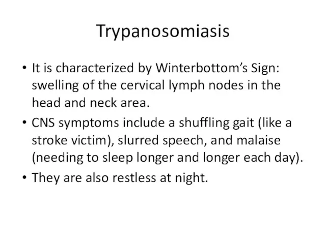
- 19. Trypanosomiasis It is characterized by Winterbottom’s Sign: swelling of the cervical lymph nodes in the head
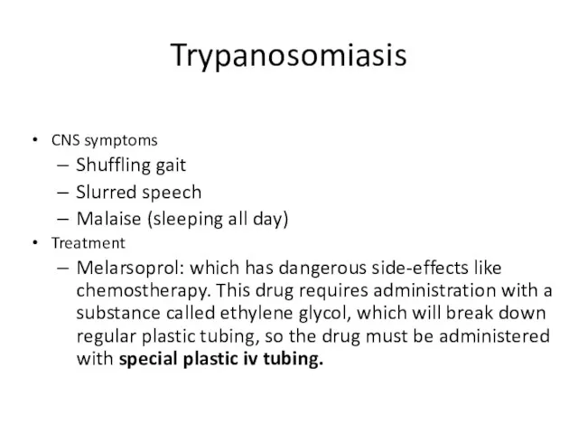
- 20. Trypanosomiasis CNS symptoms Shuffling gait Slurred speech Malaise (sleeping all day) Treatment Melarsoprol: which has dangerous
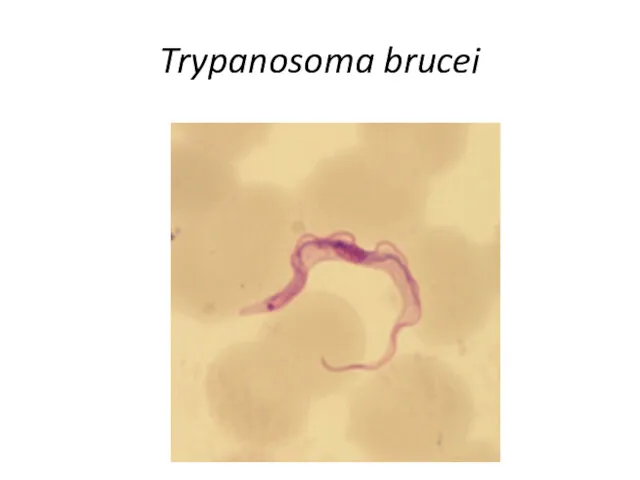
- 21. Trypanosoma brucei Trypomastigote stages are the only form found in patients. Posterior kinetoplast Centrally located nucleus
- 22. Trypanosoma brucei
- 23. Trypanosoma brucei gambiense trypomastigote
- 24. Trypanosoma
- 25. Trypanosoma brucei rhodesiense
- 27. Trypanosoma brucei UM
- 28. Tsetse Fly
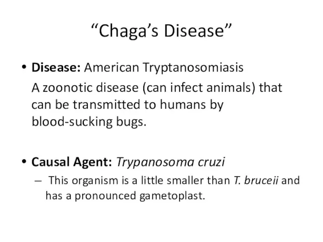
- 29. “Chaga’s Disease” Disease: American Tryptanosomiasis A zoonotic disease (can infect animals) that can be transmitted to
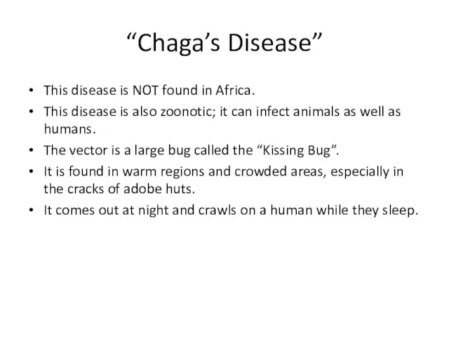
- 30. “Chaga’s Disease” This disease is NOT found in Africa. This disease is also zoonotic; it can
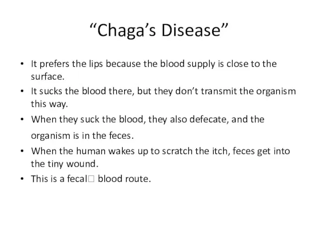
- 31. “Chaga’s Disease” It prefers the lips because the blood supply is close to the surface. It
- 32. “Chaga’s Disease” Symptoms include fever, anorexia, swollen lymph nodes, hepatosplenomegally (enlarged liver and spleen), and myocarditis
- 33. Trypanosoma life cycle
- 34. Trypanosoma cruzi Insect vector is the “kissing” bug. It takes a blood meal and releases trypomastigotes
- 35. Trypanosoma cruzi Geographic Distribution: The Americas from the southern United States to southern Argentina. Mostly in
- 36. Trypanosoma cruzi
- 37. Trypanosoma cruzi
- 38. Trypanosoma cruzi large kinetoplast
- 39. Trypanosoma cruzi Triatomine bug, Trypanosoma cruzi vector, defecating on the wound after taking a blood meal.
- 40. Kissing Bug
- 41. Romana’s sign Swollen eye, seen in Chagra’s disease.
- 42. TERMS Promastigote: has single flagella Amastigote: has no flagella Kinetoplast: round mass of circular DNA
- 43. Class Kinetoplastida Order Trypanosomatida Family Trypanosomatidae Genus Leishmania
- 44. Leishmania donovani Disease: Leishmaniasis Vector-borne disease transmitted by sandflies.
- 45. Leishmania Life Cycle Kinetoplast It starts out as a spindle-shaped, single flagellated cell called a promastigote
- 46. Leishmania rosette In prepared slides you can see promastigotes align their nose in a circle, called
- 47. Leishmaniasis rosette
- 48. Leishmania Life Cycle It reproduces in the gut of a female sandfly, and migrates to her
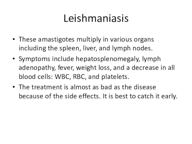
- 49. Leishmaniasis These amastigotes multiply in various organs including the spleen, liver, and lymph nodes. Symptoms include
- 50. Leishmania Life Cycle The female sandflies inject the infective stage, promastigotes, during blood meals. Macrophages phagocytize
- 51. Leishmania life cycle
- 52. Leishmania donovani (Promastigote) Single flagellum found in sand flies
- 53. Leishmaniasis Amastogotes Amastogotes with nucleus and kinetoplast Macrophage rupturing
- 54. Leishmania Amastigotes
- 55. Sandfly This looks like a mosquito, except its body is hairy and the wings are feathery.
- 56. Leishmaniasis Geographic Distribution: More than 90 percent of the world's cases of visceral leishmaniasis are in
- 57. Leishmaniasis There are three forms of Leishmaniasis: Cutaneous Mucocutaneus Visceral
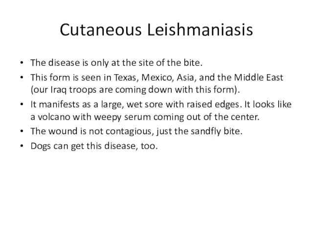
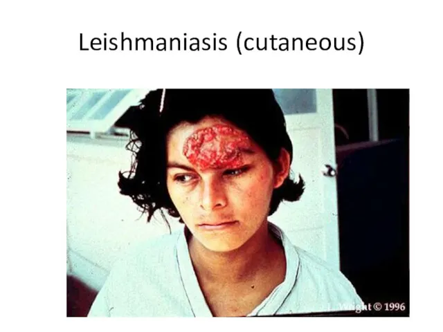
- 58. Cutaneous Leishmaniasis The disease is only at the site of the bite. This form is seen
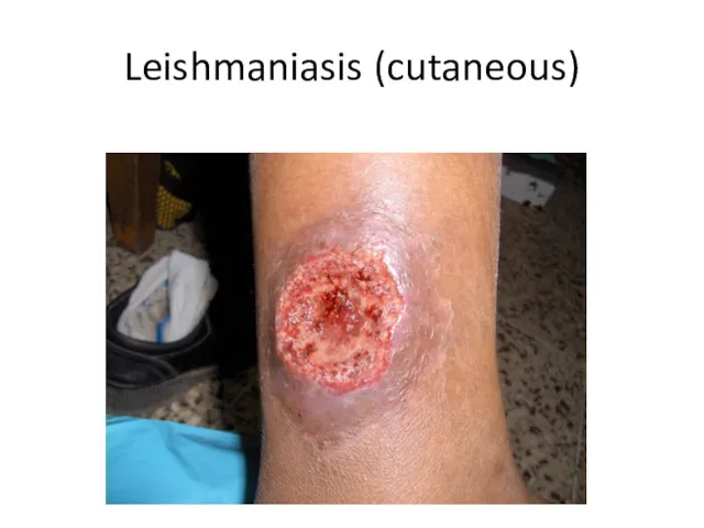
- 59. Leishmaniasis (cutaneous)
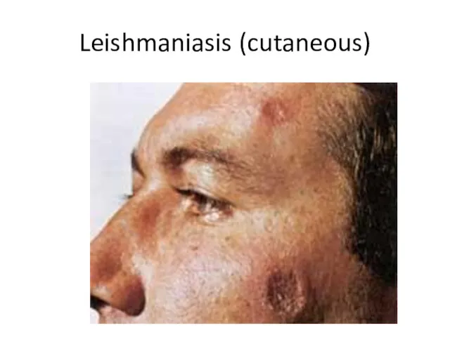
- 60. Leishmaniasis (cutaneous)
- 61. Leishmaniasis (cutaneous)
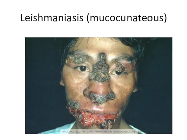
- 62. Leishmaniasis (mucocunateous) This is when the disease located in the mucous membranes of the nose and
- 63. Leishmaniasis (mucocunateous)
- 64. Leishmaniasis (visceral) This is the most serious form. It occurs especially in immunocompromised people, especially HIV
- 65. Kala Azar Hepatosplenomegaly
- 66. Kala Azar (duodenum)
- 67. Phylum Metamonada
- 68. Phylum Metemonada Order Diplomonada Family Hexamitidae Genus Giardia ARCHAEZOA DISEASES
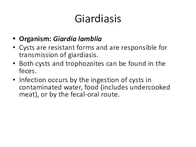
- 69. Giardiasis Organism: Giardia lamblia Cysts are resistant forms and are responsible for transmission of giardiasis. Both
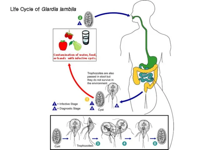
- 70. Life Cycle of Giardia lamblia
- 71. Giardia lamblia In the small intestine, excystation releases trophozoites (each cyst produces two trophozoites). Trophozoites multiply,
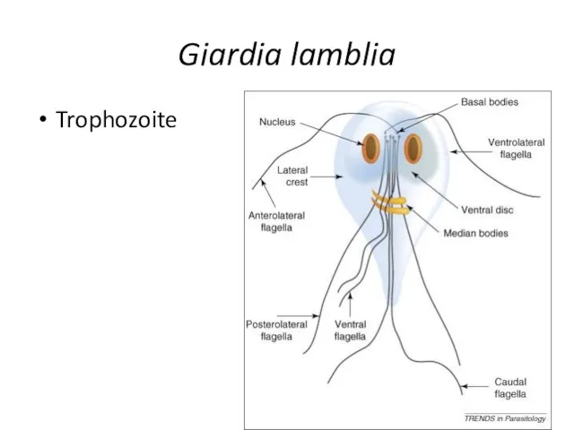
- 72. Giardia lamblia Trophozoite form: piroform (pear or teardrop shape), looks like a happy face. Discovered by
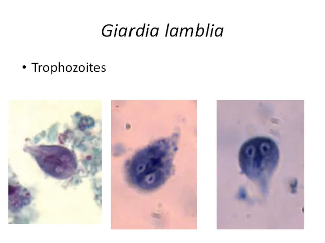
- 73. Giardia lamblia Trophozoite
- 74. Giardia lamblia Trophozoites
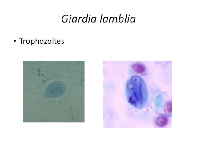
- 75. Giardia lamblia Trophozoites
- 76. Giardia lamblia trophozoite
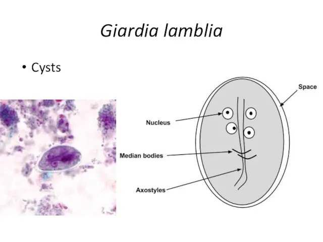
- 77. Giardia lamblia Cysts
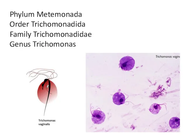
- 78. Phylum Metemonada Order Trichomonadida Family Trichomonadidae Genus Trichomonas
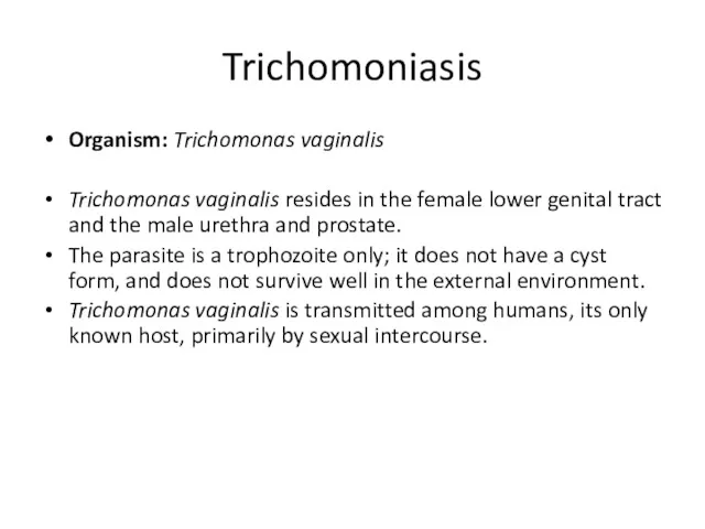
- 79. Trichomoniasis Organism: Trichomonas vaginalis Trichomonas vaginalis resides in the female lower genital tract and the male
- 80. Trichomonas vaginalis life cycle
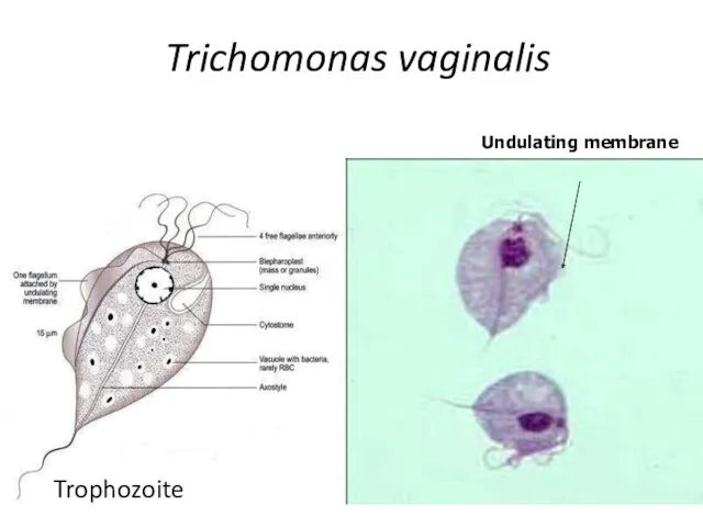
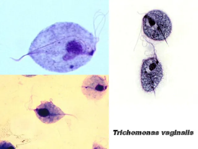
- 81. Trichomonas vaginalis Undulating membrane Trophozoite
- 82. Trichomonas vaginalis
- 84. Скачать презентацию
CHARACTERISTICS OF PROTOZOA
1. Unicellular
2. Chemoheterotrophs (get their energy by breaking down
CHARACTERISTICS OF PROTOZOA
1. Unicellular
2. Chemoheterotrophs (get their energy by breaking down
CHARACTERISTICS OF PROTOZOA
4. The vegetative form is the TROPHOZOA (tropho =
CHARACTERISTICS OF PROTOZOA
4. The vegetative form is the TROPHOZOA (tropho =
CHARACTERISTICS OF PROTOZOA
6. Some produce cysts.
These are not tissue cysts
CHARACTERISTICS OF PROTOZOA
6. Some produce cysts.
These are not tissue cysts
TERMS: Host Types
The definitive host is the one in which the
TERMS: Host Types
The definitive host is the one in which the
TERMS: Host Types
Its sexual life cycle also starts in the human,
TERMS: Host Types
Its sexual life cycle also starts in the human,
TERMS
Trophozoite: any stage in a protozoa’s life cycle which can ingest
TERMS
Trophozoite: any stage in a protozoa’s life cycle which can ingest

Phylum Euglenozoa
Phylum Euglenozoa
MASTIGOPHORA DISEASES
Trypanosomiasis
Leishmaniasis
MASTIGOPHORA DISEASES
Trypanosomiasis
Leishmaniasis
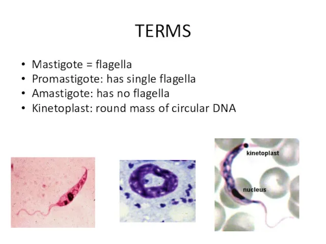
TERMS
Mastigote = flagella
Promastigote: has single flagella
Amastigote: has no flagella
Kinetoplast: round mass
TERMS
Mastigote = flagella
Promastigote: has single flagella
Amastigote: has no flagella
Kinetoplast: round mass
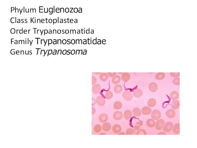
Phylum Euglenozoa
Class Kinetoplastea
Order Trypanosomatida
Family Trypanosomatidae
Genus Trypanosoma
Phylum Euglenozoa
Class Kinetoplastea
Order Trypanosomatida
Family Trypanosomatidae
Genus Trypanosoma
Trypanosomiasis
African Trypanosomiasis
(African Sleeping Sickness)
American Trypanosomiasis
(Chaga’s Disease)
Trypanosomiasis
African Trypanosomiasis
(African Sleeping Sickness)
American Trypanosomiasis
(Chaga’s Disease)
“African Sleeping Sickness”
Disease: African Tryptanosomiasis
Causal Agents:
Trypanosoma brucei gambiense
Trypanosoma brucei rhodesiense
“African Sleeping Sickness”
Disease: African Tryptanosomiasis
Causal Agents:
Trypanosoma brucei gambiense
Trypanosoma brucei rhodesiense
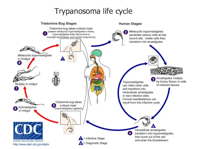
Trypanosoma life cycle
Trypanosoma life cycle
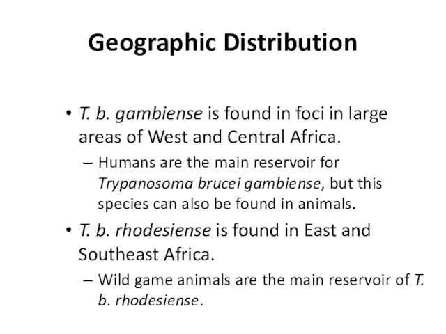
Geographic Distribution
T. b. gambiense is found in foci in large areas
Geographic Distribution
T. b. gambiense is found in foci in large areas
Trypanosomiasis
Trypanosomiasis has a biological vector, the tsetse (pronounced “set-see”) fly.
Wild animals
Trypanosomiasis
Trypanosomiasis has a biological vector, the tsetse (pronounced “set-see”) fly.
Wild animals
Trypanosomiasis
The tsetse fly bites a human and injects the trypanomastigotes into
Trypanosomiasis
The tsetse fly bites a human and injects the trypanomastigotes into
Trypanosomiasis
It is characterized by Winterbottom’s Sign: swelling of the cervical lymph
Trypanosomiasis
It is characterized by Winterbottom’s Sign: swelling of the cervical lymph
Trypanosomiasis
CNS symptoms
Shuffling gait
Slurred speech
Malaise (sleeping all day)
Treatment
Melarsoprol: which has dangerous side-effects
Trypanosomiasis
CNS symptoms
Shuffling gait
Slurred speech
Malaise (sleeping all day)
Treatment
Melarsoprol: which has dangerous side-effects
Trypanosoma brucei
Trypomastigote stages are the only form found in patients.
Posterior kinetoplast
Centrally
Trypanosoma brucei
Trypomastigote stages are the only form found in patients.
Posterior kinetoplast
Centrally
Trypanosoma brucei
Trypanosoma brucei
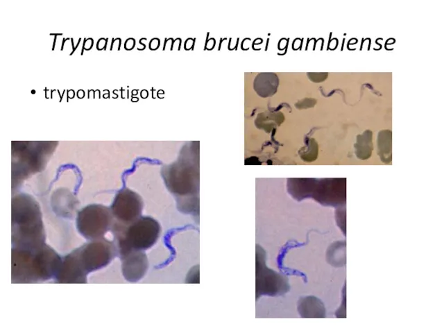
Trypanosoma brucei gambiense
trypomastigote
Trypanosoma brucei gambiense
trypomastigote
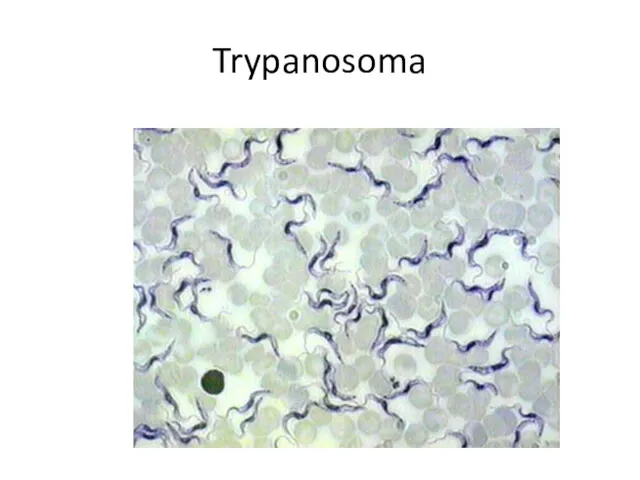
Trypanosoma
Trypanosoma
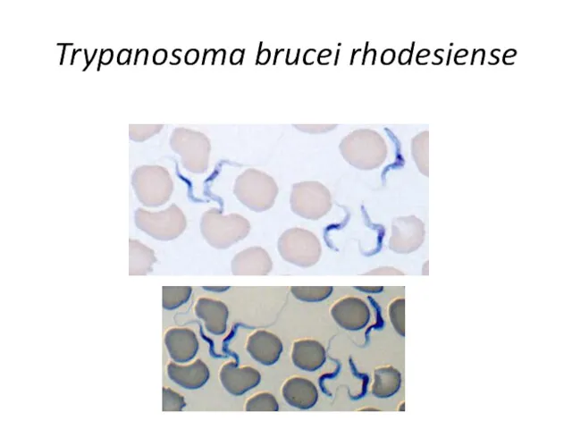
Trypanosoma brucei rhodesiense
Trypanosoma brucei rhodesiense

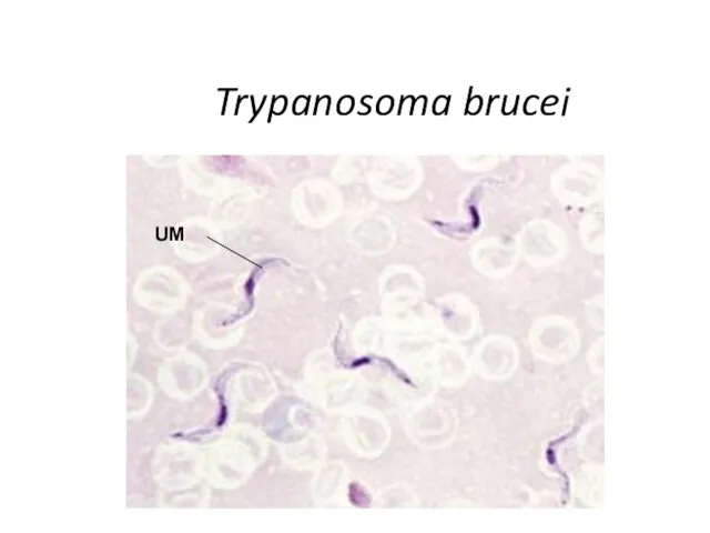
Trypanosoma brucei
UM
Trypanosoma brucei
UM
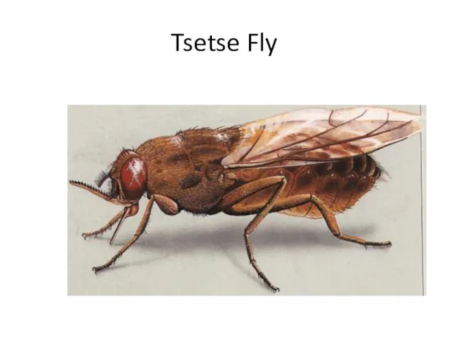
Tsetse Fly
Tsetse Fly
“Chaga’s Disease”
Disease: American Tryptanosomiasis
A zoonotic disease (can infect animals) that can
“Chaga’s Disease”
Disease: American Tryptanosomiasis
A zoonotic disease (can infect animals) that can
“Chaga’s Disease”
This disease is NOT found in Africa.
This disease is
“Chaga’s Disease”
This disease is NOT found in Africa.
This disease is
“Chaga’s Disease”
It prefers the lips because the blood supply is close
“Chaga’s Disease”
It prefers the lips because the blood supply is close
“Chaga’s Disease”
Symptoms include fever, anorexia, swollen lymph nodes, hepatosplenomegally (enlarged liver
“Chaga’s Disease”
Symptoms include fever, anorexia, swollen lymph nodes, hepatosplenomegally (enlarged liver

Trypanosoma life cycle
Trypanosoma life cycle
Trypanosoma cruzi
Insect vector is the “kissing” bug. It takes a blood
Trypanosoma cruzi
Insect vector is the “kissing” bug. It takes a blood
Trypanosoma cruzi
Geographic Distribution:
The Americas from the southern United States to southern
Trypanosoma cruzi
Geographic Distribution: The Americas from the southern United States to southern
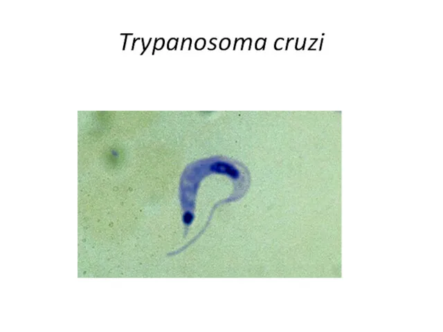
Trypanosoma cruzi
Trypanosoma cruzi
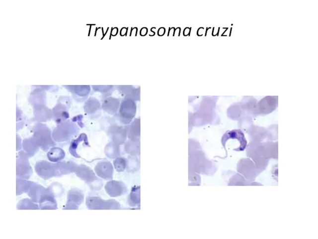
Trypanosoma cruzi
Trypanosoma cruzi
Trypanosoma cruzi
large kinetoplast
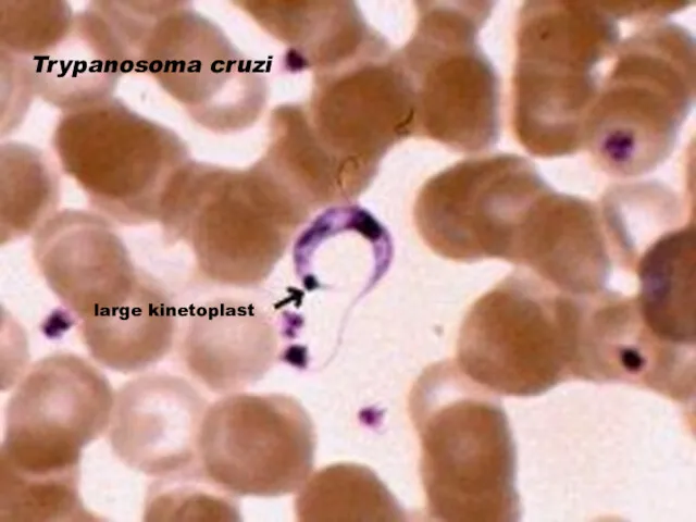
Trypanosoma cruzi
large kinetoplast
Trypanosoma cruzi
Triatomine bug, Trypanosoma cruzi vector, defecating on the wound after
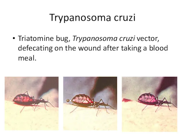
Trypanosoma cruzi
Triatomine bug, Trypanosoma cruzi vector, defecating on the wound after
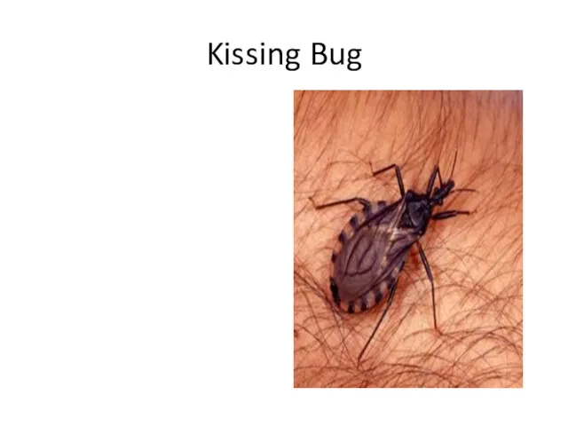
Kissing Bug
Kissing Bug
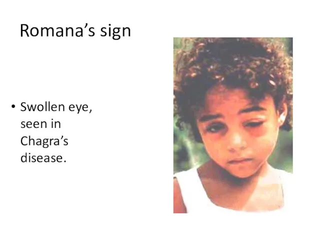
Romana’s sign
Swollen eye, seen in Chagra’s disease.
Romana’s sign
Swollen eye, seen in Chagra’s disease.
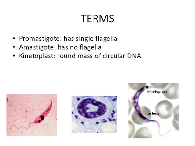
TERMS
Promastigote: has single flagella
Amastigote: has no flagella
Kinetoplast: round mass of circular
TERMS
Promastigote: has single flagella
Amastigote: has no flagella
Kinetoplast: round mass of circular
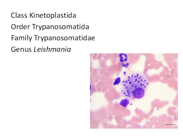
Class Kinetoplastida
Order Trypanosomatida
Family Trypanosomatidae
Genus Leishmania
Class Kinetoplastida
Order Trypanosomatida
Family Trypanosomatidae
Genus Leishmania
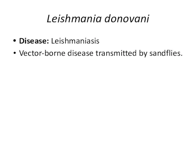
Leishmania donovani
Disease: Leishmaniasis
Vector-borne disease transmitted by sandflies.
Leishmania donovani
Disease: Leishmaniasis
Vector-borne disease transmitted by sandflies.
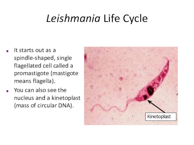
Leishmania Life Cycle
Kinetoplast
It starts out as a spindle-shaped, single flagellated cell
Leishmania Life Cycle
Kinetoplast
It starts out as a spindle-shaped, single flagellated cell
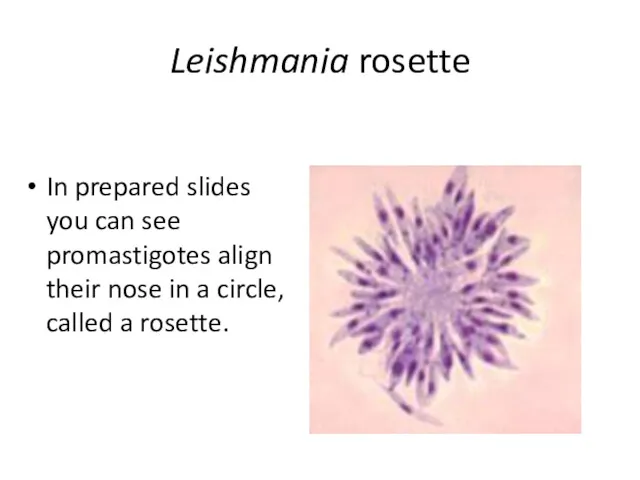
Leishmania rosette
In prepared slides you can see promastigotes align their nose
Leishmania rosette
In prepared slides you can see promastigotes align their nose
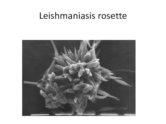
Leishmaniasis rosette
Leishmaniasis rosette
Leishmania Life Cycle
It reproduces in the gut of a female sandfly,
Leishmania Life Cycle
It reproduces in the gut of a female sandfly,
Leishmaniasis
These amastigotes multiply in various organs including the spleen, liver, and
Leishmaniasis
These amastigotes multiply in various organs including the spleen, liver, and
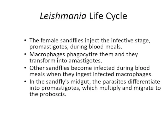
Leishmania Life Cycle
The female sandflies inject the infective stage, promastigotes, during
Leishmania Life Cycle
The female sandflies inject the infective stage, promastigotes, during
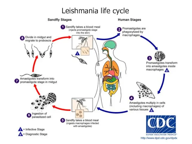
Leishmania life cycle
Leishmania life cycle
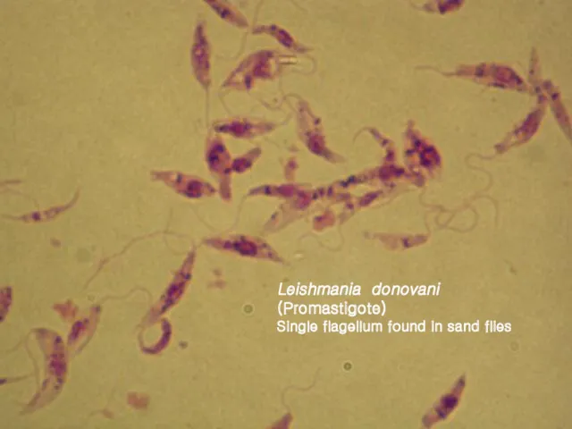
Leishmania donovani (Promastigote)
Single flagellum found in sand flies
Leishmania donovani (Promastigote)
Single flagellum found in sand flies
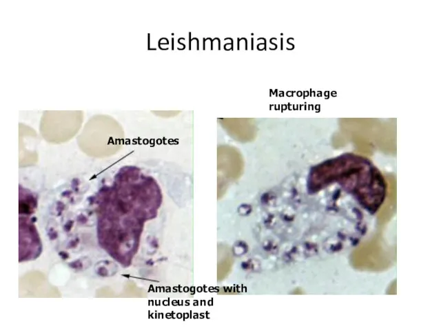
Leishmaniasis
Amastogotes
Amastogotes with nucleus and kinetoplast
Macrophage rupturing
Leishmaniasis
Amastogotes
Amastogotes with nucleus and kinetoplast
Macrophage rupturing
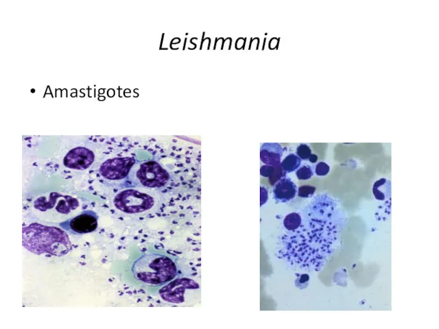
Leishmania
Amastigotes
Leishmania
Amastigotes
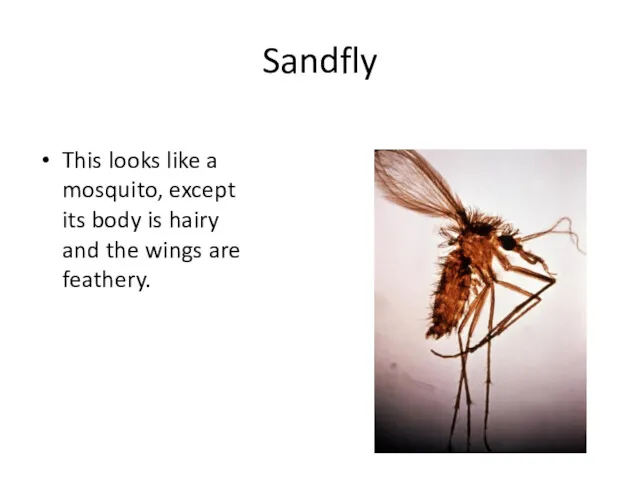
Sandfly
This looks like a mosquito, except its body is hairy and
Sandfly
This looks like a mosquito, except its body is hairy and
Leishmaniasis
Geographic Distribution:
More than 90 percent of the world's cases of visceral
Leishmaniasis
Geographic Distribution: More than 90 percent of the world's cases of visceral
Leishmaniasis
There are three forms of Leishmaniasis:
Cutaneous
Mucocutaneus
Visceral
Leishmaniasis
There are three forms of Leishmaniasis:
Cutaneous
Mucocutaneus
Visceral
Cutaneous Leishmaniasis
The disease is only at the site of the bite.
Cutaneous Leishmaniasis
The disease is only at the site of the bite.
Leishmaniasis (cutaneous)
Leishmaniasis (cutaneous)
Leishmaniasis (cutaneous)
Leishmaniasis (cutaneous)
Leishmaniasis (cutaneous)
Leishmaniasis (cutaneous)
Leishmaniasis (mucocunateous)
This is when the disease located in the mucous membranes
Leishmaniasis (mucocunateous)
This is when the disease located in the mucous membranes
Leishmaniasis (mucocunateous)
Leishmaniasis (mucocunateous)
Leishmaniasis (visceral)
This is the most serious form. It occurs especially in
Leishmaniasis (visceral)
This is the most serious form. It occurs especially in
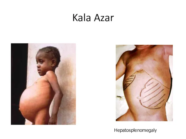
Kala Azar
Hepatosplenomegaly
Kala Azar
Hepatosplenomegaly
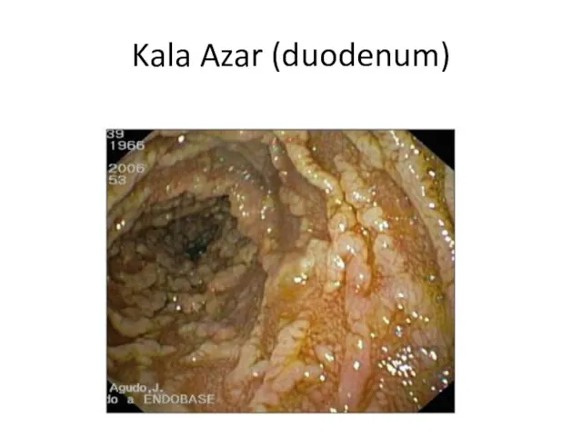
Kala Azar (duodenum)
Kala Azar (duodenum)
Phylum Metamonada
Phylum Metamonada
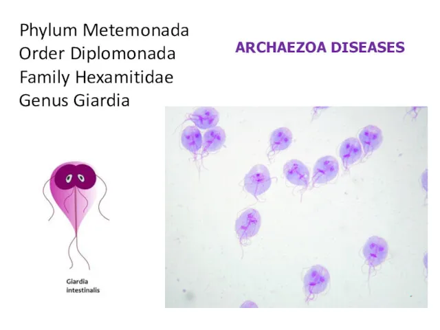
Phylum Metemonada
Order Diplomonada
Family Hexamitidae
Genus Giardia
ARCHAEZOA DISEASES
Phylum Metemonada
Order Diplomonada
Family Hexamitidae
Genus Giardia
ARCHAEZOA DISEASES
Giardiasis
Organism: Giardia lamblia
Cysts are resistant forms and are responsible for transmission
Giardiasis
Organism: Giardia lamblia
Cysts are resistant forms and are responsible for transmission
Life Cycle of Giardia lamblia
Life Cycle of Giardia lamblia
Giardia lamblia
In the small intestine, excystation releases trophozoites (each cyst produces
Giardia lamblia
In the small intestine, excystation releases trophozoites (each cyst produces
Giardia lamblia
Trophozoite form: piroform (pear or teardrop shape), looks like a
Giardia lamblia
Trophozoite form: piroform (pear or teardrop shape), looks like a
Giardia lamblia
Trophozoite
Giardia lamblia
Trophozoite
Giardia lamblia
Trophozoites
Giardia lamblia
Trophozoites
Giardia lamblia
Trophozoites
Giardia lamblia
Trophozoites
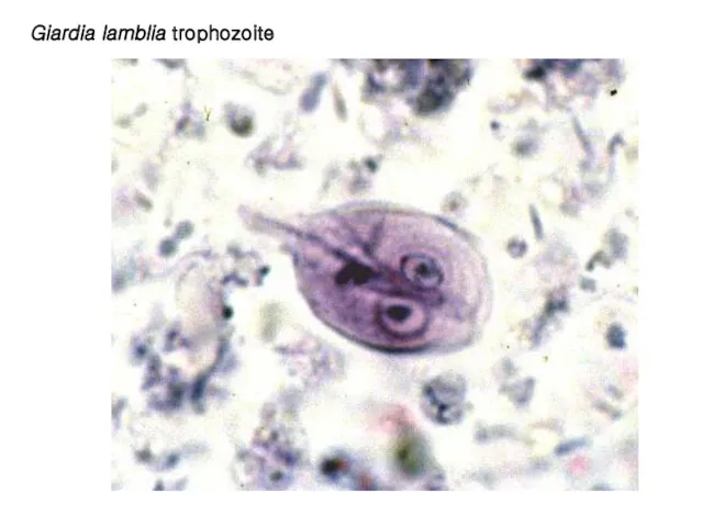
Giardia lamblia trophozoite
Giardia lamblia trophozoite
Giardia lamblia
Cysts
Giardia lamblia
Cysts
Phylum Metemonada
Order Trichomonadida
Family Trichomonadidae
Genus Trichomonas
Phylum Metemonada
Order Trichomonadida
Family Trichomonadidae
Genus Trichomonas
Trichomoniasis
Organism: Trichomonas vaginalis
Trichomonas vaginalis resides in the female lower genital tract
Trichomoniasis
Organism: Trichomonas vaginalis
Trichomonas vaginalis resides in the female lower genital tract
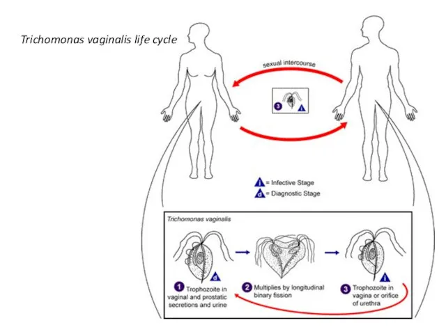
Trichomonas vaginalis life cycle
Trichomonas vaginalis life cycle
Trichomonas vaginalis
Undulating membrane
Trophozoite
Trichomonas vaginalis
Undulating membrane
Trophozoite
Trichomonas vaginalis
Trichomonas vaginalis
 Метаботропные рецепторы. Подсемейство рецепторов
 Дальневосточный леопард
 Разнообразие грибов. Значение грибов в природе и жизни человека
 Занимательный час зоологии. Презентация.
 Строение и функции ядра. Формы хранения генетического материала
 Действие пестицидов на биоценозы. (Лекция 10)
 Рамапитек. Антропогенез. Эволюция человека. Часть 2
 Служебные собаки
 Physiology of Bacteria
 Селекция: методы и достижения
 Растениеводство
 Косточковые плоды
 Корень. Корневые систем. Значение корня
 Понятие о катаболизме и анаболизме. Основы питания. Незаменимые пищевые факторы. Биоэнергетика. Структурная организация ЦПЭ
 Анатомо-физиологические особенности кожи и миофасциальной структуры лица
 Редкие и исчезающие виды растений и животных Восточно-Казахстанской области
 Головной мозг. Строение и функции его основных отделов.
 Микориза (грибокорень)
 Memory plasticity
 Систематика живого мира
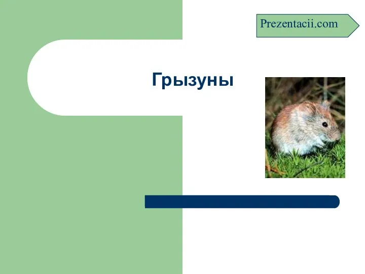 Грызуны
 Сравнительная характеристика однодольных и двудольных растений
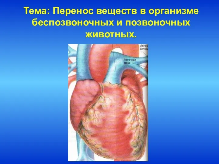 Перенос веществ в организме беспозвоночных и позвоночных животных
 Семенные растения
 Химический состав клетки
 Морфология побега растений. Разнообразие побегов
 Мой домашний питомец
 Научно-исследовательский проект на тему: Комнатные растения