- Skeletal tissues

Содержание
- 2. BONE TISSUE This is a specialized type of connective tissue with high mineralization of the intercellular
- 3. BONE TISSUE PRYMARY RETICULOFIBROSIS SECONDARY LAMELLAR
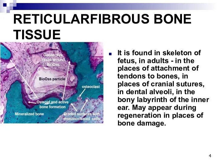
- 4. RETICULARFIBROUS BONE TISSUE It is found in skeleton of fetus, in adults - in the places
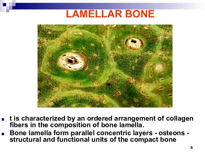
- 5. LAMELLAR BONE t is characterized by an ordered arrangement of collagen fibers in the composition of
- 6. CELLS OF BONE OSTEOGENIC DIFFERON Osteogenic cell - osteoblast - osteocyte HEMATOGENOUS DIFFERON PHSC - Promonocyte
- 7. OSTEOBLASTS - cells building bone tissue They are located on the surface of bone spicules in
- 8. FUNCTION OF OSTEOBLAST Create a bone in two stages: 1. Actively synthesize the organic bone matrix
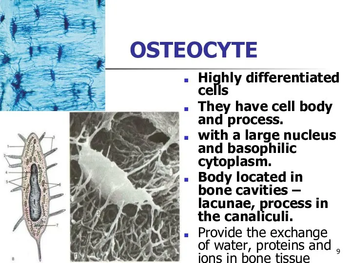
- 9. OSTEOCYTE Highly differentiated cells They have cell body and process. with a large nucleus and basophilic
- 10. 1- nucleus 2- cytoplasm; 3-process; 4- lacuna; 5- osseomucoid; 6- ossein fibers Compact substance of the
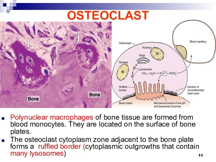
- 11. OSTEOCLAST Polynuclear macrophages of bone tissue are formed from blood monocytes. They are located on the
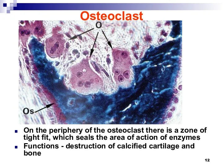
- 12. Osteoclast On the periphery of the osteoclast there is a zone of tight fit, which seals
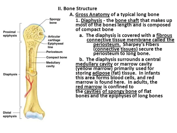
- 13. Long bone as an organ Consists of: - head of the long bone - epiphysis -
- 16. The structure of the diaphysis of the long bone histologically consists of three layers: 1. The
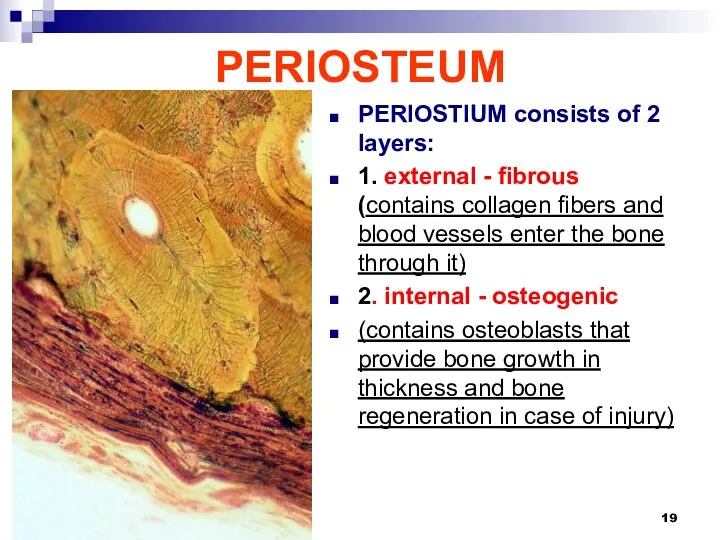
- 19. PERIOSTEUM PERIOSTIUM consists of 2 layers: 1. external - fibrous (contains collagen fibers and blood vessels
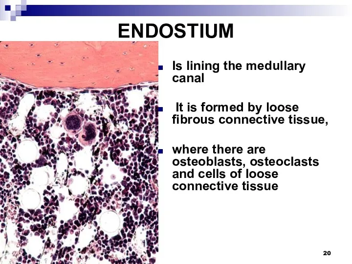
- 20. ENDOSTIUM Is lining the medullary canal It is formed by loose fibrous connective tissue, where there
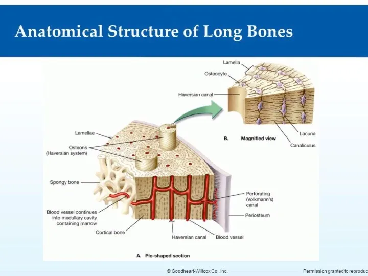
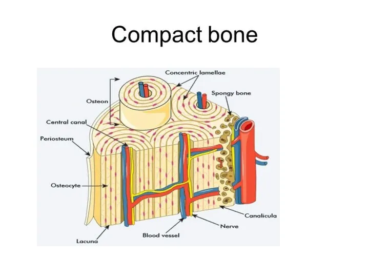
- 21. COMPACT BONE Consists of three layers of bone lamella 1. External circumferential (general) bone lamella 2.
- 22. OSTEON (haversian system) Is structural and functional unit of bone consists of bone lamella concentrically stacked
- 23. ЛАКУАНАРНО-КАНАЛЬЦЕВАЯ СИСТЕМА
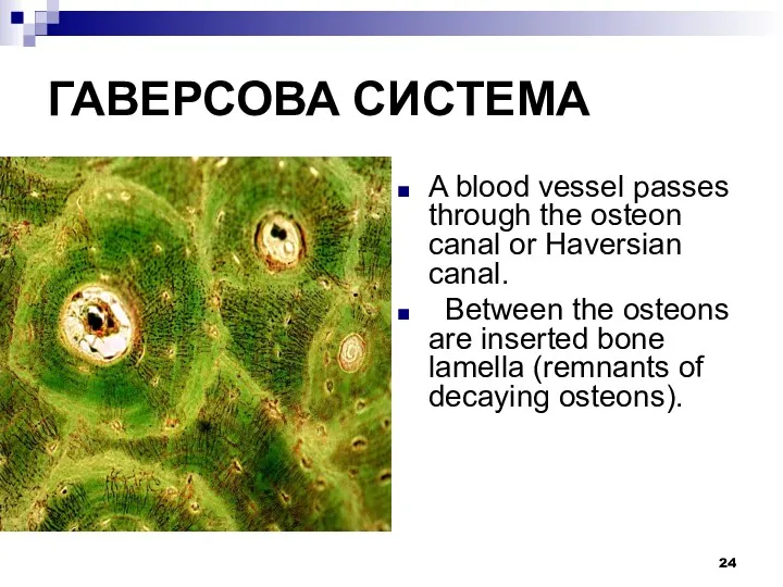
- 24. ГАВЕРСОВА СИСТЕМА A blood vessel passes through the osteon canal or Haversian canal. Between the osteons
- 25. OSTEOGENESIS PRENATAL BONE FORMATION BEGINS ON 1 MONTH OF PRENATAL DEVELOPMENT CONTINUES UNTIL 25 YEARS 1.
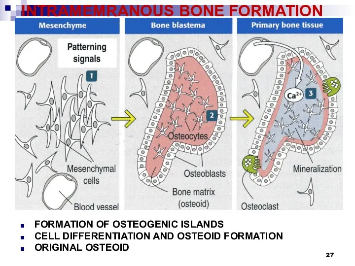
- 26. INTRAMEMRANOUS BONE FORMATION 1. osteogenic islet formation -mesenchymal cells in places of future flat bones condense
- 27. INTRAMEMRANOUS BONE FORMATION FORMATION OF OSTEOGENIC ISLANDS CELL DIFFERENTIATION AND OSTEOID FORMATION ORIGINAL OSTEOID
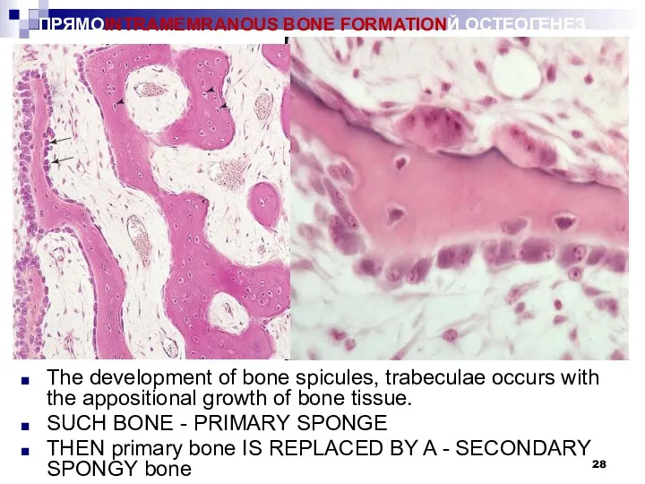
- 28. ПРЯМОINTRAMEMRANOUS BONE FORMATIONЙ ОСТЕОГЕНЕЗ The development of bone spicules, trabeculae occurs with the appositional growth of
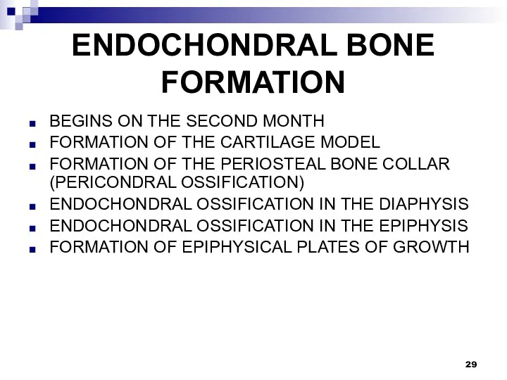
- 29. ENDOCHONDRAL BONE FORMATION BEGINS ON THE SECOND MONTH FORMATION OF THE CARTILAGE MODEL FORMATION OF THE
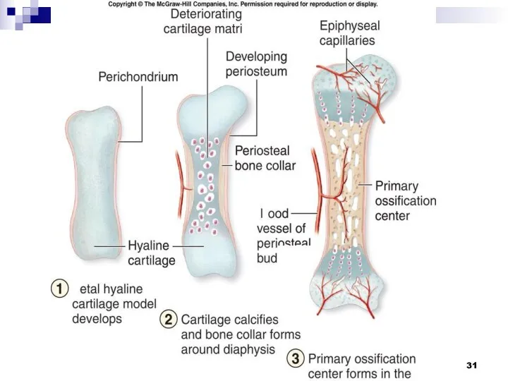
- 30. ENDOCHONDRAL BONE FORMATION 1. the formation of a cartilage model (hyaling) of the future bone; 2.
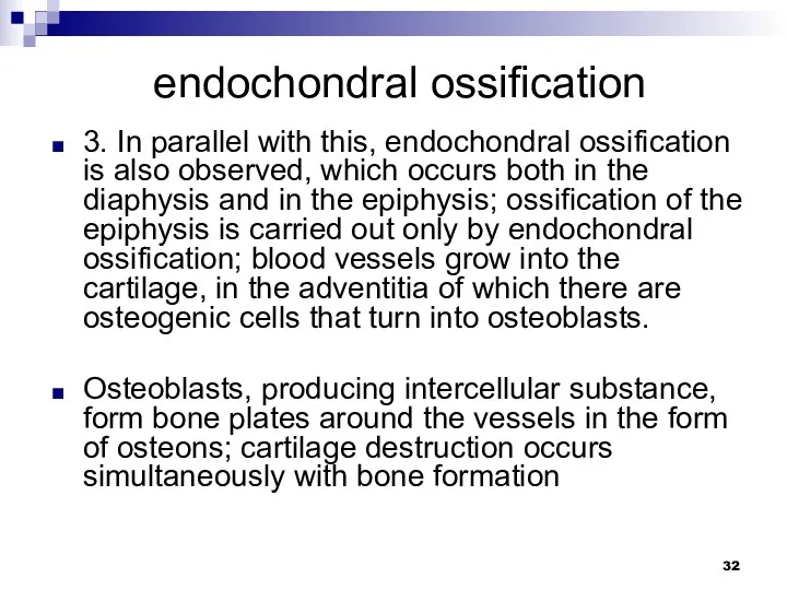
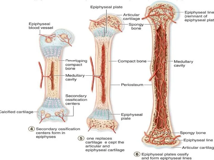
- 32. endochondral ossification 3. In parallel with this, endochondral ossification is also observed, which occurs both in
- 43. Скачать презентацию
BONE TISSUE
This is a specialized type of connective tissue with
BONE TISSUE
This is a specialized type of connective tissue with
BONE TISSUE
PRYMARY
RETICULOFIBROSIS
SECONDARY LAMELLAR
BONE TISSUE
PRYMARY
RETICULOFIBROSIS
SECONDARY LAMELLAR
RETICULARFIBROUS BONE TISSUE
It is found in skeleton of fetus, in adults
RETICULARFIBROUS BONE TISSUE
It is found in skeleton of fetus, in adults
LAMELLAR BONE
t is characterized by an ordered arrangement of collagen fibers
LAMELLAR BONE
t is characterized by an ordered arrangement of collagen fibers
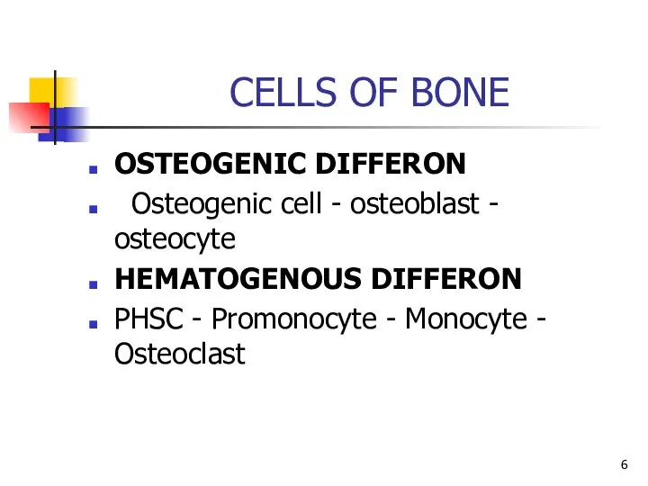
CELLS OF BONE
OSTEOGENIC DIFFERON
Osteogenic cell - osteoblast - osteocyte
HEMATOGENOUS
CELLS OF BONE
OSTEOGENIC DIFFERON
Osteogenic cell - osteoblast - osteocyte
HEMATOGENOUS
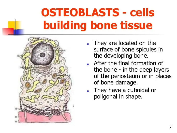
OSTEOBLASTS - cells building bone tissue
They are located on the surface
OSTEOBLASTS - cells building bone tissue
They are located on the surface
FUNCTION OF OSTEOBLAST
Create a bone in two stages:
1. Actively synthesize the
FUNCTION OF OSTEOBLAST
Create a bone in two stages:
1. Actively synthesize the
OSTEOCYTE
Highly differentiated cells
They have cell body and process.
with a large nucleus
OSTEOCYTE
Highly differentiated cells
They have cell body and process.
with a large nucleus
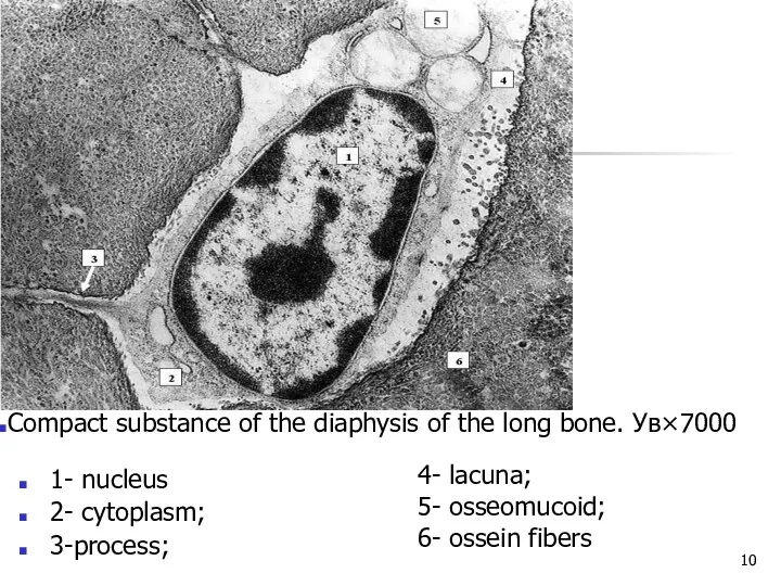
1- nucleus
2- cytoplasm;
3-process;
4- lacuna;
5- osseomucoid;
6- ossein fibers
Compact substance of the diaphysis
1- nucleus
2- cytoplasm;
3-process;
4- lacuna;
5- osseomucoid;
6- ossein fibers
Compact substance of the diaphysis
OSTEOCLAST
Polynuclear macrophages of bone tissue are formed from blood monocytes. They
OSTEOCLAST
Polynuclear macrophages of bone tissue are formed from blood monocytes. They
Osteoclast
On the periphery of the osteoclast there is a zone of
Osteoclast
On the periphery of the osteoclast there is a zone of
Long bone as an organ
Consists of:
- head of the long bone
Long bone as an organ
Consists of:
- head of the long bone
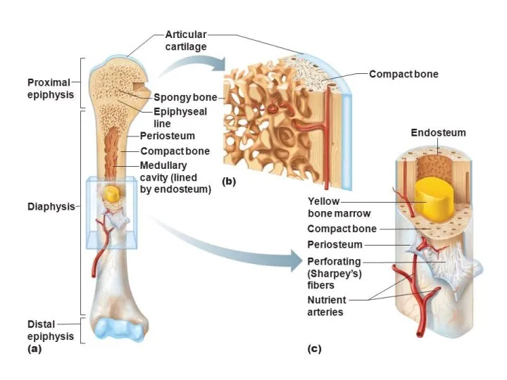
The structure of the diaphysis of the long bone
histologically consists of
The structure of the diaphysis of the long bone
histologically consists of
PERIOSTEUM
PERIOSTIUM consists of 2 layers:
1. external - fibrous (contains collagen fibers
PERIOSTEUM
PERIOSTIUM consists of 2 layers:
1. external - fibrous (contains collagen fibers
ENDOSTIUM
Is lining the medullary canal
It is formed by loose fibrous
ENDOSTIUM
Is lining the medullary canal
It is formed by loose fibrous

COMPACT BONE
Consists of three layers of bone lamella
1. External circumferential (general)
COMPACT BONE
Consists of three layers of bone lamella
1. External circumferential (general)
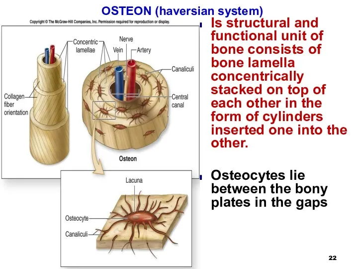
OSTEON (haversian system)
Is structural and functional unit of bone consists
OSTEON (haversian system)
Is structural and functional unit of bone consists
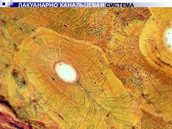
ЛАКУАНАРНО-КАНАЛЬЦЕВАЯ СИСТЕМА
ЛАКУАНАРНО-КАНАЛЬЦЕВАЯ СИСТЕМА
ГАВЕРСОВА СИСТЕМА
A blood vessel passes through the osteon canal or Haversian
ГАВЕРСОВА СИСТЕМА
A blood vessel passes through the osteon canal or Haversian
OSTEOGENESIS PRENATAL
BONE FORMATION BEGINS ON 1 MONTH OF PRENATAL DEVELOPMENT
CONTINUES
OSTEOGENESIS PRENATAL
BONE FORMATION BEGINS ON 1 MONTH OF PRENATAL DEVELOPMENT
CONTINUES
INTRAMEMRANOUS BONE FORMATION
1. osteogenic islet formation -mesenchymal cells in places of
INTRAMEMRANOUS BONE FORMATION
1. osteogenic islet formation -mesenchymal cells in places of
INTRAMEMRANOUS BONE FORMATION
FORMATION OF OSTEOGENIC ISLANDS
CELL DIFFERENTIATION AND OSTEOID FORMATION
ORIGINAL OSTEOID
INTRAMEMRANOUS BONE FORMATION
FORMATION OF OSTEOGENIC ISLANDS
CELL DIFFERENTIATION AND OSTEOID FORMATION
ORIGINAL OSTEOID
ПРЯМОINTRAMEMRANOUS BONE FORMATIONЙ ОСТЕОГЕНЕЗ
The development of bone spicules, trabeculae occurs with
ПРЯМОINTRAMEMRANOUS BONE FORMATIONЙ ОСТЕОГЕНЕЗ
The development of bone spicules, trabeculae occurs with
ENDOCHONDRAL BONE FORMATION
BEGINS ON THE SECOND MONTH
FORMATION OF THE CARTILAGE MODEL
FORMATION
ENDOCHONDRAL BONE FORMATION
BEGINS ON THE SECOND MONTH
FORMATION OF THE CARTILAGE MODEL
FORMATION
ENDOCHONDRAL BONE FORMATION
1. the formation of a cartilage model (hyaling)
ENDOCHONDRAL BONE FORMATION
1. the formation of a cartilage model (hyaling)
endochondral ossification
3. In parallel with this, endochondral ossification is also observed,
endochondral ossification
3. In parallel with this, endochondral ossification is also observed,
 Водоросли. 6 класс
 Генетика пола
 Биохимические сдвиги в организме при мышечной работе
 Тип Моллюски
 Гигиена зрения. Предупреждение глазных болезней
 Функціональна анатомія верхньої кінцівки
 Чага гриб - сенсация
 Строение растительной клетки
 Сердечно-сосудистая система. Физиологические свойства и функции сердца
 Роль биологических исследований в современной медицине
 Шляпочные грибы
 Основные процессы жизнедеятельности растений
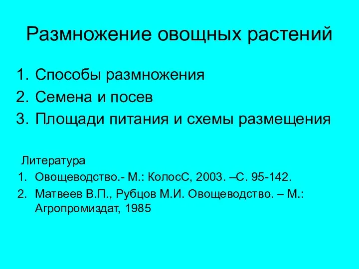 Овощеводство. Размножение овощных растений
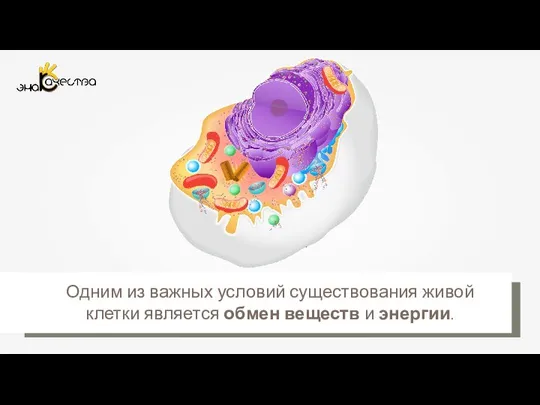 Обмен веществ и энергии в клетке
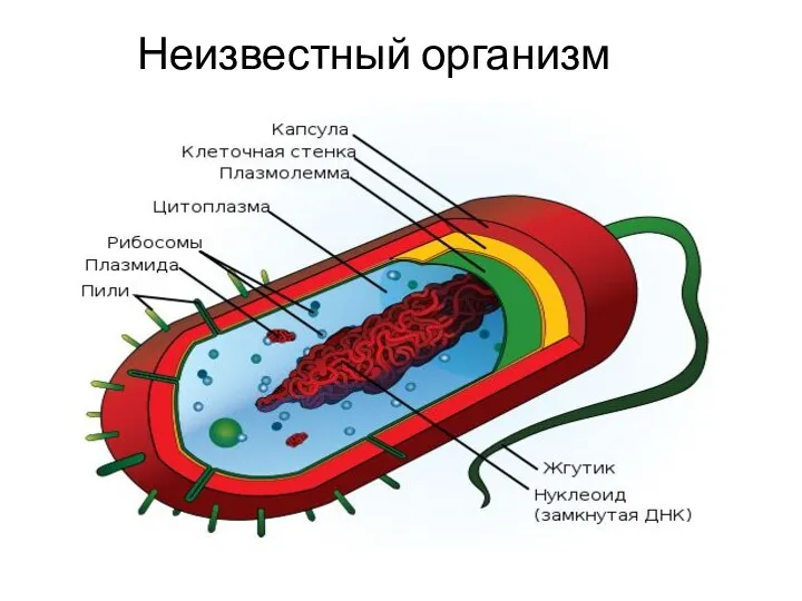 Бактерии. Строение бактериальной клетки. Формы бактерий. Распространение и условия обитания
 Нервная система. Рефлекс. Инстинкт
 Изучение микроскопического строения клетки и тканей
 Морфология микроорганизмов. Лекция 1. Морфология грибов
 Клетка и методы цитологии
 Ағзаларды клондау
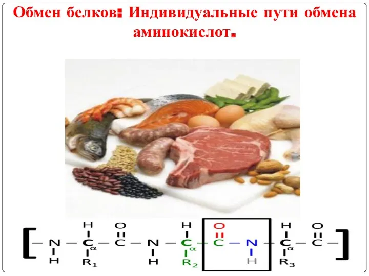 Обмен белков: Индивидуальные пути обмена аминокислот
 Транскрипция. Биосинтез белка
 Введение в курс Концепции современного естествознания
 ЕГЭ. Биология 2007 год
 Класс млекопитающие. (7)
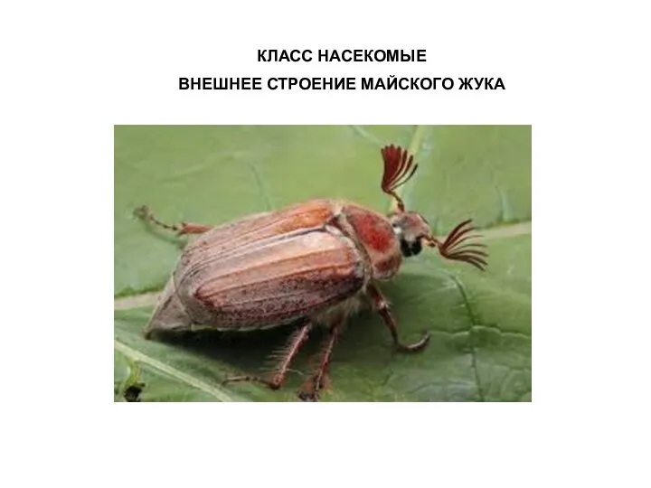 Класс Насекомые. Внешнее строение майского жука
 Расы человека
 презентация по биологии на тему Современное состояние и охрана растительности для 11 класса