- Соединительныя ткани

Содержание
- 2. Функции соединительных тканей 1. Опорная (капсулы органов, сухожилии, фасции, скелет) 2. Трофическая (обмен веществ между кровью
- 3. Рыхлая волокнистая соединительная ткань (РВСТ) Loose collagen connective tissue (LCCT) Клетки Cells Эластические волокна Elastic fibers
- 4. The fundamental (amorphous) substance – a gel colloid system from water, salts and organic substances: glycoproteins,
- 5. Основное вещество Fundamental (amorphous) substance Неорганические вещества - salts Вода - water Органические вещества - organic
- 6. 1 группа – хондроитинсульфаты А,В,С 2 группа – дерматансульфаты 3 группа – кератансульфаты 4 группа –
- 7. Hyaluronic acid is hydrophilic, well connects water, stimulates a metabolism, phagocytosis, duplication and mobility of cells.
- 8. Коллагеновые волокна (из белка коллагена) Collagen fibers (from protein collagen) Collagenic fibers are very strong, oxiphilic,
- 9. Collagenic fibers are constructed of tropocollagen-protein which consists of triplets of amino-acids - in each triplet
- 10. ФИБРОБЛАСТ Тропоколлаген Tropocollagen Прото- фибрилла Образование коллагеновых волокон Collagen fibers formation Fibroblast nucleus cytoplasm Intercellular substance
- 11. Фибрилла (тропоколлаген) Fibril (tropocollagen) Молекула тропоколлагена tropocollagen molecule Зазоры заполнены гликозамино- гликанами (Intervals fill in GAG)
- 12. Типы коллагена Types of collagen 1 тип – в соединительной ткани, в костях, зубах 1 type
- 13. Ретикулярные волокна (коллаген 3 типа) Reticular fibers (collagen 3 type) Ядра клеток (nucleous of cells) (импрегнация
- 14. Эластические волокна (из белка эластина) Анастомозы волокон Elastic fibers (protein elastin) Anastomosis of fibers are more
- 15. Клетки соединительной ткани: Фибробласты Тканевые макрофаги 3. Тучные клетки 4. Малодиффе- ренцированные клетки 5. Липоциты 6.
- 16. Рыхлая волокнистая соединительная ткань Тучные клетки Labrocytes Липоцит Lipocytes Плазмоциты Plasmocytes Лейкоциты Leucocytes Капилляры Hemocfpillary Адвентициальная
- 17. Плазмоциты Plasmocytes Светлый дворик light court yard Lymphocytes, eosinophis, basophils prevalence at the immune answer, neutrophils
- 18. Виды фибробластов: Types of fibroblasts: Малодифференцированные Little differentiated (divide and differentiate) 2. Зрелые Mature (40-50 мкм)
- 19. Фибробласты Fibroblasts Фиброциты Fibrocytes Митохондрия mitochondrion грЭПС endoplasmic reticulum Комплекс Гольджи Golgi Complex Комплекс Гольджи Golgi
- 20. Тучные клетки – тканевые базофилы (лаброциты) Labrocytes – basophilic granulocytes Тучные клетки Labrocytes Адипоциты Lipocytes Дегрануляция
- 21. Макрофаги – гистиоциты Macrophages - histiocytes dense oval nucleus, cytoplasm is foamy, a lot of lysosomes
- 22. Бактерии (bacteria) Псевдоподии макрофага Pseudopodia of macrophage
- 23. Мононуклеарная фагоцитарная система Гистиоциты Альвеолярные макрофаги лёгких Звёздчатые клетки Купфера печени Береговые клетки лимфоузлов Макрофаги ретикулярной
- 25. Скачать презентацию
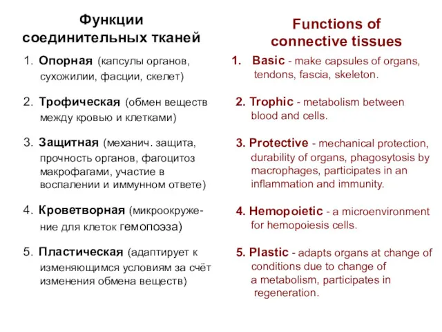
Функции соединительных тканей
1. Опорная (капсулы органов, сухожилии, фасции, скелет)
2. Трофическая (обмен
Функции соединительных тканей
1. Опорная (капсулы органов, сухожилии, фасции, скелет)
2. Трофическая (обмен
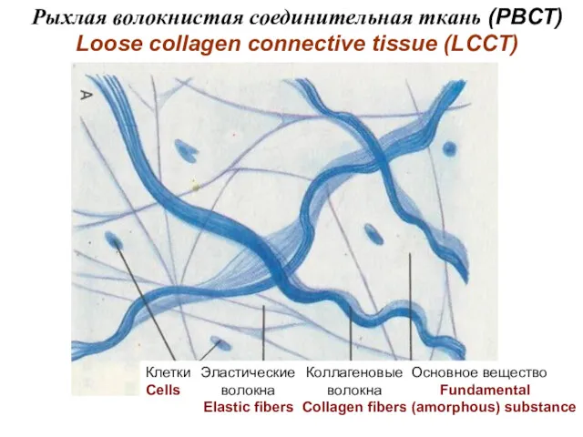
Рыхлая волокнистая соединительная ткань (РВСТ)
Loose collagen connective tissue (LCCT)
Клетки
Cells
Эластические
волокна
Elastic fibers
Коллагеновые
волокна
Collagen
Рыхлая волокнистая соединительная ткань (РВСТ)
Loose collagen connective tissue (LCCT)
Клетки
Cells
Эластические
волокна
Elastic fibers
Коллагеновые
волокна
Collagen
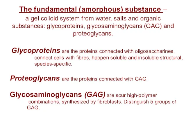
The fundamental (amorphous) substance –
a gel colloid system from water,
The fundamental (amorphous) substance – a gel colloid system from water,
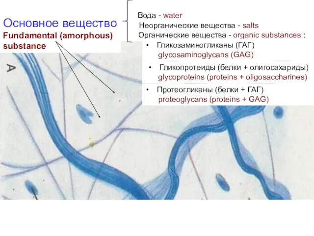
Основное вещество
Fundamental (amorphous)
substance
Неорганические вещества - salts
Вода - water
Органические
Основное вещество
Fundamental (amorphous)
substance
Неорганические вещества - salts
Вода - water
Органические
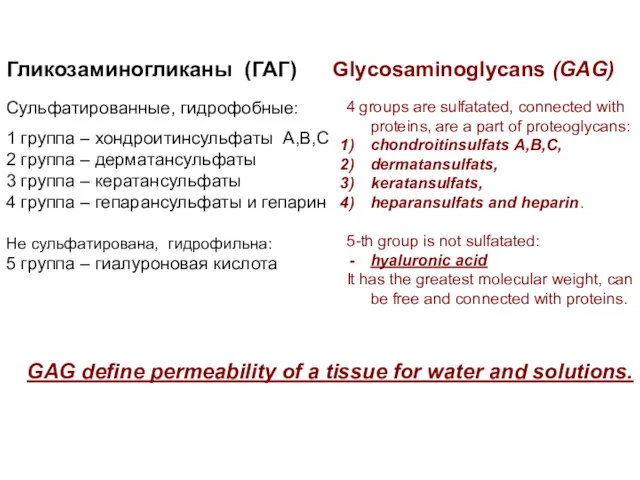
1 группа – хондроитинсульфаты А,В,С
2 группа – дерматансульфаты
3 группа – кератансульфаты
4
1 группа – хондроитинсульфаты А,В,С
2 группа – дерматансульфаты
3 группа – кератансульфаты
4
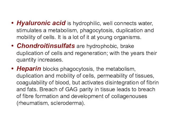
Hyaluronic acid is hydrophilic, well connects water, stimulates a metabolism, phagocytosis,
Hyaluronic acid is hydrophilic, well connects water, stimulates a metabolism, phagocytosis,
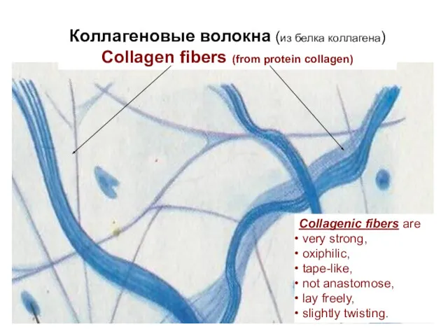
Коллагеновые волокна (из белка коллагена)
Collagen fibers (from protein collagen)
Collagenic fibers are
Коллагеновые волокна (из белка коллагена)
Collagen fibers (from protein collagen)
Collagenic fibers are
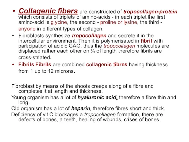
Collagenic fibers are constructed of tropocollagen-protein which consists of triplets of
Collagenic fibers are constructed of tropocollagen-protein which consists of triplets of
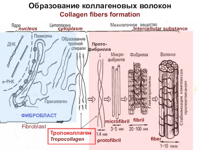
ФИБРОБЛАСТ
Тропоколлаген
Tropocollagen
Прото-
фибрилла
Образование коллагеновых волокон
Collagen fibers formation
Fibroblast
nucleus
cytoplasm
Intercellular substance
protofibril
microfibril
fibril
fiber
ФИБРОБЛАСТ
Тропоколлаген
Tropocollagen
Прото-
фибрилла
Образование коллагеновых волокон
Collagen fibers formation
Fibroblast
nucleus
cytoplasm
Intercellular substance
protofibril
microfibril
fibril
fiber
Фибрилла (тропоколлаген)
Fibril (tropocollagen)
Молекула тропоколлагена
tropocollagen molecule
Зазоры заполнены
гликозамино-
гликанами
(Intervals fill in GAG)
64 нм
Фибрилла (тропоколлаген)
Fibril (tropocollagen)
Молекула тропоколлагена
tropocollagen molecule
Зазоры заполнены
гликозамино-
гликанами
(Intervals fill in GAG)
64 нм
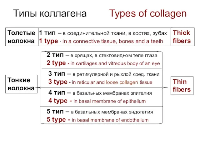
Типы коллагена Types of collagen
1 тип – в соединительной ткани,
Типы коллагена Types of collagen
1 тип – в соединительной ткани,
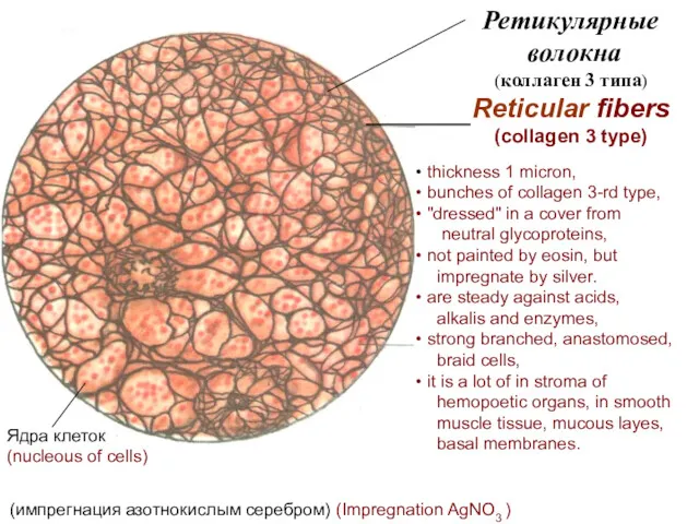
Ретикулярные
волокна
(коллаген 3 типа)
Reticular fibers
(collagen 3 type)
Ядра клеток (nucleous
Ретикулярные
волокна
(коллаген 3 типа)
Reticular fibers
(collagen 3 type)
Ядра клеток (nucleous
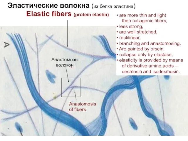
Эластические волокна (из белка эластина)
Анастомозы
волокон
Elastic fibers (protein elastin)
Anastomosis
of fibers
are
Эластические волокна (из белка эластина)
Анастомозы
волокон
Elastic fibers (protein elastin)
Anastomosis
of fibers
are
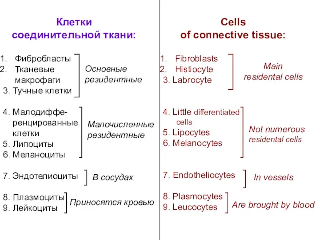
Клетки
соединительной ткани:
Фибробласты
Тканевые
макрофаги
3. Тучные клетки
4. Малодиффе-
ренцированные
клетки
5.
Клетки
соединительной ткани:
Фибробласты
Тканевые
макрофаги
3. Тучные клетки
4. Малодиффе-
ренцированные
клетки
5.
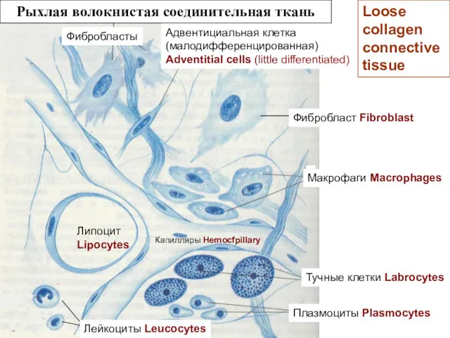
Рыхлая волокнистая соединительная ткань
Тучные клетки Labrocytes
Липоцит
Lipocytes
Плазмоциты Plasmocytes
Лейкоциты Leucocytes
Капилляры Hemocfpillary
Адвентициальная клетка
Рыхлая волокнистая соединительная ткань
Тучные клетки Labrocytes
Липоцит
Lipocytes
Плазмоциты Plasmocytes
Лейкоциты Leucocytes
Капилляры Hemocfpillary
Адвентициальная клетка
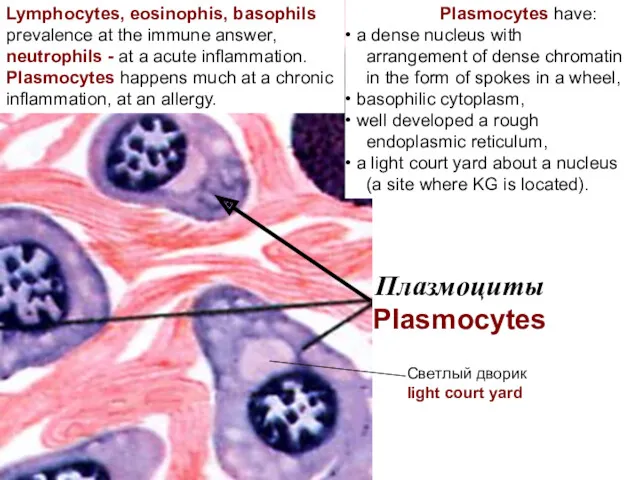
Плазмоциты
Plasmocytes
Светлый дворик
light court yard
Lymphocytes, eosinophis, basophils
prevalence at the immune answer,
neutrophils
Плазмоциты
Plasmocytes
Светлый дворик
light court yard
Lymphocytes, eosinophis, basophils
prevalence at the immune answer,
neutrophils
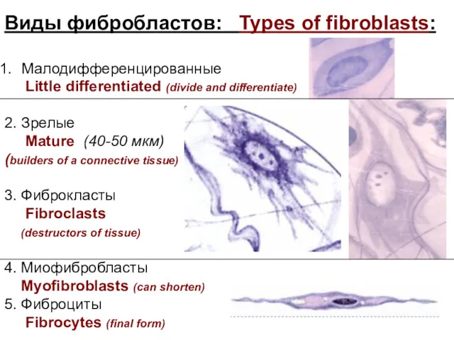
Виды фибробластов: Types of fibroblasts:
Малодифференцированные
Little differentiated (divide and differentiate)
2. Зрелые
Виды фибробластов: Types of fibroblasts:
Малодифференцированные
Little differentiated (divide and differentiate)
2. Зрелые
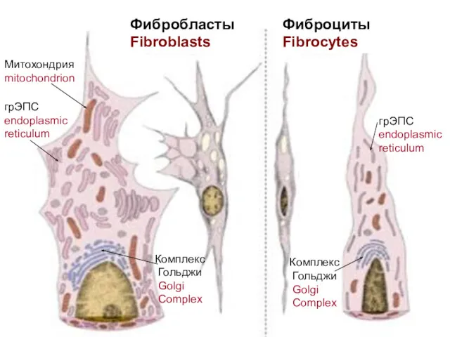
Фибробласты
Fibroblasts
Фиброциты
Fibrocytes
Митохондрия
mitochondrion
грЭПС
endoplasmic
reticulum
Комплекс
Гольджи
Golgi
Complex
Комплекс
Гольджи
Golgi
Complex
грЭПС
endoplasmic
Фибробласты
Fibroblasts
Фиброциты
Fibrocytes
Митохондрия
mitochondrion
грЭПС
endoplasmic
reticulum
Комплекс
Гольджи
Golgi
Complex
Комплекс
Гольджи
Golgi
Complex
грЭПС
endoplasmic
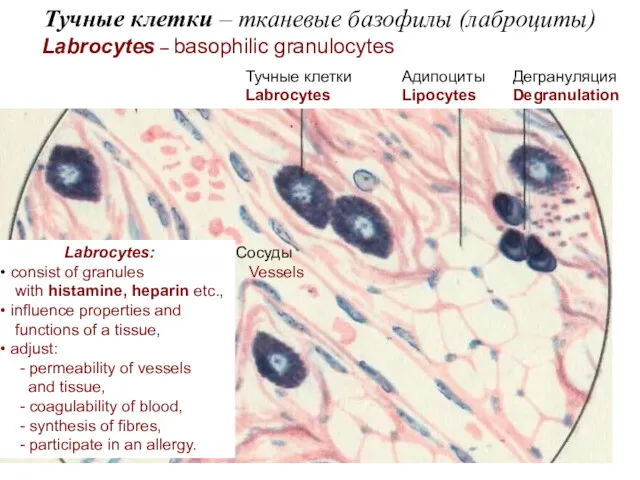
Тучные клетки – тканевые базофилы (лаброциты)
Labrocytes – basophilic granulocytes
Тучные клетки
Labrocytes
Адипоциты
Lipocytes
Дегрануляция
Degranulation
Сосуды
Тучные клетки – тканевые базофилы (лаброциты)
Labrocytes – basophilic granulocytes
Тучные клетки
Labrocytes
Адипоциты
Lipocytes
Дегрануляция
Degranulation
Сосуды
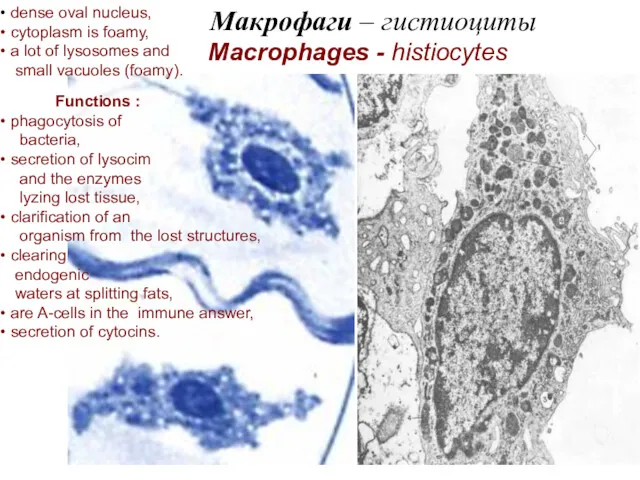
Макрофаги – гистиоциты
Macrophages - histiocytes
dense oval nucleus,
Макрофаги – гистиоциты
Macrophages - histiocytes
dense oval nucleus,
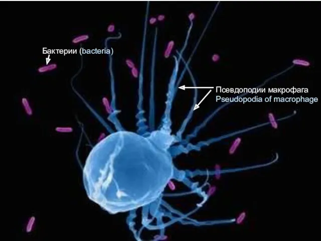
Бактерии (bacteria)
Псевдоподии макрофага
Pseudopodia of macrophage
Бактерии (bacteria)
Псевдоподии макрофага
Pseudopodia of macrophage
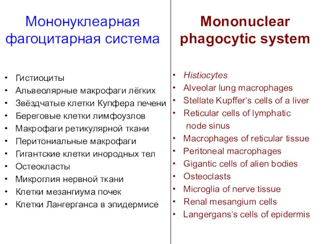
Мононуклеарная
фагоцитарная система
Гистиоциты
Альвеолярные макрофаги лёгких
Звёздчатые клетки Купфера печени
Береговые клетки лимфоузлов
Макрофаги ретикулярной
Мононуклеарная
фагоцитарная система
Гистиоциты
Альвеолярные макрофаги лёгких
Звёздчатые клетки Купфера печени
Береговые клетки лимфоузлов
Макрофаги ретикулярной
 Female culex
 Гели косметические
 Поле и его обитатели
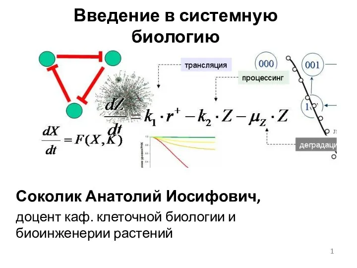 Особенности экспериментальных данных в биологии, применение математических методов, технологий
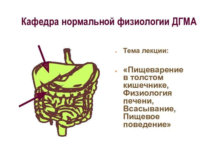 Пищеварение в толстом кишечнике, Физиология печени, Всасывание, Пищевое поведение
 Термодинамика биологических процессов(new)
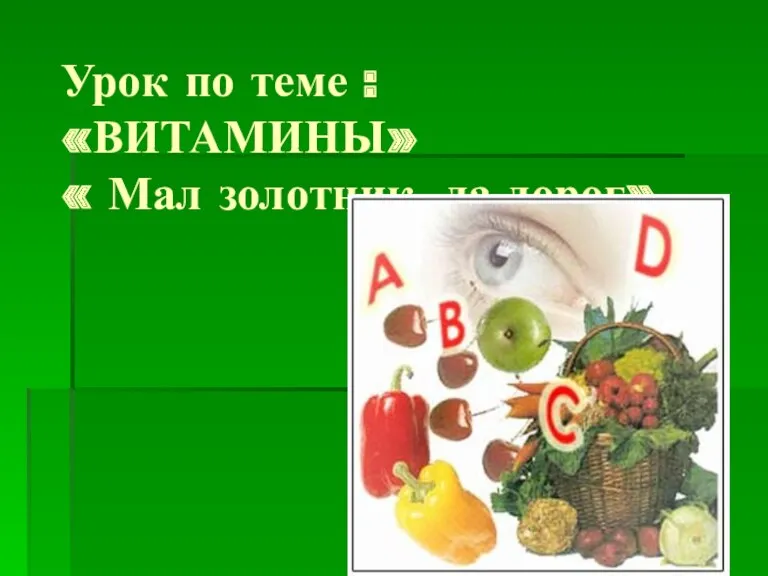 Витамины: Мал золотник, да дорог
 Двойное оплодотворение у цветковых растений
 Тварини - частина живої природи
 Строение и жизненные циклы первичнополостных червей. Тип nematoda
 Методы исследования в биологии
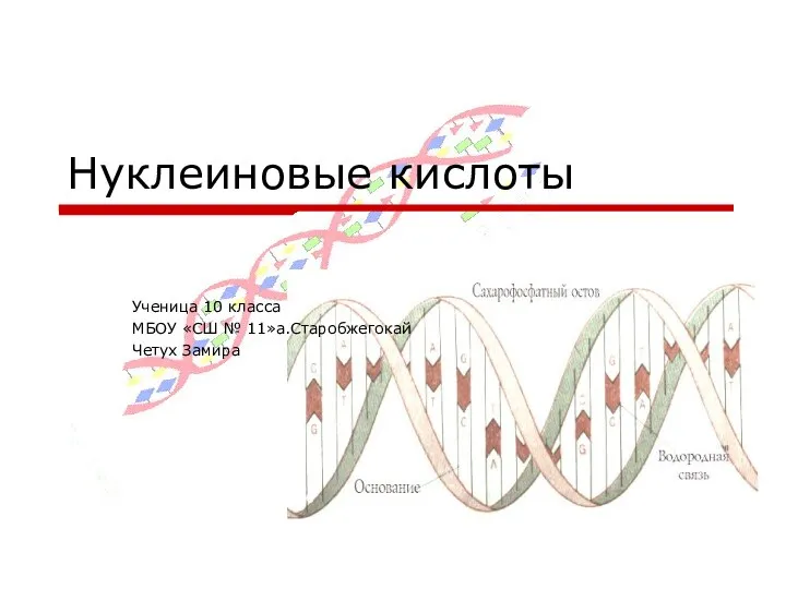 Презентация по биологии ДНК и РНК
 Этапы решения задач по родословной
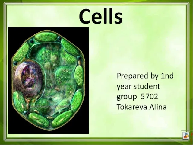 Cells. Muscle cells
 Развитие опорно-двигательного аппарата
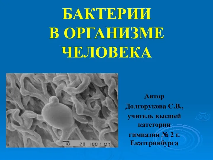 Бактерии в организме человека
 Сны и сновидения
 Влияние фитонцидов различных растений на жизнедеятельность колорадского жука
 Условия, необходимые для прорастания семян
 Дәнді дақылдардың вирустық аурулары
 Строение и процессы жизнедеятельности птиц
 Огород без химии, или бюджетные удобрения
 The Link Reaction and Krebs Cycle
 Циклы развития растений
 Интерактивная модель по биологии Строение животной клетки для 7 (8) классов
 Подготовка учащихся к ЕГЭ по биологии
 Таксы - охотничья порода собак
 Особенности действия элементарных эволюционных факторов в человеческих популяциях