- Urinary system

Содержание
- 2. А: 1 - канальцы пронефроса; 2 - проток пронефроса; 3 - канальцы мезонефроса; 4 - клоака;
- 6. RENAL CORTEX
- 7. Photomicrograph of renal cortex. A macula densa is clearly seen (arrow) at the vascular pole of
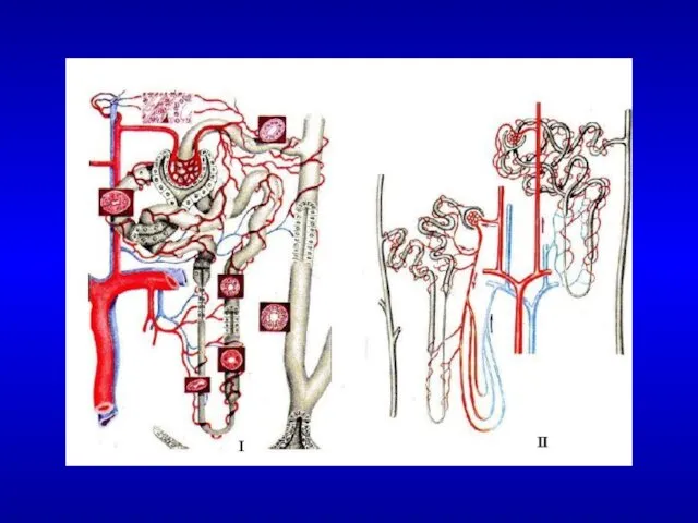
- 8. The renal corpuscle. The upper part of the drawing shows the vascular pole, with afferent and
- 9. Photomicrograph of an afferent arteriole entering a renal corpuscle. The wall of this arteriole shows the
- 10. Schematic representation of a glomerular capillary with the visceral layer of Bowman’s capsule (formed of podocytes).
- 12. Mesangial cell located between capillaries enveloped by the basement membrane. Mesangial cell located between capillaries enveloped
- 14. Renal cortex section showing a proximal convoluted tubule (PCT) with its large cuboidal cells presenting a
- 15. 1-descending thick limb of the Henle loop 2-ascending thick limb of the Henle loop 3-blood vessels
- 16. Collecting tubule
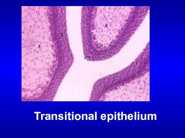
- 18. THE URINARY BLADDER Transitional epithelium Submucosa layer Muscle layer
- 19. Transitional epithelium
- 21. Скачать презентацию
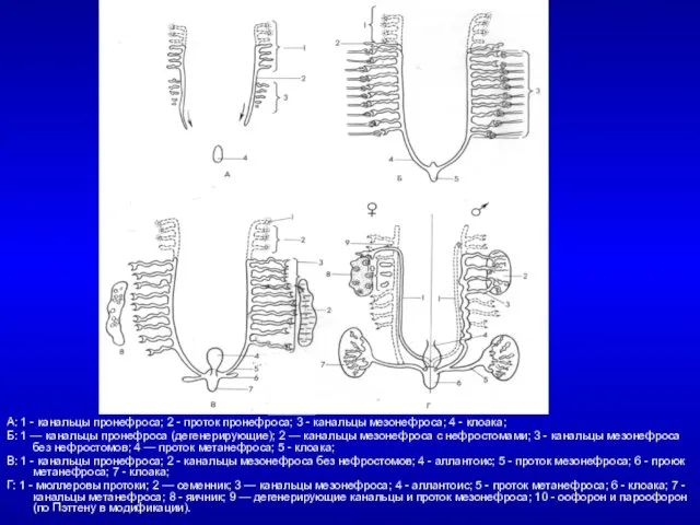
А: 1 - канальцы пронефроса; 2 - проток пронефроса; 3 -
А: 1 - канальцы пронефроса; 2 - проток пронефроса; 3 -
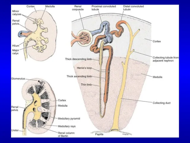
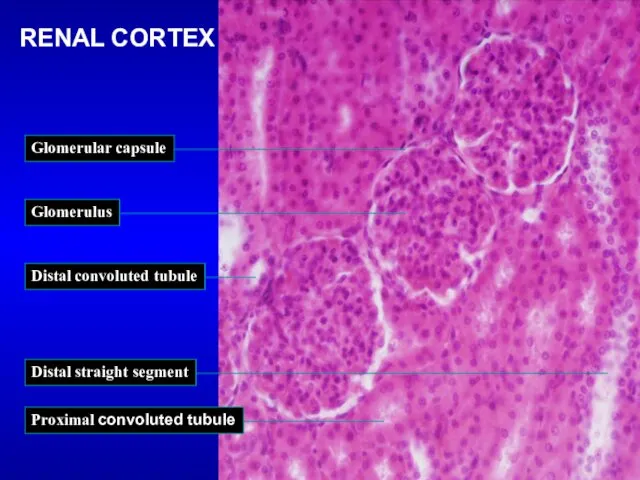
RENAL CORTEX
RENAL CORTEX
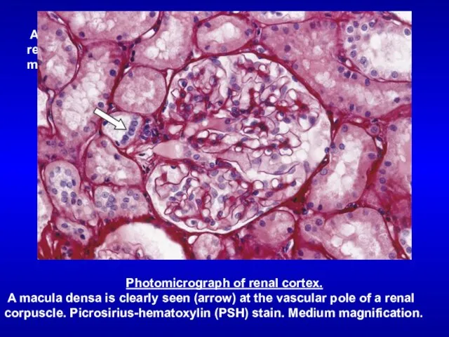
Photomicrograph of renal cortex.
A macula densa is clearly seen (arrow)
Photomicrograph of renal cortex.
A macula densa is clearly seen (arrow)
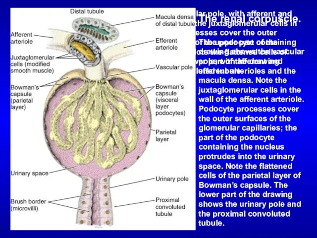
The renal corpuscle.
The upper part of the drawing shows the vascular
The renal corpuscle.
The upper part of the drawing shows the vascular
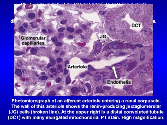
Photomicrograph of an afferent arteriole entering a renal corpuscle. The wall
Photomicrograph of an afferent arteriole entering a renal corpuscle. The wall
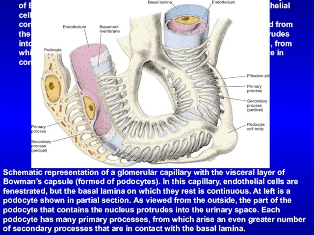
Schematic representation of a glomerular capillary with the visceral layer of
Schematic representation of a glomerular capillary with the visceral layer of
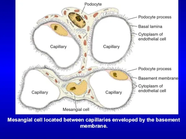
Mesangial cell located between capillaries enveloped by the basement membrane.
Mesangial cell
Mesangial cell located between capillaries enveloped by the basement membrane.
Mesangial cell
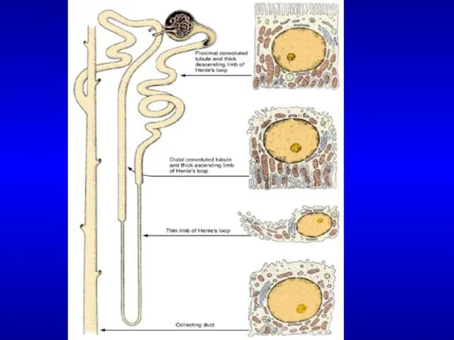
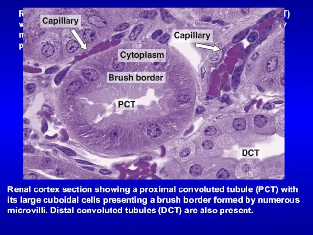
Renal cortex section showing a proximal convoluted tubule (PCT) with its
Renal cortex section showing a proximal convoluted tubule (PCT) with its
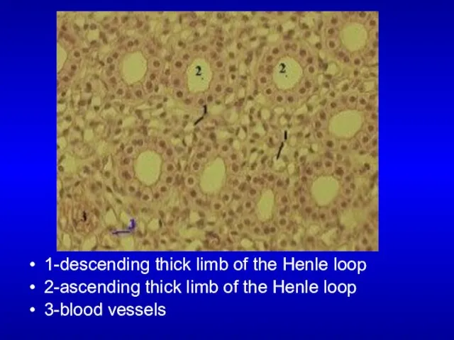
1-descending thick limb of the Henle loop
2-ascending thick limb of the
1-descending thick limb of the Henle loop
2-ascending thick limb of the
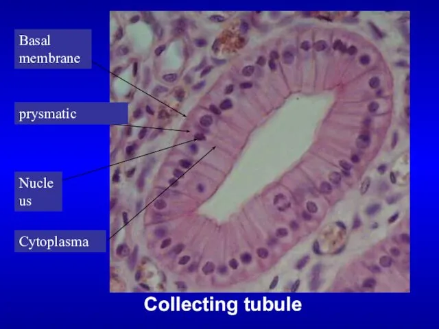
Collecting tubule
Collecting tubule

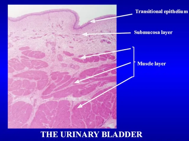
THE URINARY BLADDER
Transitional epithelium
Submucosa layer
Muscle layer
THE URINARY BLADDER
Transitional epithelium
Submucosa layer
Muscle layer
Transitional epithelium
Transitional epithelium
 Смешанный лес
 Первоцветы в природе
 Использование цифрового микроскопа на уроках
 Времена года
 Введение в центральную нервную систему
 Углерод
 Акция Каждой пичужке кормушка
 Морфология бактерий, грибов, спирохет, актиномицетов, микоплазм, риккетсий и хламидий
 Типы развития насекомых
 Класс насекомые. Общая характеристика
 Збудливі тканини. Нервова, м’язова, залозиста
 СӨЖ Тақырыбы : Жыныссыз және жынысты көбею және олардың түрлері
 Transcription_and_translation
 Экстерьер корпуса
 Балдырлар. Жалпы сипаттамасы. Бір жасушалы балдырлар
 Біологічні особливості і агротехніка вирощування бобових овочевих культур і цукрової кукурудзи
 Загадки мозга. Как мы считаем. Модель кодирования чисел
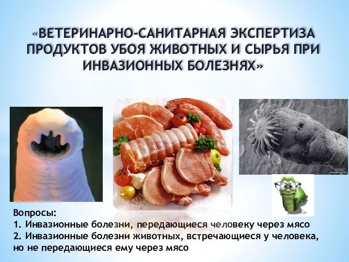 Ветеринарно-санитарная экспертиза продуктов убоя животных и сырья при инвазионных болезнях
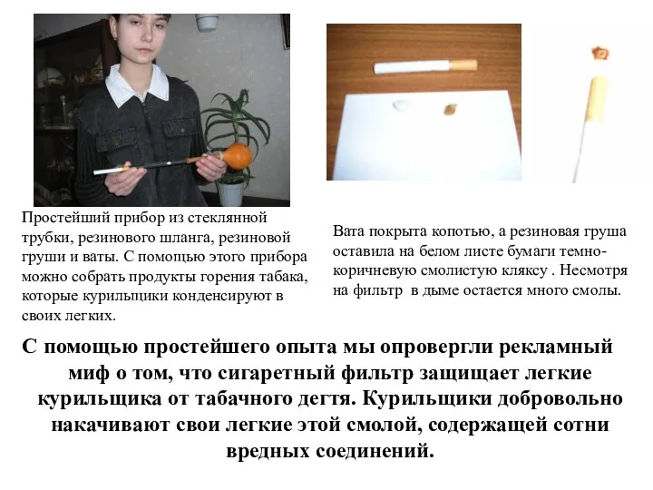 Вред курения.
 Видеоурок (открытый урок)
 Бездомные животные - проблема всех и каждого
 Зелёная аптека
 Биомембраны. Основные функции биомембран
 Важность охраны живого мира.
 Зоомагазин и его бизнес-план
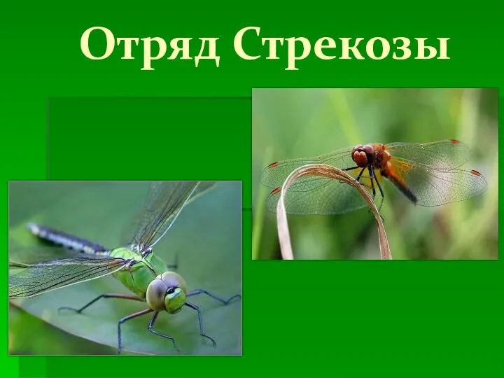 Отряд Стрекозы
 Мышечные ткани
 Удивительные животные