Microflora and sanitary-indicative bacteria of the soil, water, air the methods of studying презентация
- Microflora and sanitary-indicative bacteria of the soil, water, air the methods of studying

Содержание
- 2. Microorganisms are widespread. Microbes are distributed everywhere in the environment surrounding us. They are found in
- 3. The environment is a transmission factor of infectious diseases. Potentially pathogenic and pathogenic microorganisms get to
- 4. Sanitary microbiology is a science that studies the microflora of the environment and its harmful effect
- 5. Direct detection of pathogenic microorganisms in the different objects of environment, in general, is complicated because
- 6. Total viable count (TVC) is used for evaluation of total microbial contamination. TVC is the number
- 7. Sanitary-indicative microorganisms (SIMs) or sanitary-indicative bacteria are used for indirect evaluation of possible presence of pathogens
- 8. Presence of Escherichia coli and Enterococcus faecalis on environmental objects is indicative of fecal contamination. Escherichia
- 9. Simultaneous isolation of Staphylococcus aureus and hemolytic streptococci indicates possible contamination by oral droplets. Staphylococcus aureus
- 10. If the amount of SIMs increases in environmental objects, the probability of the presence of pathogenic
- 11. WATER MICROFLORA Pseudomonas fluorescens, Micrococcus roseus etc., are among the specific aquatic aerobic microorganisms. Anaerobic bacteria
- 12. The microflora of rivers depends on the degree of pollution and the quality of purification of
- 13. https://dornsife.usc.edu/labs/laketyrrell/research/
- 14. Water is an important factor for the transmission of a number of infectious diseases (enteric fever,
- 15. How do we monitor the sanitary quality of water? There are many kinds of pathogens that
- 16. The sanitary - bacteriological investigation of water includes: 1) determination of total viable count in 1
- 17. Giardia lamblia T4 bacteriophages infecting a live E. coli bacteria cell http://www.hyglos.de/en/technology.html Desulfovibrio vulgaris is the
- 18. 1. Sampling: 500 ml (tap water and purified water), 20 ml (water for injection), 100 ml
- 19. If there are more than 300 colonies do 10-fold dilutions (1: 10; 1: 100, etc.). When
- 20. Gram negative asporogenous small rods that do not have oxidase activity and ferment lactose to acid
- 21. MEMBRANE FILTRATION METHOD The investigated water (3 x 100 ml) are filtered through the three bacterial
- 22. MEMBRANE FILTRATION METHOD From 2 to 3 red-colored colonies are used for preparation of smear and
- 23. MEMBRANE FILTRATION METHOD The index of CFU (colony forming units) of coliforms in 100 ml water
- 24. What are the standards for drinking water? The USEPA issued revised Primary Drinking Water Standards in
- 25. STANDARDS The drinking water should not have more than 50 microbes in 1 ml. The microbial
- 26. Soil Microflora Soil fertility depends not only on the presence of inorganic and organic substances, but
- 27. Soil microflora consists bacteria (nitrifying, nitrogen-fixing, denitrifying), cellulose-splitting and sulfur bacteria, pigmented microbes fungi, protozoa, etc.
- 28. The greatest amount of microbes (1 000000 per cubic cm) is found in the top layer
- 29. The number of microorganisms in the soil depends on the extent of contamination with faeces and
- 30. Usually the soil is an unfavourable habitat for most pathogenic species of bacteria, rickettsiae, viruses, fungi,
- 31. Taking into consideration the definite epidemiological role played by the soil in spreading some infectious diseases
- 32. The sanitary-indicative bacteria of the soil are 1) E. coli/Enterococcus faecalis E. Coli (Gram Stain) Enterococcus
- 33. The sanitary-indicative bacteria of the soil are 2) Citrobacter spp. /Enterobacter spp.
- 34. The sanitary-indicative bacteria of the soil are 3) Clostridium perfringens Clostridium perfringens (Gram Stain)
- 35. More accurate evaluation is performed using coli-index — number of Enterobacteriaceae (so called coliform bacteria) found
- 36. Determination of Soil TVC For this purpose it is necessary to select most typical area not
- 37. Determination of Soil TVC 1. Prepare 10-fold dilutions (1:10, 1: 100, etc.) in an isotonic sterile
- 38. Determination of Perfringens-titer Seeding onto the Wilson-Blair medium: black colonies are formed and the gas breaks
- 39. AIR MICROFLORA The composition of the microbes of the air is quite variable. Then more dust,
- 40. The number of microbes in factories and homes is associated closely with the sanitary hygienic conditions
- 41. Pathogenic Species of Microbes Mycobacterium tuberculosis (Gram Stain) Anthrax bacilli (Gram Stain) Francisella tularensis (Gram Stain)
- 42. The air is an unfavourable medium for microbes. The absence of nutrient substances, the presence of
- 43. The laboratory investigation of air is carried out to determine the qualitative and quantitative composition of
- 44. Sanitary-indicative bacteria of air of closed buildings are 1) Streptococcus viridans 2) Streptococcus haemolyticus 3) Staphylococcus
- 45. The sanitary - bacteriological investigation of air includes: 1) determination the total viable count (TVC) in
- 46. Plate method (sedimentation method) The Petri’s dishes with meat-peptone agar or another special nutrient media for
- 47. ASPIRATION METHOD Krotov’s apparatus is used for bacteriological air research. It give us the possibility to
- 48. ASPIRATION METHOD For example, 250 colonies are revealed on the surface of dish after 2-minutes exposure
- 49. Determination of Staphylococci and Streptococci Using Krotov’s apparatus 250 L of air are seeded on the
- 50. Staphylococci and Streptococci colonies Staphylococcus aureus colony morhology on TSA. Cultivation 24 hours in an aerobic
- 52. Скачать презентацию
 Биологические ресурсы, их рациональное использование
 Глобальные экологические проблемы
 Учебная экологическая тропа
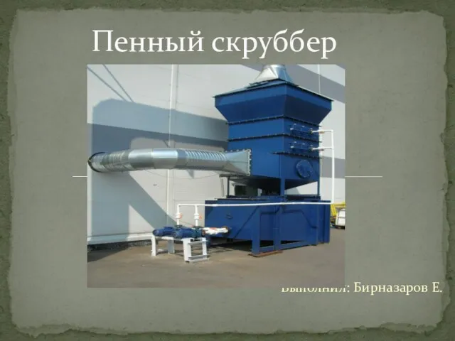 Пенный скруббер
 Судың экологиялық фактор ретінде маңызы
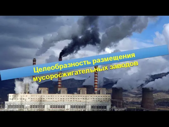 Целесообразность размещения мусоросжигательных заводов
 Биологиялық дозиметрия (электрондық парамагниттік резонанс және тағы басқалары) және олардың. (Тақырып 3)
 Пермакультура. Академия Управления городской средой, градостроительства и печати
 Ноосфера. Концепция ноосферы
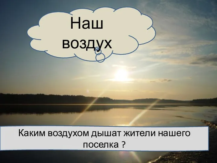 презентация наш воздухкасающийся проблемы
 Экология как наука: ее предмет и задачи. Понятие и виды природопользования
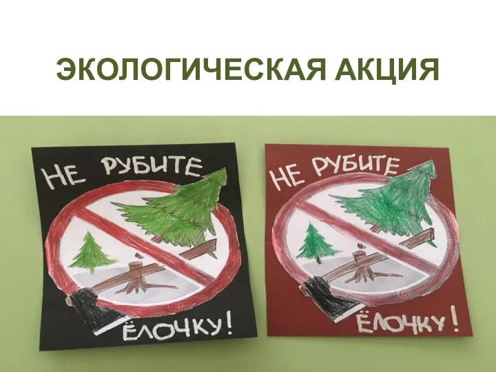 Экологическая акция
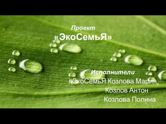 Проект ЭкоСемьЯ
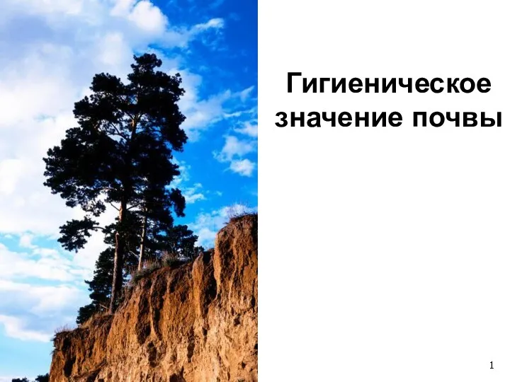 Гигиеническое значение почвы
 Час Земли
 Топырақ қорғау кешені. (Дәріс 4)
 Социально – экологический проект Думай глобально, действуй локально
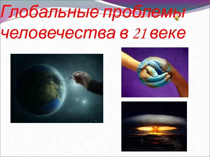 Глобальные проблемы человечества в 21 веке
 Збалансоване природокористування
 Атмосфераның ластануы. Атмосфералық ауаның ластануының зардаптары
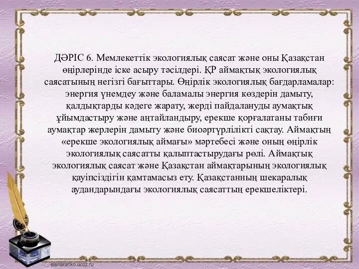 Мемлекеттік экологиялық саясат және оны қазақстан өңірлерінде іске асыру тәсілдері. Дәріс 6
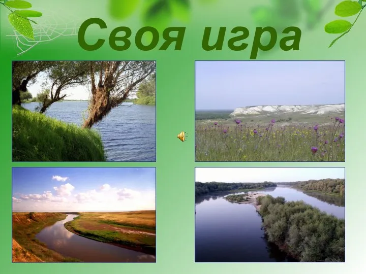 Своя игра по краеведению
 Наш дом - Земля
 Значение воды на Земле
 Червона книга світу. Тварини
 Ауаның қасиеті және маңызы
 Сортируй мусор правильно. Интерактивная карта
 Система нормирования воздействия на окружающую среду в 2019 году