- X-Ray Machine

Содержание
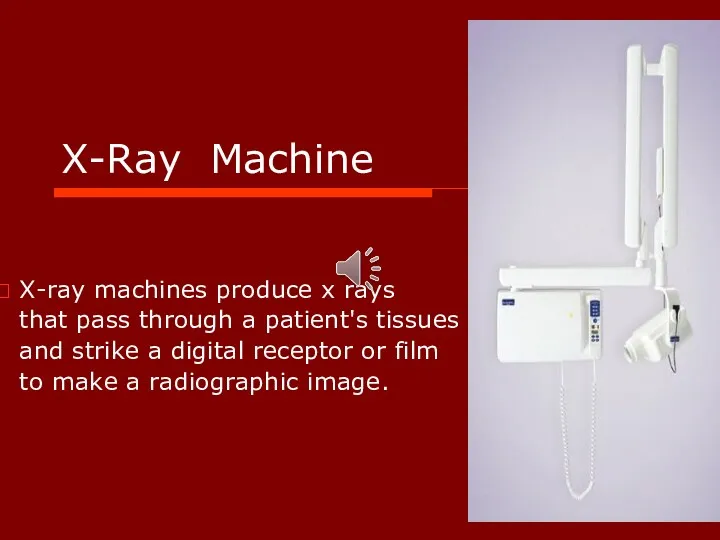
- 2. X-Ray Machine X-ray machines produce x rays that pass through a patient's tissues and strike a
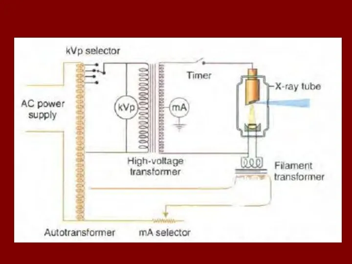
- 3. primary components of an x-ray machine : x-ray tube power supply The x-ray tube is positioned
- 4. An electrical insulating material, usually oil, surrounds the tube and transformers. Often, the tube is recessed
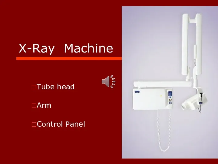
- 5. X-Ray Machine Tube head Arm Control Panel
- 6. Tube Head X-Ray Tube Power Supply
- 7. Power supply Heat the cathode filament to generate electrons. High potential difference accelerate electrons from cathode
- 8. Cathode Filament: - tungsten + 1% thorium Focusing cup - molybdenum
- 9. Filament The source of electrons within the x-ray tube The filament is heated to incandescence by
- 10. Focusing cup Negatively charged concave reflector made of molybdenum. The parabolic shape of the focusing cup
- 11. X-Ray Tube Glass envelope
- 12. X-Ray Tube Glass envelope Evacuated to prevent collision of the fast-moving electrons with gas molecules, which
- 13. Anode Tungsten Target Copper stem
- 14. Anode Purpose of target: Conversion of energy to X-ray is inefficient
- 15. Ideal Target High atomic number(74) High melting point(3422 ˚C) High thermal conductivity(173 W, mˉ¹,Kˉ¹) Low vapor
- 16. Focal Spot The area on the target to which the focusing cup directs the electrons and
- 18. Focal Spot Size : is important to image quality - sharpness - heat:1.stationary anode 2.rotating anode
- 20. Methods of dissipating the heat from focal spot : Anode Angle of target Copper stem Insulating
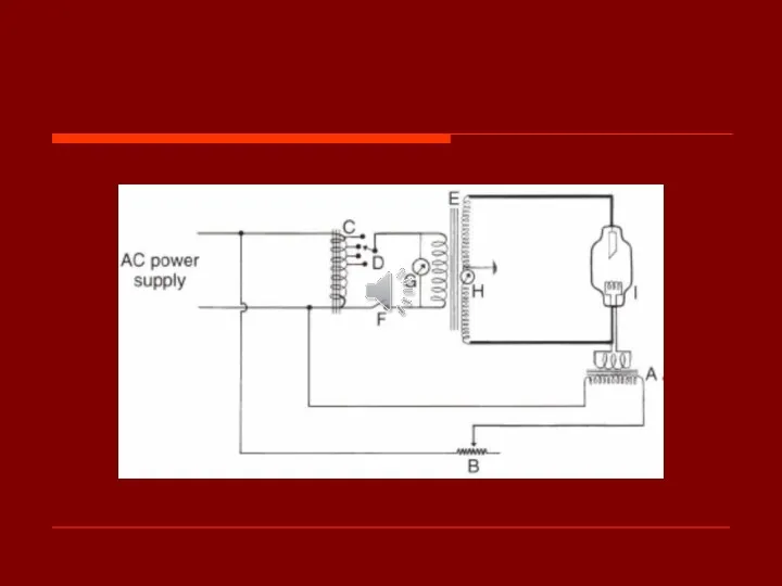
- 21. Power supply Primary functions: Low voltage: emit electrons High voltage: accelerate electrons Head of x-ray machine:
- 23. Tube Current Filament step-down transformer (filament transformer)(10v) mA selector or filament current control: - actually tube
- 24. When the hot filament releases electrons, it creates a cloud of electrons around the filament, a
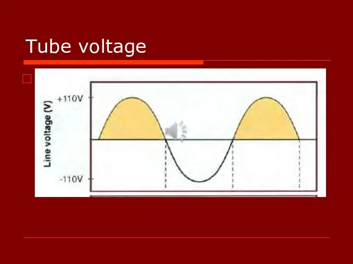
- 25. Tube voltage Why High voltage? Autotransformer: The actual voltage used on an x-ray machine is adjusted
- 27. Tube voltage Because the polarity of the line current alternates (60 cycles per second), the polarity
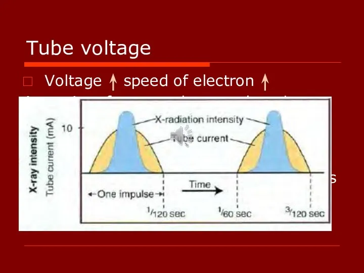
- 28. Tube voltage Voltage speed of electron intensity of x-ray pulses tends to be sharply peaked at
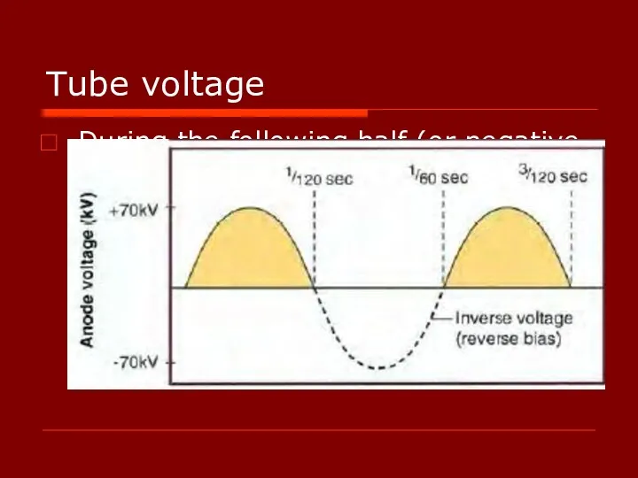
- 29. Tube voltage During the following half (or negative half) of each cycle, the filament becomes positive,
- 30. Tube voltage Self-rectified or Half-wave rectified: The alternating high voltage is applied directly across the x-ray
- 31. Tube voltage Replace the conventional 60-cycle AC, half-wave rectified power supply with a full-wave rectified, high-frequency
- 32. Tube voltage Intraoral,Panoramic, and Cephalometric machines operate between 50 and 90 kVp, whereas cone-beam computed tomographic
- 33. Timer Duration of x-ray exposure/ into the high-voltage circuit Length of high-voltage To minimize filament damage
- 34. Tube Rating : longest exposure time HU = (kVp x mA) x seconds The heat storage
- 35. Duty Cycle : frequency of exposures - anode size - cooling methods
- 36. Production of X-Rays Most energy : Heat
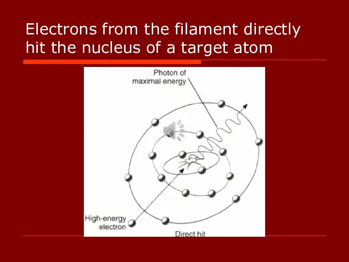
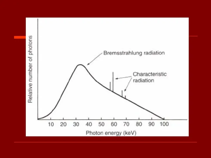
- 37. Bremsstrahlung Radiation (برم اشترالانگ) The sudden stopping or slowing of high-speed electrons by tungsten nuclei “breaking
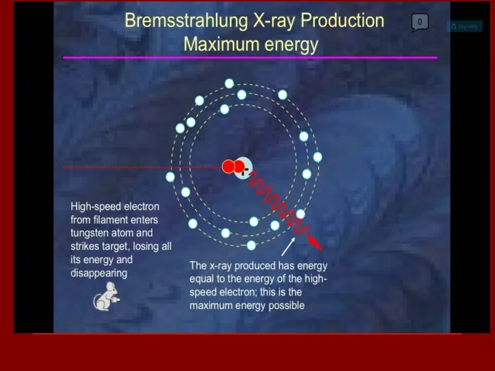
- 38. Electrons from the filament directly hit the nucleus of a target atom
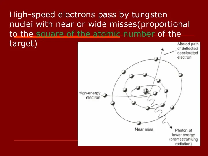
- 40. High-speed electrons pass by tungsten nuclei with near or wide misses(proportional to the square of the
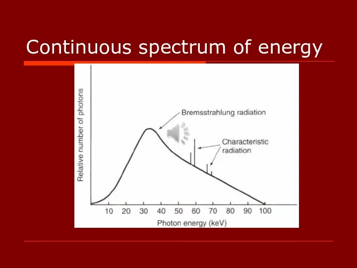
- 42. Continuous spectrum of energy
- 43. Bremsstrahlung Radiation The continuously varying voltage difference between the target and filament causes the electrons striking
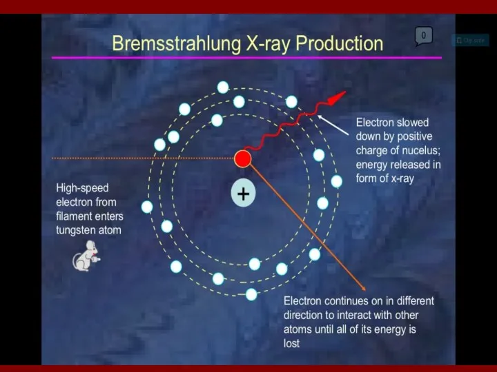
- 44. Bremsstrahlung Radiation The bombarding electrons pass at varying distances around tungsten nuclei and are thus deflected
- 45. Bremsstrahlung Radiation Most electrons participate in the target before losing all their kinetic energy. As a
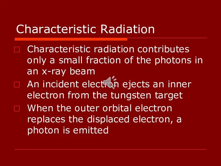
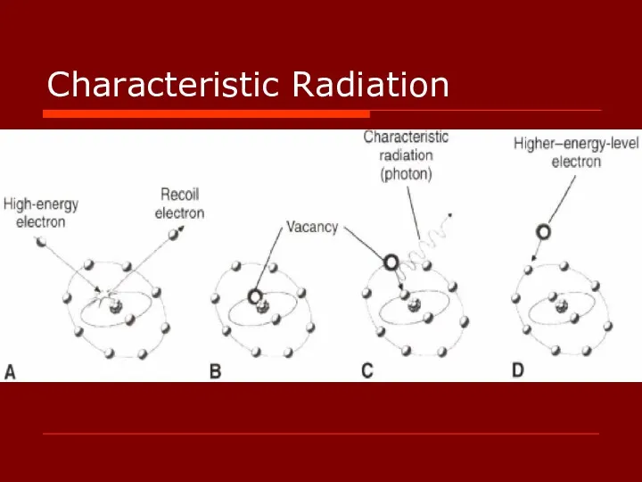
- 46. Characteristic Radiation Characteristic radiation contributes only a small fraction of the photons in an x-ray beam
- 47. Characteristic Radiation
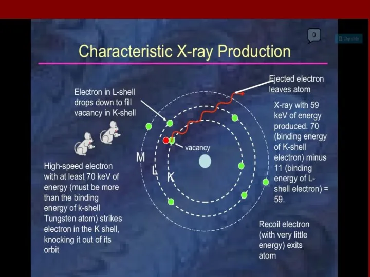
- 49. Small fraction Discrete spectrum Difference of energy levels of electron orbitals Characteristic of target atoms
- 52. Скачать презентацию
X-Ray Machine
X-ray machines produce x rays
that pass through a
X-Ray Machine
X-ray machines produce x rays
that pass through a
primary components of an x-ray machine :
x-ray tube
power
primary components of an x-ray machine :
x-ray tube
power
An electrical insulating material, usually oil, surrounds the tube and transformers.
An electrical insulating material, usually oil, surrounds the tube and transformers.
X-Ray Machine
Tube head
Arm
Control Panel
X-Ray Machine
Tube head
Arm
Control Panel
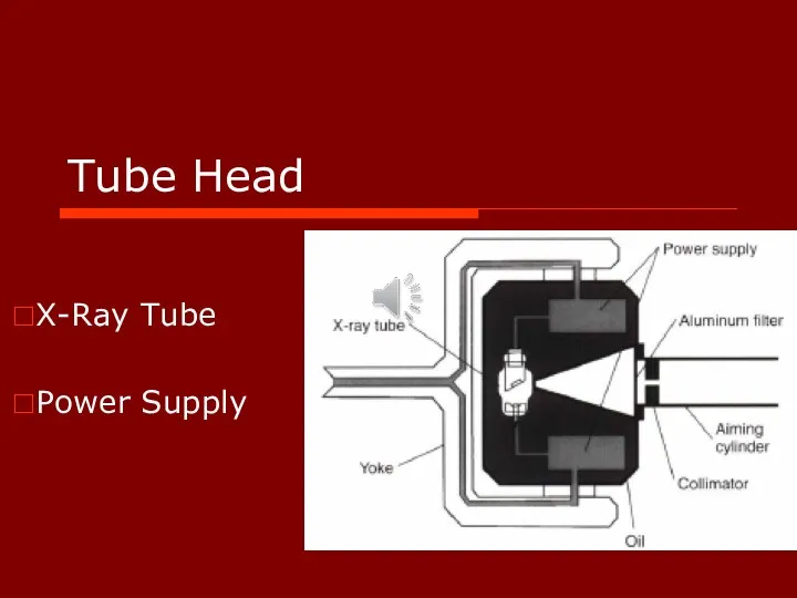
Tube Head
X-Ray Tube
Power Supply
Tube Head
X-Ray Tube
Power Supply
Power supply
Heat the cathode filament to generate electrons.
High potential difference accelerate
Power supply
Heat the cathode filament to generate electrons.
High potential difference accelerate
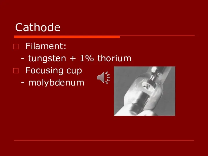
Cathode
Filament:
- tungsten + 1% thorium
Focusing cup
- molybdenum
Cathode
Filament:
- tungsten + 1% thorium
Focusing cup
- molybdenum
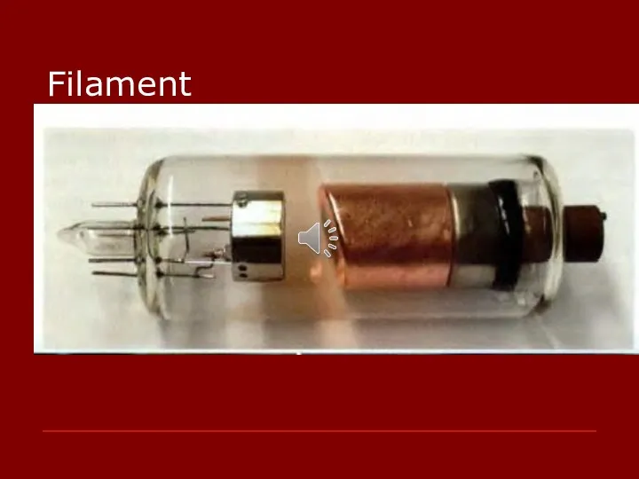
Filament
The source of electrons within the x-ray tube
The filament is
Filament
The source of electrons within the x-ray tube
The filament is
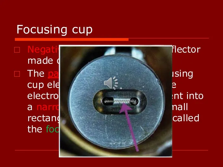
Focusing cup
Negatively charged concave reflector made of molybdenum.
The parabolic
Focusing cup
Negatively charged concave reflector made of molybdenum.
The parabolic
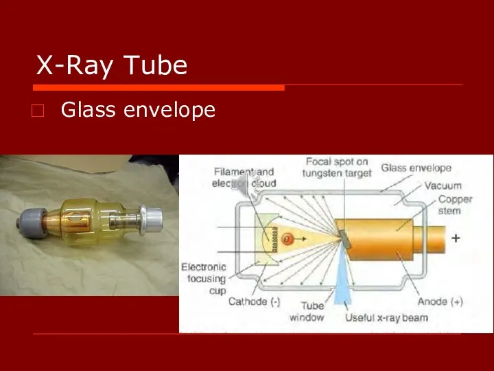
X-Ray Tube
Glass envelope
X-Ray Tube
Glass envelope
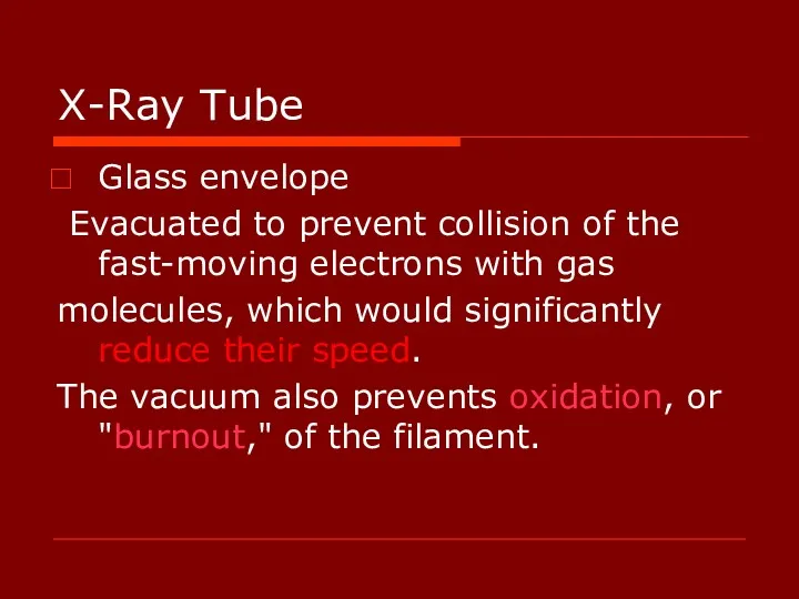
X-Ray Tube
Glass envelope
Evacuated to prevent collision of the fast-moving electrons
X-Ray Tube
Glass envelope
Evacuated to prevent collision of the fast-moving electrons
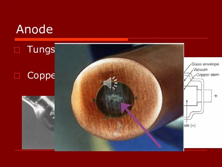
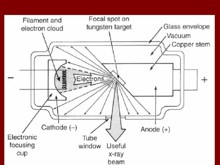
Anode
Tungsten Target
Copper stem
Anode
Tungsten Target
Copper stem
Anode
Purpose of target:
Conversion of energy to X-ray is inefficient
Anode
Purpose of target:
Conversion of energy to X-ray is inefficient
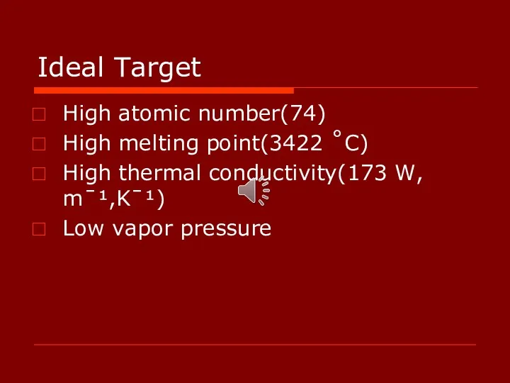
Ideal Target
High atomic number(74)
High melting point(3422 ˚C)
High thermal conductivity(173 W, mˉ¹,Kˉ¹)
Low
Ideal Target
High atomic number(74)
High melting point(3422 ˚C)
High thermal conductivity(173 W, mˉ¹,Kˉ¹)
Low
Focal Spot
The area on the target to which the focusing cup
Focal Spot
The area on the target to which the focusing cup
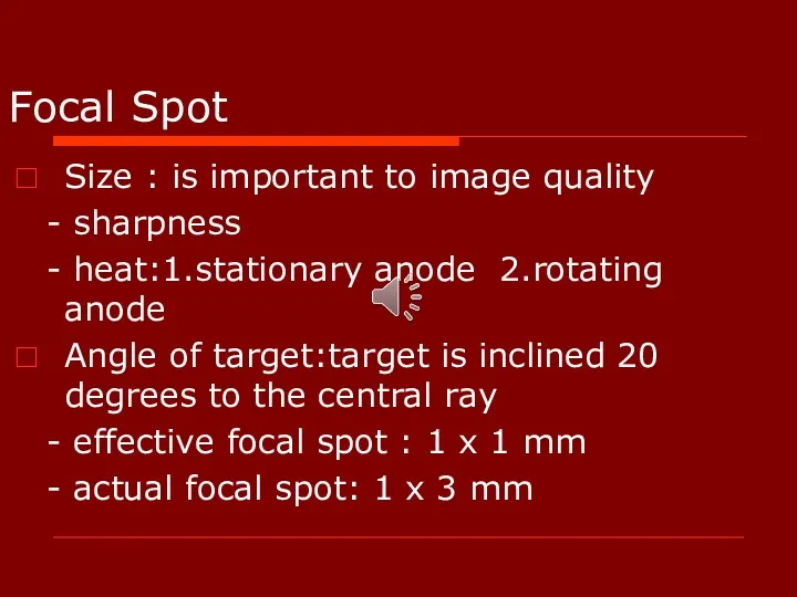
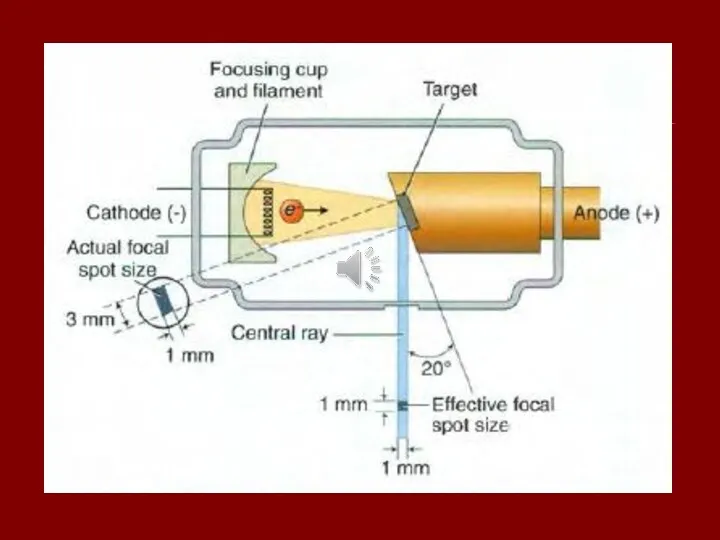
Focal Spot
Size : is important to image quality
- sharpness
-
Focal Spot
Size : is important to image quality
- sharpness
-
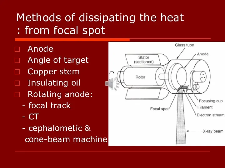
Methods of dissipating the heat from focal spot :
Anode
Angle of target
Copper
Methods of dissipating the heat from focal spot :
Anode
Angle of target
Copper
Power supply
Primary functions:
Low voltage: emit electrons
High voltage: accelerate electrons
Head of x-ray
Power supply
Primary functions:
Low voltage: emit electrons
High voltage: accelerate electrons
Head of x-ray
Tube Current
Filament step-down transformer (filament transformer)(10v)
mA selector or filament current control:
Tube Current
Filament step-down transformer (filament transformer)(10v)
mA selector or filament current control:
When the hot filament releases electrons, it creates a cloud of
When the hot filament releases electrons, it creates a cloud of
Tube voltage
Why High voltage?
Autotransformer:
The actual voltage used on an x-ray
Tube voltage
Why High voltage?
Autotransformer:
The actual voltage used on an x-ray
Tube voltage
Because the polarity of the line current alternates (60 cycles
Tube voltage
Because the polarity of the line current alternates (60 cycles
Tube voltage
Voltage speed of electron
intensity of x-ray pulses tends to
Tube voltage
Voltage speed of electron
intensity of x-ray pulses tends to
Tube voltage
During the following half (or negative half) of each cycle,
Tube voltage
During the following half (or negative half) of each cycle,
Tube voltage
Self-rectified or Half-wave rectified:
The alternating high voltage is applied directly
Tube voltage
Self-rectified or Half-wave rectified:
The alternating high voltage is applied directly
Tube voltage
Replace the conventional 60-cycle AC, half-wave rectified power supply with
Tube voltage
Replace the conventional 60-cycle AC, half-wave rectified power supply with
Tube voltage
Intraoral,Panoramic, and Cephalometric machines operate between 50 and 90 kVp,
Tube voltage
Intraoral,Panoramic, and Cephalometric machines operate between 50 and 90 kVp,
Timer
Duration of x-ray exposure/ into the high-voltage circuit
Length of high-voltage
To
Timer
Duration of x-ray exposure/ into the high-voltage circuit
Length of high-voltage
To
Tube Rating : longest exposure time
HU = (kVp x mA)
Tube Rating : longest exposure time
HU = (kVp x mA)
Duty Cycle : frequency of exposures
- anode size
- cooling
Duty Cycle : frequency of exposures
- anode size
- cooling
Production of X-Rays
Most energy : Heat
Production of X-Rays
Most energy : Heat
Bremsstrahlung Radiation
(برم اشترالانگ)
The sudden stopping or slowing of high-speed electrons by
Bremsstrahlung Radiation
(برم اشترالانگ)
The sudden stopping or slowing of high-speed electrons by
Electrons from the filament directly hit the nucleus of a target
Electrons from the filament directly hit the nucleus of a target
High-speed electrons pass by tungsten nuclei with near or wide misses(proportional
High-speed electrons pass by tungsten nuclei with near or wide misses(proportional
Continuous spectrum of energy
Continuous spectrum of energy
Bremsstrahlung Radiation
The continuously varying voltage difference between the target and filament
Bremsstrahlung Radiation
The continuously varying voltage difference between the target and filament
Bremsstrahlung Radiation
The bombarding electrons pass at varying distances around tungsten nuclei
Bremsstrahlung Radiation
The bombarding electrons pass at varying distances around tungsten nuclei
Bremsstrahlung Radiation
Most electrons participate in the target before losing all their
Bremsstrahlung Radiation
Most electrons participate in the target before losing all their
Characteristic Radiation
Characteristic radiation contributes only a small fraction of the photons
Characteristic Radiation
Characteristic radiation contributes only a small fraction of the photons
Characteristic Radiation
Characteristic Radiation
Small fraction
Discrete spectrum
Difference of energy levels of electron orbitals
Characteristic of target
Small fraction
Discrete spectrum
Difference of energy levels of electron orbitals
Characteristic of target
 Измерение удельной теплоемкости твердого тела
 Вихретоковый неразрушающий контроль
 Преломление света
 Постоянный ток. Занятие 1
 Механические свойства твердых тел
 викторина Юный физик
 Көч – җисемнәрнең үзара тәэсир итешү үлчәме ул
 Применение в фармацевтическом анализе рефрактометрии, поляриметрии, полярографии
 Введение в спектральный анализ. Природа и свойства электромагнитного излучения
 Основы Молекулярно-Кинетической Теории (физика 10 класс)
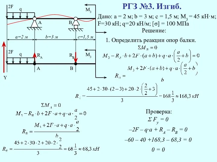 Определить реакцию опоры балки
 Вектор магнитной индукции. Линии магнитной индукции
 Презентация по физике для 7 класса по теме Простые механизмы
 Получение и передача переменного электрического тока. Трансформатор
 Электрические явления. Законы постоянного тока
 Простые механизмы и их использование в машинах
 Викторина на тему: Дисперсия света
 Простые механизмы. Рычаг
 Фізика. Середня швидкість
 Электронный парамагнитный резонанс
 Повышение качества обработки колец подшипников
 Лампа накаливания
 Сила трения
 Архимед күші
 Рентгеновское излучение
 Laws of Thermodynamics
 Проводники в электростатическом поле. Конденсаторы. Энергия электрического поля
 Свойства топлив. Теплота сгорания топлив. Урок № 4