- Atlas of 3D Ultrasound

Содержание
- 2. Contents History of Ultrasound Principle of 3D Ultrasound 3D Utilities Clinical Advantages of 3D Ultrasound 3D
- 3. History of Ultrasound
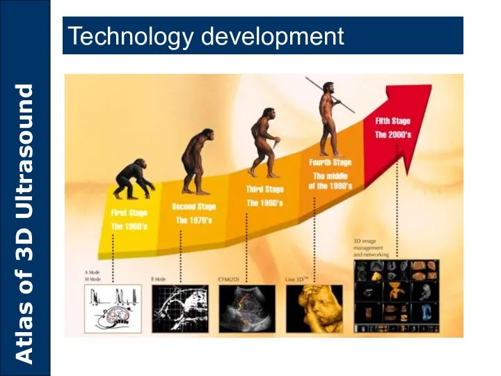
- 4. Technology development
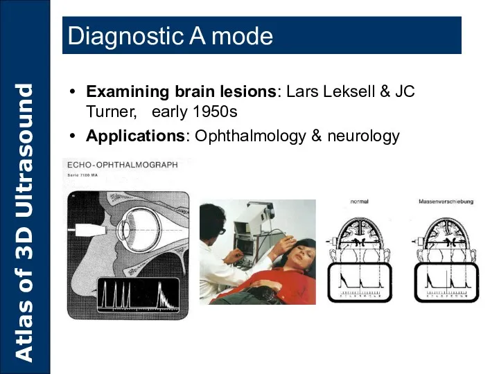
- 5. Diagnostic A mode Examining brain lesions: Lars Leksell & JC Turner, early 1950s Applications: Ophthalmology &
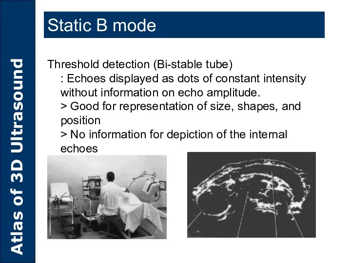
- 6. Static B mode Threshold detection (Bi-stable tube) : Echoes displayed as dots of constant intensity without
- 7. The first use of Sector Probe : The element installed on the tip of the probe
- 8. Electronic Probe : Obtain an image using each of a element with the front of Probe
- 9. 2D 3D 4D Concept of 3D
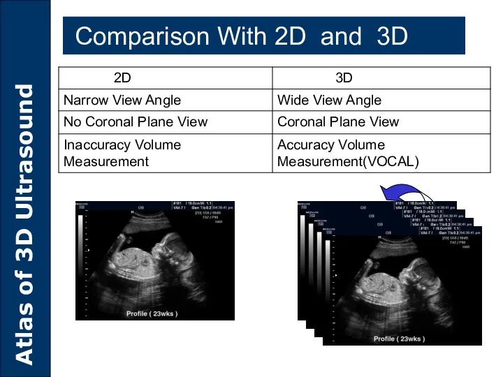
- 10. Real Time 2D(B-Mode) -NO Coronal Plane. -Limitation of View Angle Comparison With 2D and 3D
- 11. 3D Scan Freely Scan Arbitrary Plane C Plane(Coronal) Comparison With 2D and 3D
- 12. Comparison With 2D and 3D
- 13. Static 3D
- 14. Live 3D
- 15. Classification of 3D US Workstation 3D Post processing 3D with VHS output Poor image resolution and
- 16. Principles of 3D Ultrasound
- 17. How 3D image is produced? Data acquisition Scan conversion Volume rendering
- 18. Data Process 3D Scan Conversion 3D Image
- 19. 3D convex Probe 3D Linear Probe 3D Vaginal Probe Step 1: Probe selection Data Acquisition I
- 20. Step 2: Data scanning 3D Convex Probe Data Acquisition I
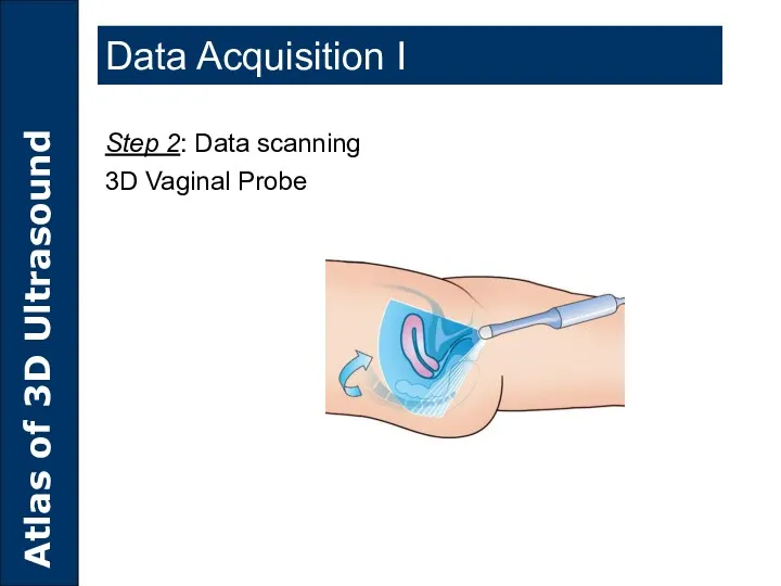
- 21. Data Acquisition I Step 2: Data scanning 3D Vaginal Probe
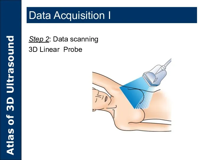
- 22. Data Acquisition I Step 2: Data scanning 3D Linear Probe
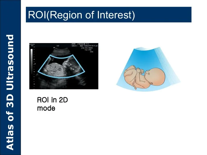
- 23. ROI(Region of Interest) ROI in 2D mode
- 24. Step 3: Adjust ROI Size Axial Depth, Lateral View Angle, Elevation Angle ROI(Region of Interest)
- 25. Auto ROI(Only ACCUIX XQ) ROI(Region of Interest)
- 26. Data Acquisition Comparison of Sequence between adjustable ROI and Auto ROI Adjustable ROI Auto ROI(Quickly and
- 27. Data Quantity 3D Image Spatial Resolution Volume Frame Rate Scan Quality Selection (ACCUVIX XQ 4 step,
- 28. Y direction Z direction Echo= f(x,y,z) 3D Scan conversion 3D Scan변환 X direction X direction Y
- 29. Definition of Voxel A voxel is a cubic cell, which has a single value cover the
- 30. A-Plane resolution: Axial resolution x Lateral resolution B-Plane resolution: Lateral resolution x Elevation resolution C-Plane resolution:
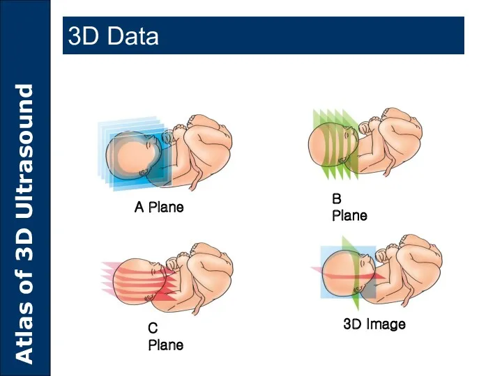
- 31. 3D Data A Plane 3D Image C Plane B Plane
- 32. Volume Rendering Rendering mode Surface mode Transparent Mode Maximum transparent mode Minimum transparent mode X-ray mode
- 33. Volume Rendering (Surface mode)
- 34. Volume Rendering (Transparent mode) Transparent mode Maximum mode The maximum gray values of the ROI characterization
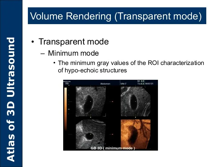
- 35. Volume Rendering (Transparent mode) Transparent mode Minimum mode The minimum gray values of the ROI characterization
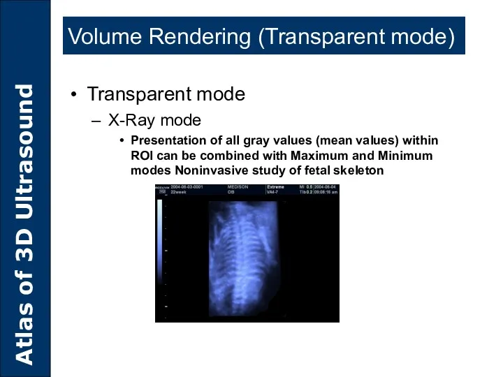
- 36. Volume Rendering (Transparent mode) Transparent mode X-Ray mode Presentation of all gray values (mean values) within
- 37. 3D Utilities
- 38. MULTI-PLANAR IMAGING VolumeCT mode VOCAL SHELL IMAGING SEE-THRU MODE 3D Utilities
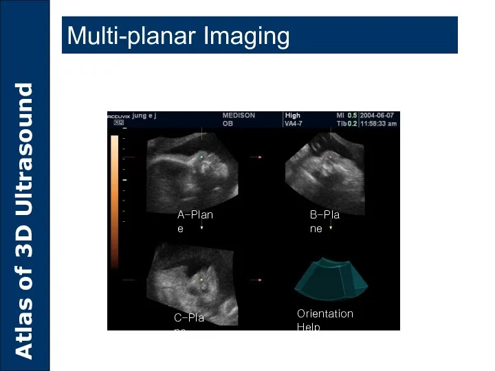
- 39. Multi-planar Imaging A-Plane B-Plane C-Plane Orientation Help
- 40. Coronal Transverse Sagital Volume CT mode
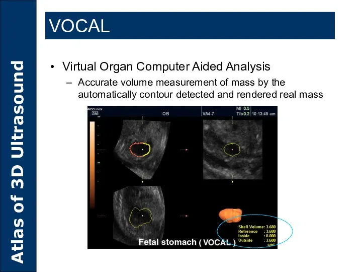
- 41. VOCAL Virtual Organ Computer Aided Analysis Accurate volume measurement of mass by the automatically contour detected
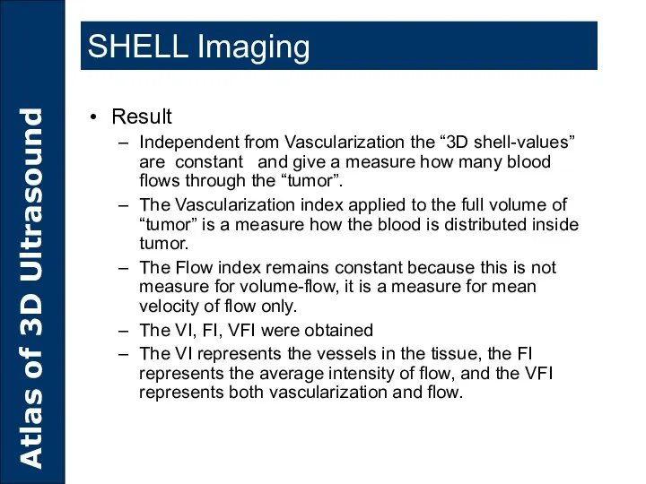
- 42. SHELL Imaging Result Independent from Vascularization the “3D shell-values” are constant and give a measure how
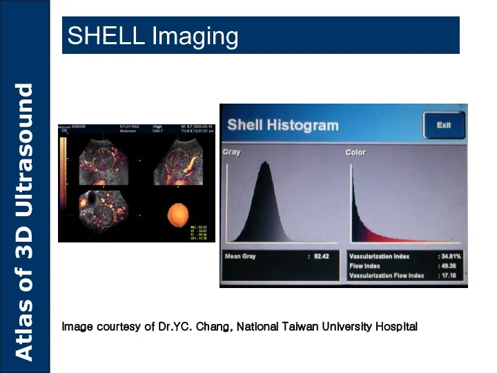
- 43. SHELL Imaging Image courtesy of Dr.YC. Chang, National Taiwan University Hospital
- 44. SEE-THRU MODE 3D imaging simultaneously for muscle tissue and blood vessels as well
- 45. Clinical Advantages of 3D Ultrasound
- 46. Multi-Planar Imaging Congenital uterine abnormal(A,B,C Plane) Fetal cleft lip and palate detection Needle Position during breast
- 47. Multi-Planar Imaging Complete diagnosis of cleft lip on 3D multi-planar view
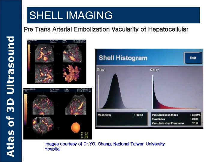
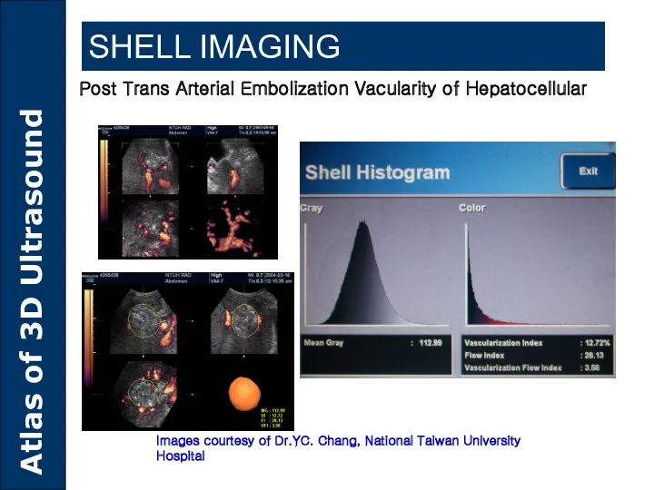
- 48. Used to measure the Pre-and Post-TAE vascularity of Hepatocellular The tumor volume, Vascularization Index(VI), Flow Index(VFI)
- 49. SHELL IMAGING Pre Trans Arterial Embolization Vacularity of Hepatocellular Images courtesy of Dr.YC. Chang, National Taiwan
- 50. SHELL IMAGING Post Trans Arterial Embolization Vacularity of Hepatocellular Images courtesy of Dr.YC. Chang, National Taiwan
- 51. SEE-THRU MODE Fetal Circulation Lateral Oblique Frontage Oblique
- 52. Congenital uterine abnormal – Bicornuate uterus Gynecology
- 53. Uterus Mass in Surface Rendering Gynecology Endometrium mass Uterus
- 54. Meningomyelocele Obstetrics
- 55. Anecephaly Obstetrics
- 56. Limbs with developmental defect and body edema Obstetrics
- 57. Cleft lip and palate Obstetrics
- 58. Limbs with developmental defect Obstetrics
- 59. Accuvix XQ 3D
- 60. Probe Selection
- 61. Freeze 2D Press button Adjustable ROI Live 3D Selection Press button Live 3D
- 62. Multi-planar image Display image format select Live 3D
- 63. Orientation Live 3D
- 64. 2D Press button Adjustable ROI Live 3D Selection Press button Freeze Live 3D
- 65. VOCAL
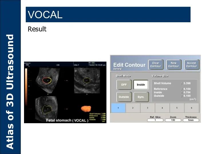
- 66. Result VOCAL
- 67. SA8000LV 3D
- 68. Probe Selection Press button Live 3D
- 69. Press button Press Keyboard Live 3D Live 3D 3D Live 3D
- 70. Live 3D
- 72. Скачать презентацию
Contents
History of Ultrasound
Principle of 3D Ultrasound
3D Utilities
Clinical Advantages of 3D Ultrasound
3D
Contents
History of Ultrasound
Principle of 3D Ultrasound
3D Utilities
Clinical Advantages of 3D Ultrasound
3D

History of Ultrasound
History of Ultrasound
Technology development
Technology development
Diagnostic A mode
Examining brain lesions: Lars Leksell & JC Turner, early
Diagnostic A mode
Examining brain lesions: Lars Leksell & JC Turner, early
Static B mode
Threshold detection (Bi-stable tube)
: Echoes displayed as dots of
Static B mode
Threshold detection (Bi-stable tube) : Echoes displayed as dots of
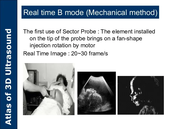
The first use of Sector Probe : The element installed on
The first use of Sector Probe : The element installed on
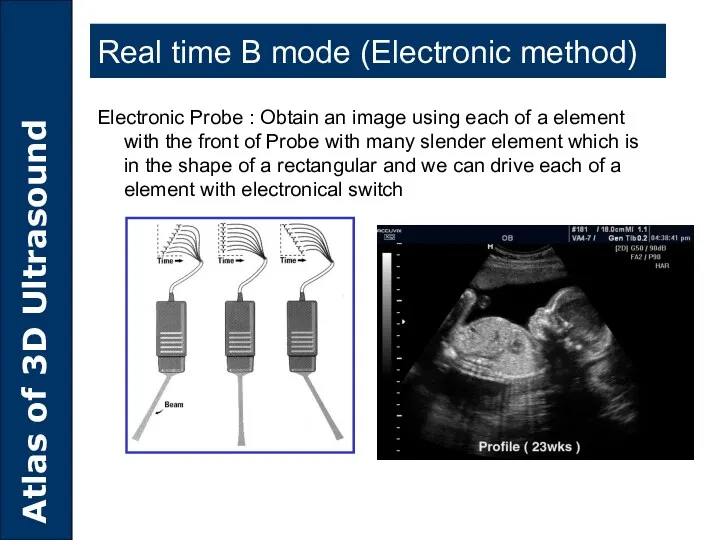
Electronic Probe : Obtain an image using each of a element
Electronic Probe : Obtain an image using each of a element
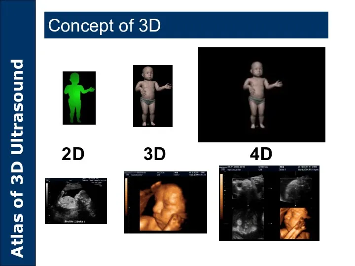
2D 3D 4D
Concept of 3D
2D 3D 4D
Concept of 3D
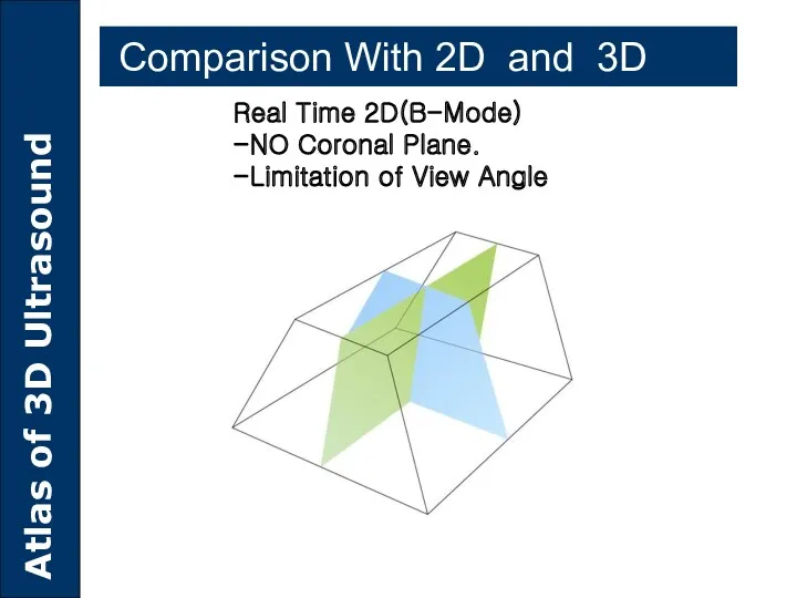
Real Time 2D(B-Mode)
-NO Coronal Plane.
-Limitation of View Angle
Comparison With 2D
Real Time 2D(B-Mode)
-NO Coronal Plane.
-Limitation of View Angle
Comparison With 2D
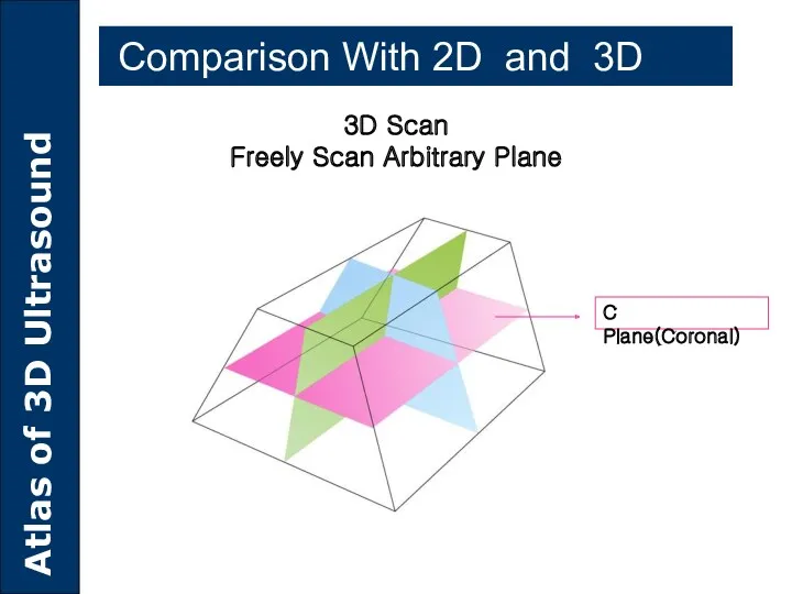
3D Scan
Freely Scan Arbitrary Plane
C Plane(Coronal)
Comparison With 2D and
3D Scan
Freely Scan Arbitrary Plane
C Plane(Coronal)
Comparison With 2D and
Comparison With 2D and 3D
Comparison With 2D and 3D
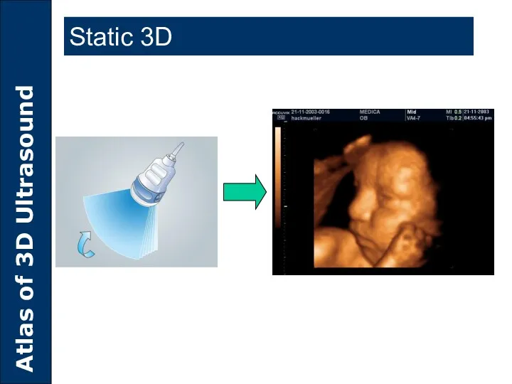
Static 3D
Static 3D
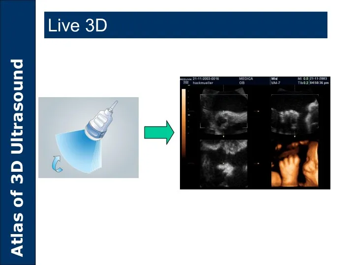
Live 3D
Live 3D
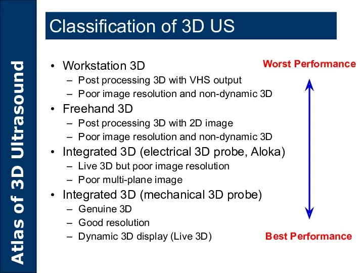
Classification of 3D US
Workstation 3D
Post processing 3D with VHS output
Poor image
Classification of 3D US
Workstation 3D
Post processing 3D with VHS output
Poor image
Principles of 3D Ultrasound
Principles of 3D Ultrasound
How 3D image is produced?
Data acquisition
Scan conversion
Volume rendering
How 3D image is produced?
Data acquisition
Scan conversion
Volume rendering
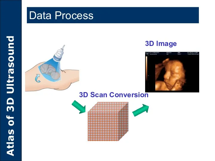
Data Process
3D Scan Conversion
3D Image
Data Process
3D Scan Conversion
3D Image
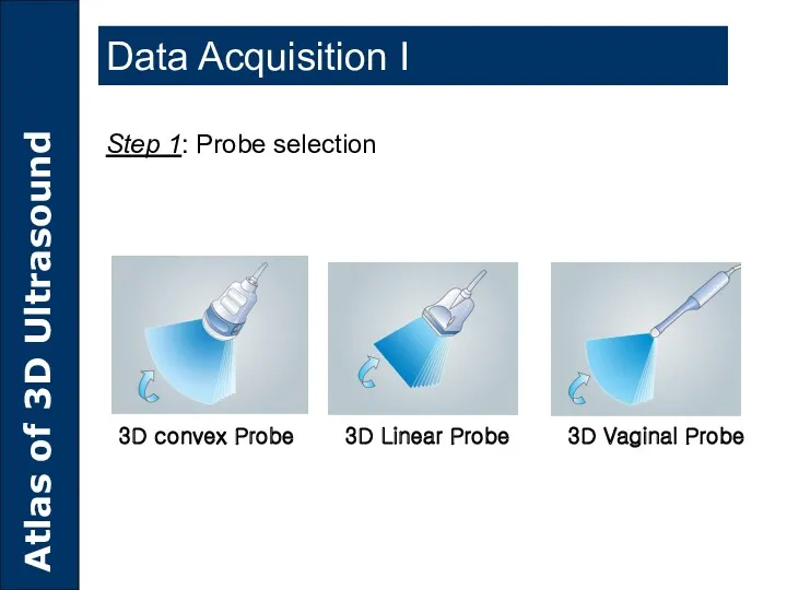
3D convex Probe 3D Linear Probe 3D Vaginal Probe
Step 1: Probe
Step 1: Probe
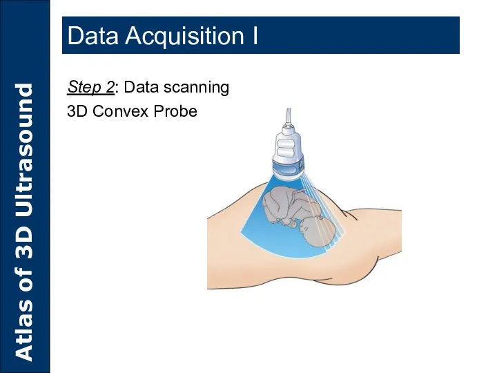
Step 2: Data scanning
3D Convex Probe
Data Acquisition I
Step 2: Data scanning
3D Convex Probe
Data Acquisition I
Data Acquisition I
Step 2: Data scanning
3D Vaginal Probe
Data Acquisition I
Step 2: Data scanning
3D Vaginal Probe
Data Acquisition I
Step 2: Data scanning
3D Linear Probe
Data Acquisition I
Step 2: Data scanning
3D Linear Probe
ROI(Region of Interest)
ROI in 2D mode
ROI(Region of Interest)
ROI in 2D mode
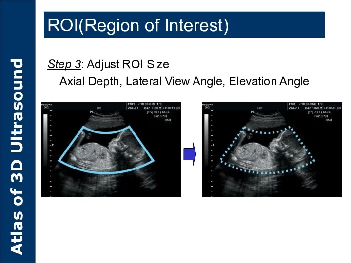
Step 3: Adjust ROI Size
Axial Depth, Lateral View Angle, Elevation
Step 3: Adjust ROI Size
Axial Depth, Lateral View Angle, Elevation
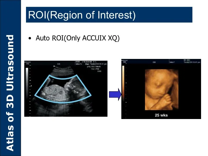
Auto ROI(Only ACCUIX XQ)
ROI(Region of Interest)
Auto ROI(Only ACCUIX XQ)
ROI(Region of Interest)
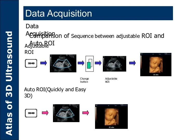
Data Acquisition
Comparison of Sequence between adjustable ROI and Auto ROI
Adjustable ROI
Auto
Data Acquisition
Comparison of Sequence between adjustable ROI and Auto ROI
Adjustable ROI
Auto
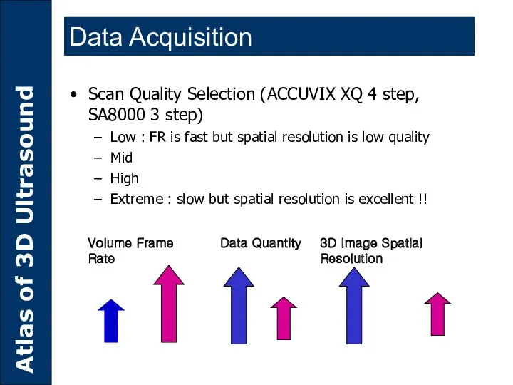
Data Quantity
3D Image Spatial Resolution
Volume Frame Rate
Scan Quality Selection (ACCUVIX XQ
Data Quantity
3D Image Spatial Resolution
Volume Frame Rate
Scan Quality Selection (ACCUVIX XQ
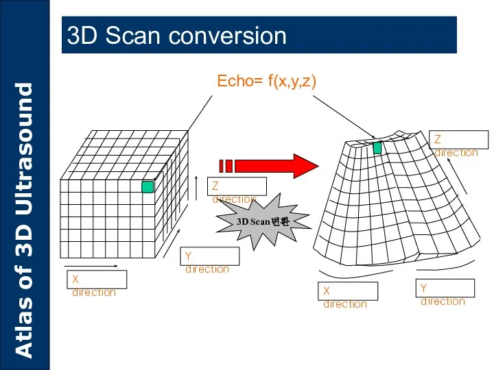
Y direction
Z direction
Echo= f(x,y,z)
3D Scan conversion
3D Scan변환
X direction
X direction
Y direction
Z direction
Y direction
Z direction
Echo= f(x,y,z)
3D Scan conversion
3D Scan변환
X direction
X direction
Y direction
Z direction
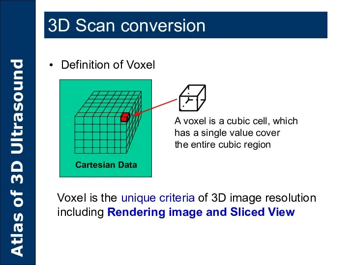
Definition of Voxel
A voxel is a cubic cell, which
has a single
Definition of Voxel
A voxel is a cubic cell, which
has a single
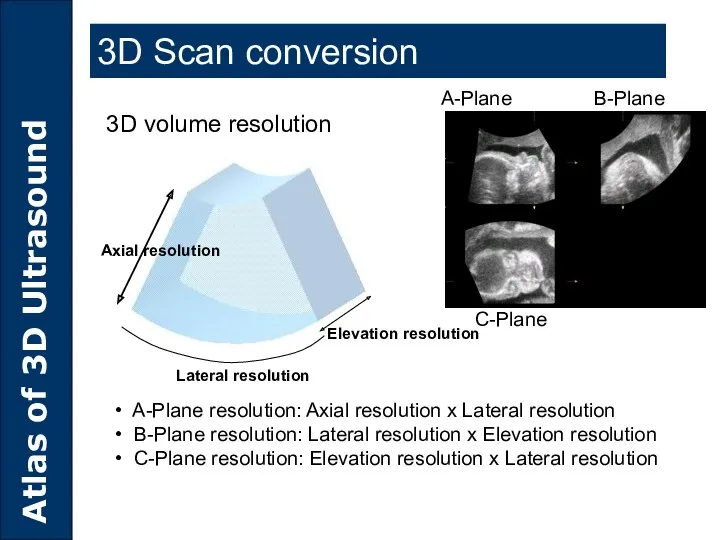
A-Plane resolution: Axial resolution x Lateral resolution
B-Plane resolution: Lateral
A-Plane resolution: Axial resolution x Lateral resolution
B-Plane resolution: Lateral
3D Data
A Plane
3D Image
C Plane
B Plane
3D Data
A Plane
3D Image
C Plane
B Plane
Volume Rendering
Rendering mode
Surface mode
Transparent Mode
Maximum transparent mode
Minimum transparent mode
X-ray mode
Volume Rendering
Rendering mode
Surface mode
Transparent Mode
Maximum transparent mode
Minimum transparent mode
X-ray mode
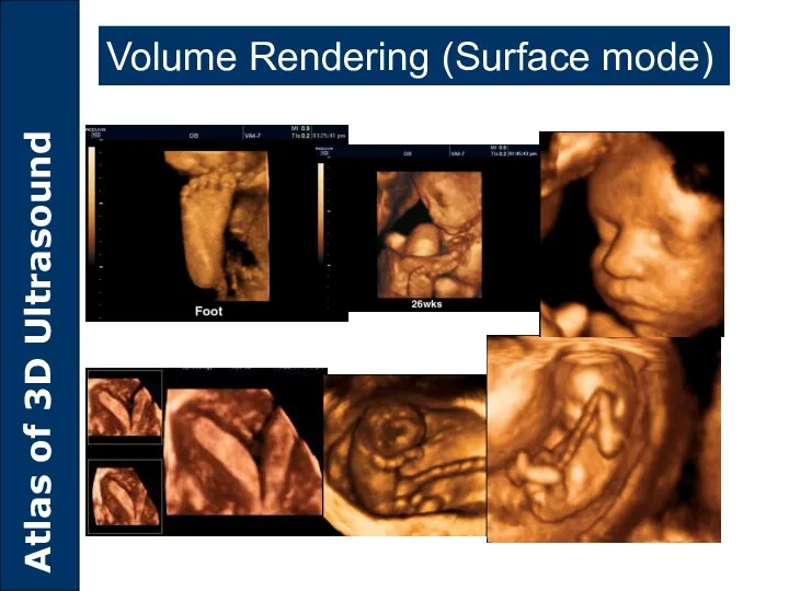
Volume Rendering (Surface mode)
Volume Rendering (Surface mode)
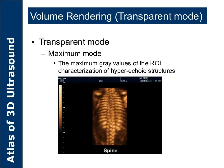
Volume Rendering (Transparent mode)
Transparent mode
Maximum mode
The maximum gray values of the
Volume Rendering (Transparent mode)
Transparent mode
Maximum mode
The maximum gray values of the
Volume Rendering (Transparent mode)
Transparent mode
Minimum mode
The minimum gray values of the
Volume Rendering (Transparent mode)
Transparent mode
Minimum mode
The minimum gray values of the
Volume Rendering (Transparent mode)
Transparent mode
X-Ray mode
Presentation of all gray values
Volume Rendering (Transparent mode)
Transparent mode
X-Ray mode
Presentation of all gray values

3D Utilities
3D Utilities
MULTI-PLANAR IMAGING
VolumeCT mode
VOCAL
SHELL IMAGING
SEE-THRU MODE
3D Utilities
MULTI-PLANAR IMAGING
VolumeCT mode
VOCAL
SHELL IMAGING
SEE-THRU MODE
3D Utilities
Multi-planar Imaging
A-Plane
B-Plane
C-Plane
Orientation Help
Multi-planar Imaging
A-Plane
B-Plane
C-Plane
Orientation Help
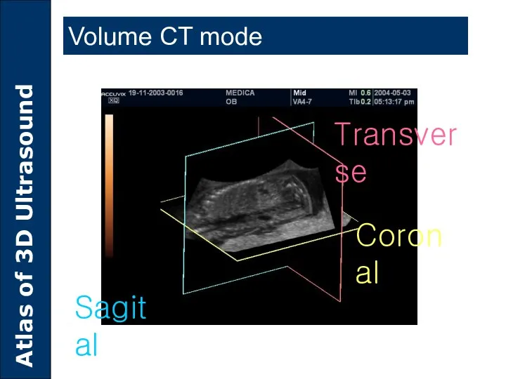
Coronal
Transverse
Sagital
Volume CT mode
Coronal
Transverse
Sagital
Volume CT mode
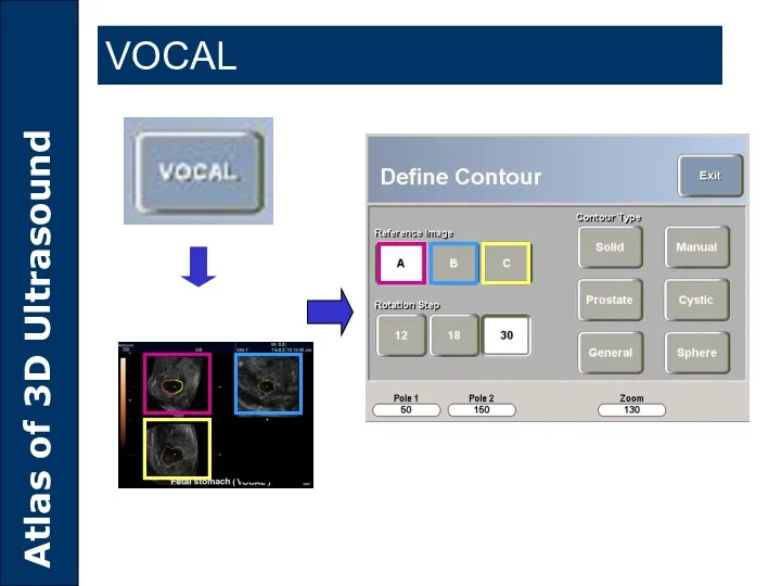
VOCAL
Virtual Organ Computer Aided Analysis
Accurate volume measurement of mass by the
VOCAL
Virtual Organ Computer Aided Analysis
Accurate volume measurement of mass by the
SHELL Imaging
Result
Independent from Vascularization the “3D shell-values” are constant and give
SHELL Imaging
Result
Independent from Vascularization the “3D shell-values” are constant and give
SHELL Imaging
Image courtesy of Dr.YC. Chang, National Taiwan University Hospital
SHELL Imaging
Image courtesy of Dr.YC. Chang, National Taiwan University Hospital
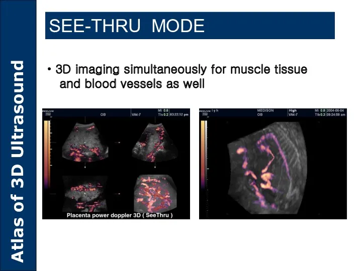
SEE-THRU MODE
3D imaging simultaneously for muscle tissue
and blood
SEE-THRU MODE
3D imaging simultaneously for muscle tissue
and blood

Clinical Advantages of 3D Ultrasound
Clinical Advantages of 3D Ultrasound
Multi-Planar Imaging
Congenital uterine abnormal(A,B,C Plane)
Fetal cleft lip and palate detection
Needle
Multi-Planar Imaging
Congenital uterine abnormal(A,B,C Plane)
Fetal cleft lip and palate detection
Needle
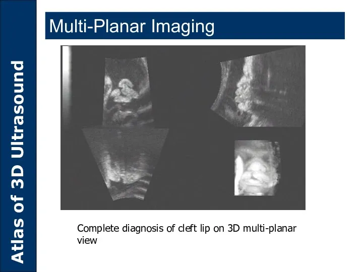
Multi-Planar Imaging
Complete diagnosis of cleft lip on 3D multi-planar view
Multi-Planar Imaging
Complete diagnosis of cleft lip on 3D multi-planar view
Used to measure the Pre-and Post-TAE vascularity of Hepatocellular
The tumor
Used to measure the Pre-and Post-TAE vascularity of Hepatocellular
The tumor
SHELL IMAGING
Pre Trans Arterial Embolization Vacularity of Hepatocellular
Images courtesy of Dr.YC.
SHELL IMAGING
Pre Trans Arterial Embolization Vacularity of Hepatocellular
Images courtesy of Dr.YC.
SHELL IMAGING
Post Trans Arterial Embolization Vacularity of Hepatocellular
Images courtesy of Dr.YC.
SHELL IMAGING
Post Trans Arterial Embolization Vacularity of Hepatocellular
Images courtesy of Dr.YC.
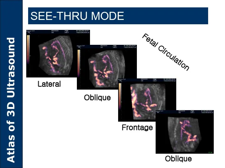
SEE-THRU MODE
Fetal Circulation
Lateral
Oblique
Frontage
Oblique
SEE-THRU MODE
Fetal Circulation
Lateral
Oblique
Frontage
Oblique
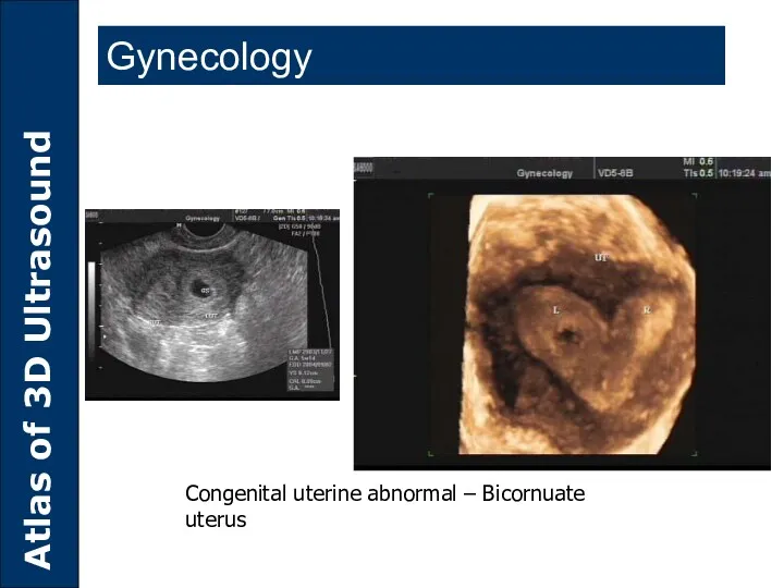
Congenital uterine abnormal – Bicornuate uterus
Gynecology
Congenital uterine abnormal – Bicornuate uterus
Gynecology
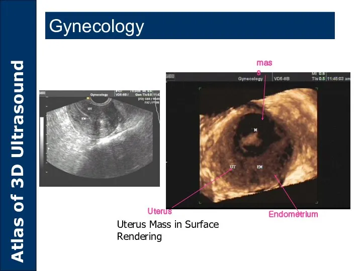
Uterus Mass in Surface Rendering
Gynecology
Endometrium
mass
Uterus
Uterus Mass in Surface Rendering
Gynecology
Endometrium
mass
Uterus
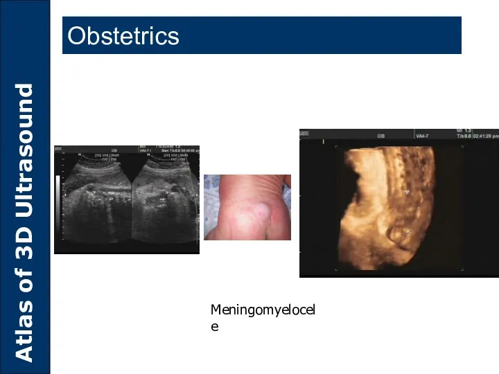
Meningomyelocele
Obstetrics
Meningomyelocele
Obstetrics
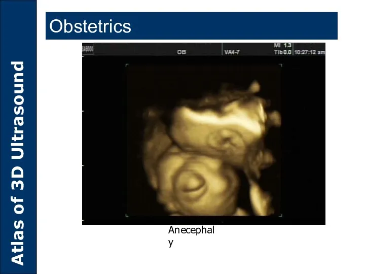
Anecephaly
Obstetrics
Anecephaly
Obstetrics
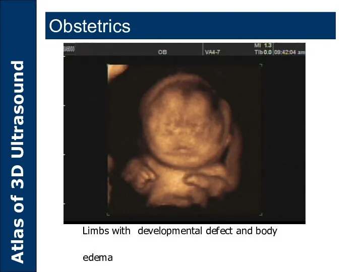
Limbs with developmental defect and body edema
Obstetrics
Limbs with developmental defect and body edema
Obstetrics
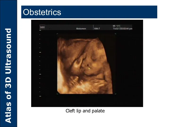
Cleft lip and palate
Obstetrics
Cleft lip and palate
Obstetrics
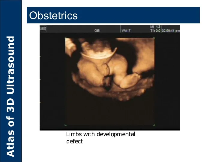
Limbs with developmental defect
Obstetrics
Limbs with developmental defect
Obstetrics

Accuvix XQ 3D
Accuvix XQ 3D
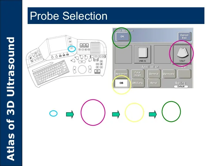
Probe Selection
Probe Selection
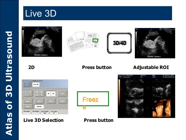
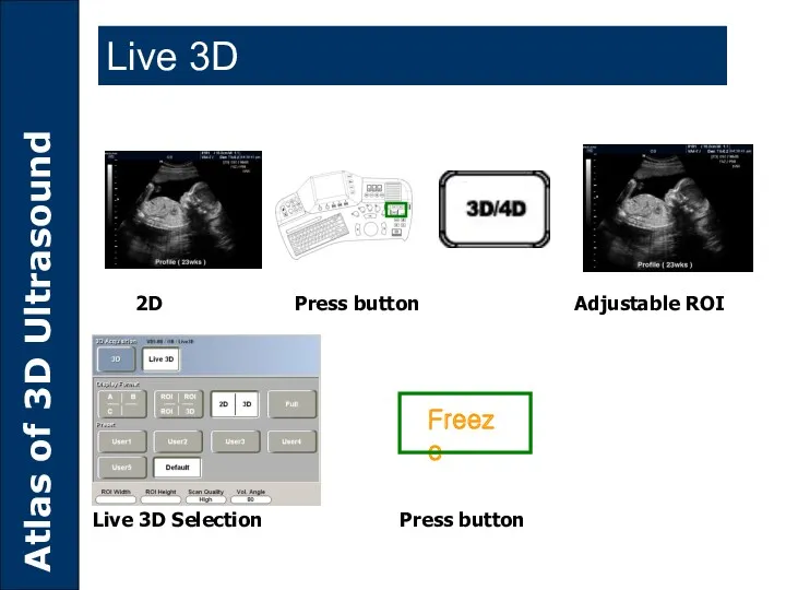
Freeze
2D Press button Adjustable ROI
Live 3D Selection Press button
Live 3D
Freeze
2D Press button Adjustable ROI
Live 3D Selection Press button
Live 3D
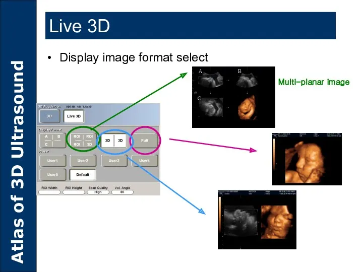
Multi-planar image
Display image format select
Live 3D
Multi-planar image
Display image format select
Live 3D
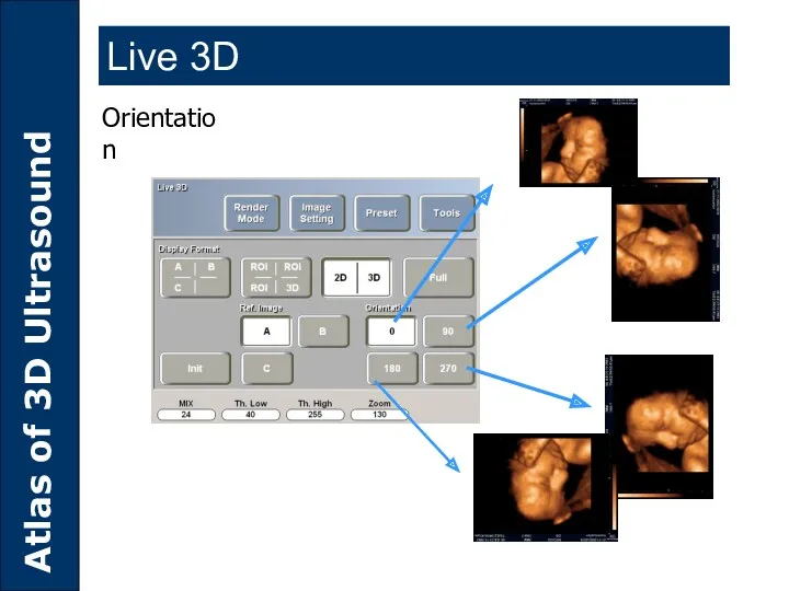
Orientation
Live 3D
Orientation
Live 3D
2D Press button Adjustable ROI
Live 3D Selection Press button
Freeze
Live 3D
2D Press button Adjustable ROI
Live 3D Selection Press button
Freeze
Live 3D
VOCAL
VOCAL
Result
VOCAL
Result
VOCAL

SA8000LV 3D
SA8000LV 3D
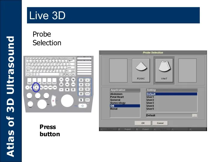
Probe Selection
Press button
Live 3D
Probe Selection
Press button
Live 3D
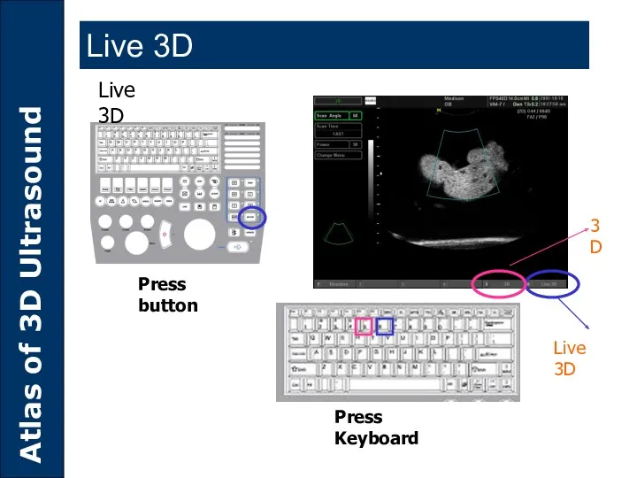
Press button
Press Keyboard
Live 3D
Live 3D
3D
Live 3D
Press button
Press Keyboard
Live 3D
Live 3D
3D
Live 3D
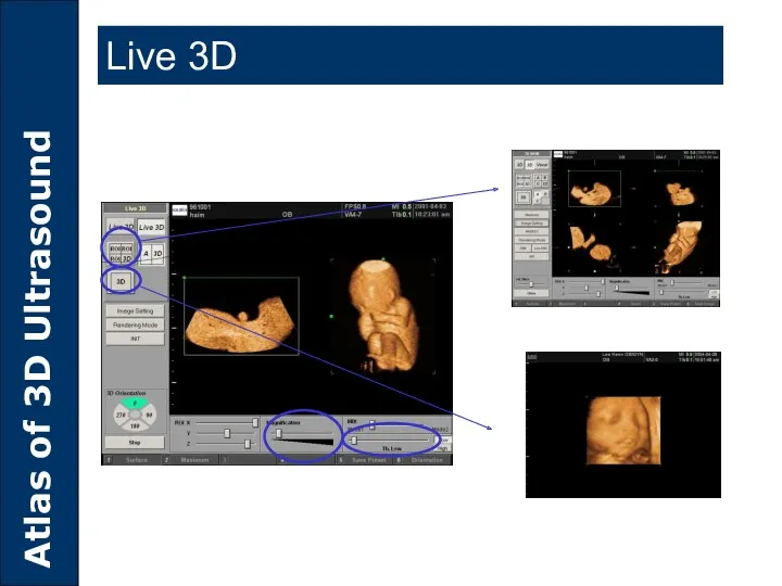
Live 3D
Live 3D
 Особенности проведения медицинских осмотров обучающихся в целях раннего выявления потребления наркотических средств
 Медикалық-генетикалық кеңес. Пренатальдық диогностика әдістері
 Полимеразная цепная реакция
 Исследование функции внешнего дыхания мое
 Гипертоническая болезнь (Артериальная гипертония)
 Проблемы адаптации и здоровья в образовании
 Современные концепции психотерапии в клинической психологии
 Lacalut. Стоматологи рекомендуют
 Вступ в курс інфектології. Поняття про інфекційні хвороби. Принципи діагностики, лікування, профілактики
 Новокузнецкая городская клиническая больница №2
 Осложнения после вакцинации
 Фиксация съёмных ортопедических конструкций. Виды кламмеров
 Сердечно-легочная и церебральная реанимация
 Аккредитации специалистов
 Экзема дегеніміз - эпидермис пен дерманың қабынуы
 Введение в медицинскую рецептуру
 Особенности организации работы детских поликлиник
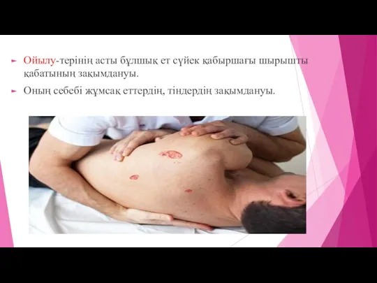 Жиектер
 Облачные технологии управления. Модуль скорая помощь
 History of the Kanash Central District Hospital
 Из опыта работы по организации подготовки и защиты ВКР по специальности СД
 Нормальный Менструальный цикл. Эндометриоз
 Лечение гриппа
 Программа подготовки медицинского персонала к проведению предрейсовых, послерейсовых и текущих медицинских осмотров
 Описание клинического случая
 Адамның психикалық қызметтерінің ерекшеліктері (зейін,түйсік,ой,сана,сөз)
 Барєрні методи контрацепції
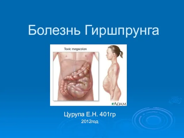 Болезнь Гиршпрунга