- Cerebral-vascular diseases

Содержание
- 2. atheromatosis and athero-caltsinosis of arteries with stenosis circular hyalinosis with the critical narrowing of heart or
- 3. 1.Vascular-discirculation encephalopathy: Ischemic Hypertensive 2. Cerebral haemorrhage: Intracerebral Ssubarachnoidal 3.Brain stroke (ischemic, hemorrhagic, ischemic infarction with
- 4. It is a diffuse defeat of brain neurons with diffuse small-part character necrosis of neurons and
- 5. stenosis of cerebral arteries thrombosis of the atherosclerotic plaque protracted spasm of vessels Reasons of the
- 6. Laminar necrosis - ischemic changers of pyramidal cell layers of the cerebral cortex. Adaptive (around-neuronal) satellitosis
- 7. Acute Sub acute Chronic with relapses (at seniors with the expressed atherosclerosis) Outcomes of IE -
- 8. It is hypertensive hyaline arteriolar sclerosis. At the moment of crisis a fibrinoid necrosis of the
- 9. Haemorrhage begins in the upper 1/3 of Pons (in the zone of cardio-respiratory centers). Displacement of
- 10. Lacunar infarcts ("lacunae") are little infarcts, a few mm across, typically in the deep structures of
- 11. death in the acute period the progressive disorders of memory, sensitiveness, motions and etc Outcomes of
- 12. “Brain Stroke“ - it is a sudden onset of a permanent, localized neurologic deficit, may result
- 13. ischemic infarction (75%) - develops at the obstructive thrombosis or thrombi-emboli ischemic infarction with hemorrhages (5-10%)
- 14. Thrombotic infarcts Embolic infarcts Subclavian steal syndrome (Robin Hood syndrome), in which a patient with occlusive
- 15. ischemic ( 1-3 days) - there is an area of ischemia after the occlusion of artery
- 16. Brain hemorrhage Sudden arising up of the volume in one hemisphere of brain brings to the
- 17. "Hypertension" - arterial pressure higher then 180mmHg item the break of artery, or aneurism, or vascular
- 18. Intra-brain - in area of under-cortex ganglier and visual hillock, rarely in the cerebellum and trunk
- 19. The sub-arahnoidal hemorrhage - reasons of development Break off innate or acquired aneurism. Vascular malformations -
- 20. Acute IHD: angina pectoris, acute coronal insufficiensy, acute myocardial infarction, repeated myocardial infarction, Sudden cardiac death
- 21. It is disease that is conditioned by the relative or absolute insufficiency of coronal blood supplying
- 22. It is disparity between necessities of oxygen and its supplying to myocardium. Reasons of development: 1.
- 23. Stable ("classic") angina - results from increased work in a patient with coronary atherosclerosis, and relieved
- 24. It is inability to satisfy metabolic necessities of myocardium by coronal blood supplying. Reasons of development:
- 25. Reperfusion post-ischemic damage of myocardium by free radicals, ions, ets. Damage by mediators of platelets, toxins
- 26. It is ischemic partial necrosis of myocardium wall due to sudden loss of the blood supplying.
- 27. Atherosclerosis: a ruptured plaque - often with an overlying thrombus ("coronary thrombosis"); massive haemorrhage into a
- 28. According to localization and spreading: sub-epicardial, sub-endocardial, intramural, transmural According to time of development: acute primary
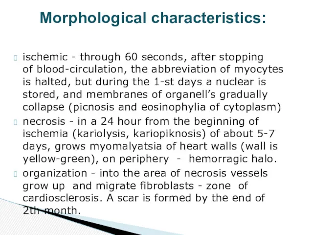
- 29. ischemic - through 60 seconds, after stopping of blood-circulation, the abbreviation of myocytes is halted, but
- 30. The nitro-blue tetrazolium technique can demonstrate early myocardial infarcts. Drop a slice of heart in the
- 31. Ischemic stage: rhythm disturbances with stopping heart work, Left-sided congestive heart failure, Cardiogenic shock, Acute coronal
- 33. Скачать презентацию
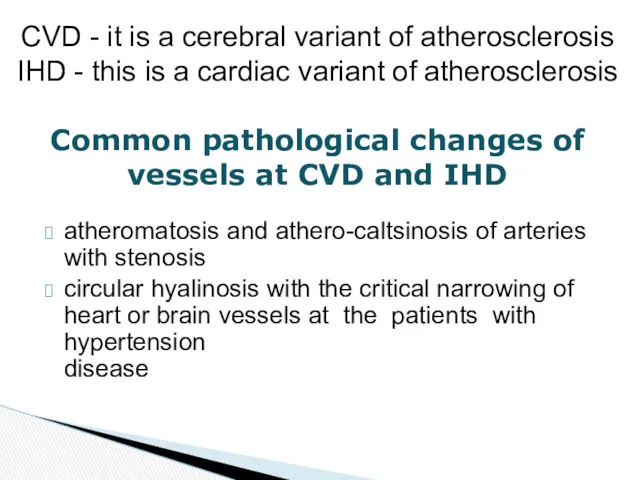
atheromatosis and athero-caltsinosis of arteries with stenosis
circular hyalinosis with the critical
atheromatosis and athero-caltsinosis of arteries with stenosis
circular hyalinosis with the critical
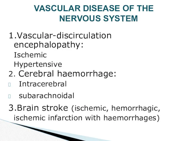
1.Vascular-discirculation encephalopathy:
Ischemic
Hypertensive
2. Cerebral haemorrhage:
Intracerebral
Ssubarachnoidal
3.Brain stroke (ischemic, hemorrhagic,
1.Vascular-discirculation encephalopathy:
Ischemic
Hypertensive
2. Cerebral haemorrhage:
Intracerebral
Ssubarachnoidal
3.Brain stroke (ischemic, hemorrhagic,
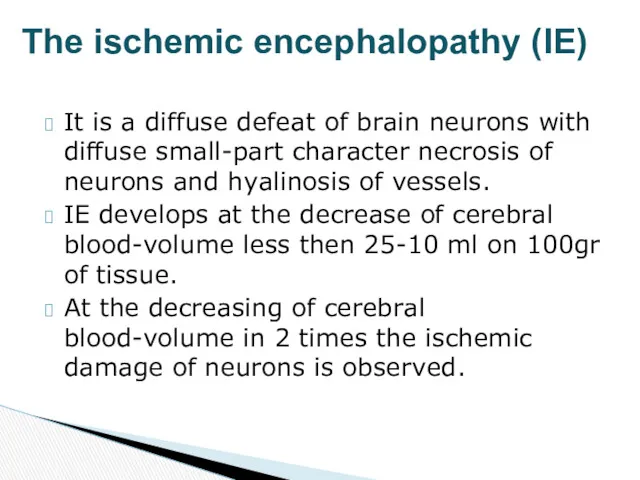
It is a diffuse defeat of brain neurons with diffuse small-part
It is a diffuse defeat of brain neurons with diffuse small-part
stenosis of cerebral arteries
thrombosis of the atherosclerotic plaque
protracted spasm of vessels
Reasons
stenosis of cerebral arteries
thrombosis of the atherosclerotic plaque
protracted spasm of vessels
Reasons
Laminar necrosis - ischemic changers of
pyramidal cell layers of the cerebral
cortex.
Adaptive
Laminar necrosis - ischemic changers of
pyramidal cell layers of the cerebral
cortex.
Adaptive
Acute
Sub acute
Chronic with relapses (at seniors with the expressed atherosclerosis)
Outcomes
Acute
Sub acute
Chronic with relapses (at seniors with the expressed atherosclerosis)
Outcomes
It is hypertensive hyaline arteriolar sclerosis. At the moment of crisis
It is hypertensive hyaline arteriolar sclerosis. At the moment of crisis
Haemorrhage begins in the upper 1/3 of Pons (in the zone
Haemorrhage begins in the upper 1/3 of Pons (in the zone
Lacunar infarcts ("lacunae") are little infarcts, a few mm across, typically
Lacunar infarcts ("lacunae") are little infarcts, a few mm across, typically

death in the acute period
the progressive disorders of memory, sensitiveness, motions
death in the acute period
the progressive disorders of memory, sensitiveness, motions

“Brain Stroke“ -
it is a sudden onset of a permanent,
“Brain Stroke“ -
it is a sudden onset of a permanent,
ischemic infarction (75%) - develops at the obstructive thrombosis or thrombi-emboli
ischemic
ischemic infarction (75%) - develops at the obstructive thrombosis or thrombi-emboli
ischemic
Thrombotic infarcts
Embolic infarcts
Subclavian steal syndrome (Robin Hood syndrome), in which a
Thrombotic infarcts
Embolic infarcts
Subclavian steal syndrome (Robin Hood syndrome), in which a
ischemic ( 1-3 days) - there is an area of ischemia
ischemic ( 1-3 days) - there is an area of ischemia
Brain hemorrhage
Sudden arising up of the volume in one hemisphere of
Brain hemorrhage
Sudden arising up of the volume in one hemisphere of
"Hypertension" - arterial pressure higher then 180mmHg item
the break of artery,
"Hypertension" - arterial pressure higher then 180mmHg item
the break of artery,
Intra-brain - in area of under-cortex ganglier and visual hillock, rarely
Intra-brain - in area of under-cortex ganglier and visual hillock, rarely
The sub-arahnoidal hemorrhage - reasons of development
Break off innate or acquired
The sub-arahnoidal hemorrhage - reasons of development
Break off innate or acquired
Acute IHD: angina pectoris, acute coronal insufficiensy, acute myocardial infarction, repeated
Acute IHD: angina pectoris, acute coronal insufficiensy, acute myocardial infarction, repeated
It is disease that is conditioned by the relative or absolute
It is disease that is conditioned by the relative or absolute
It is disparity between necessities of oxygen and its supplying to
It is disparity between necessities of oxygen and its supplying to
Stable ("classic") angina - results from increased work in a patient
Stable ("classic") angina - results from increased work in a patient
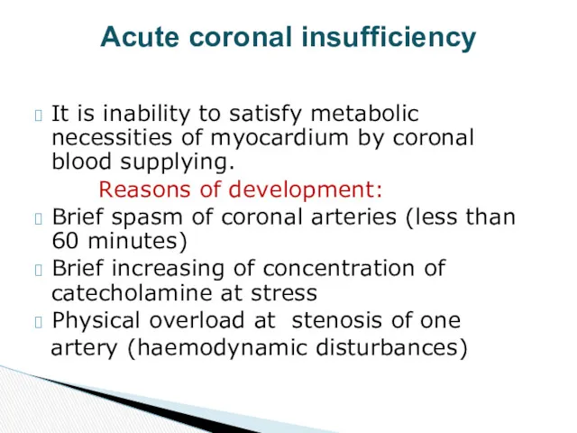
It is inability to satisfy metabolic necessities of myocardium by coronal
It is inability to satisfy metabolic necessities of myocardium by coronal
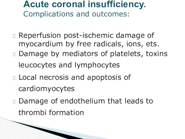
Reperfusion post-ischemic damage of myocardium by free radicals, ions, ets.
Damage by
Reperfusion post-ischemic damage of myocardium by free radicals, ions, ets.
Damage by
It is ischemic partial necrosis of myocardium wall due to sudden
It is ischemic partial necrosis of myocardium wall due to sudden
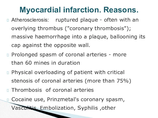
Atherosclerosis: a ruptured plaque - often with an overlying thrombus ("coronary
Atherosclerosis: a ruptured plaque - often with an overlying thrombus ("coronary
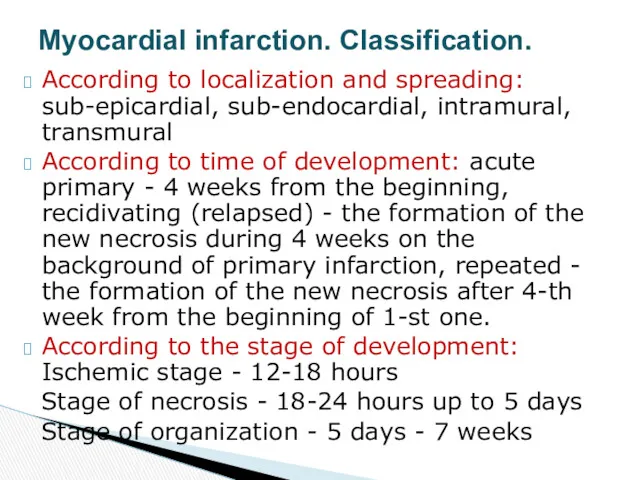
According to localization and spreading: sub-epicardial, sub-endocardial, intramural, transmural
According to time
According to localization and spreading: sub-epicardial, sub-endocardial, intramural, transmural
According to time
ischemic - through 60 seconds, after stopping
of blood-circulation, the abbreviation of
ischemic - through 60 seconds, after stopping of blood-circulation, the abbreviation of
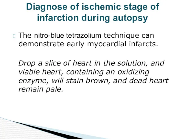
The nitro-blue tetrazolium technique can
demonstrate early myocardial infarcts.
Drop a slice
The nitro-blue tetrazolium technique can
demonstrate early myocardial infarcts.
Drop a slice
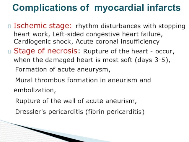
Ischemic stage: rhythm disturbances with stopping heart work, Left-sided congestive heart
Ischemic stage: rhythm disturbances with stopping heart work, Left-sided congestive heart
 Лекарственные средства, влияющие на афферентную иннервацию . Местные анестетики
 О резистентности антибиотиков в России
 Металлокерамика. Монолитная гипсовая модель
 Пародонт
 Внутрибрюшинное депонирование лекарственных смесей для собак
 Болезни органов дыхания пневмомикозы
 Генетическое консультирование, или медико-генетическое консультирование
 Холера – особо опасная инфекция
 Инфекционный мононуклеоз
 Методы диагностики в неврологии. Нейровизуализационная диагностика
 Неотложные состояния на амбулаторном стоматологическом приеме
 Противоопухолевая химиотерапия
 Новая концепция электромиографического исследования
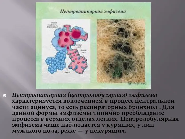 Центроацинарная эмфизема
 Антибиотики, характеристика
 Ecology of microorganisms
 Наследственность и ее формы. Наследственные болезни как вариант изменчивости
 Виды лабораториий и их назначения. Обязаности медецинского лабораторного техника. Виды исследованиий. Специальная посуда
 Эндоскопическая анатомия плевральной полости и средостения
 Иерсиниозы
 Особенности ЭКГ у детей; механизм образования тонов сердца
 Анкилоздаушы спондилоартрит және реактивті артрит
 Взаимосвязь всех видов обмена веществ. Лекция № 3
 Купирование скелетно-мышечной боли в поликлинике
 Этиология, характеристика эпидемического процесса, противоэпидемические и профилактические мероприятия при шигеллезе и холере
 Нейроциркуляторная дистония
 Иммунологическая память, иммунологическая толерантность
 Клиническая анатомия мозгового отдела головы с операциями