- Critical alteration morphology of cell death necrosis of tissue and organ

Содержание
- 2. Critical Damage and Cell Death Critical damage it is molecular-sub- cellular violation of metabolism and fading
- 3. Critical Damage and Cell Death Objects of critical damage are structures of the cells: mitochondria genetical
- 4. Reasons of development: Endogenous metabolic catastrophe: bioenergetics insufficiency of cells (hypoxia of different genesis) damage of
- 5. II. Exogenous factors of injury: infectious aggression (viruses, bacteria, fungi's) physical and/or chemical damages (gamma- and
- 6. Consequences of critical damage: ⮚ partial necrosis of cell ⮚ destruction of cells by necrosis ⮚
- 7. NECROSIS Necrosis - it is death of cells or tissues in living organism. Other forms of
- 8. NECROSIS CELL Necrosis: -coagulative -colliquative Apoptosis Immune- mediated cell death: - phagocytosis TISSUE 1. Coagulative: -
- 9. It is the premature death and destruction of cell’s organelles in the living organism under action
- 10. Classification of Cells Death, based on the mechanism of development: Necrosis of specialized cells Pathogenic inducted
- 11. Morphology of cell destruction: 1. Karyolysis - the basophilic of the chromatin may fade, a change
- 12. Post-necrotic transformation of cells in living organism Autolysis – destruction of cells by ferments of the
- 13. Pathogenic inducted apoptosis It is a "programmed by genes cell death" or cell suicide, that is
- 14. 1. Cell shrinkage. Cell is smaller in size; the cytoplasm is dense. 2. Chromatin condensation. The
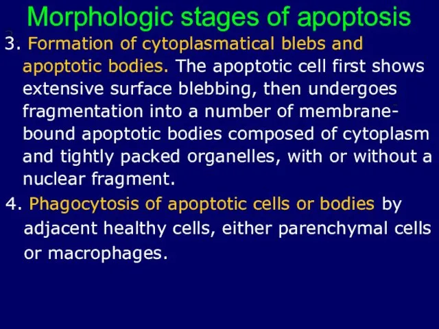
- 15. Morphologic stages of apoptosis 3. Formation of cytoplasmatical blebs and apoptotic bodies. The apoptotic cell first
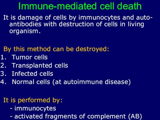
- 16. Immune-mediated cell death It is damage of cells by immunocytes and auto- antibodies with destruction of
- 17. Phases of process: Recognition of damaged cells by IS Destroying (killing) of cells Types of Immune-mediated
- 18. Cells, that perform immune killing: T-lymphocytes killers natural killers (NK-cells) Zero-cells (K-cell) macrophages Mechanisms of killing:
- 19. NECROSIS OF ORGANS It is destruction of all components of organs (specialized cells, vessels, stroma, intercellular
- 20. Reasons of development of necrosis: ⮚protracted ischemia ⮚stopping of arterial blood supplying ⮚damage by the mechanical
- 21. CLASSIFICATION OF NECROSIS OF ORGANS I According to morphology: Dry necrosis (coagulative) – coagulations of proteins
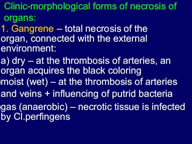
- 22. Clinic-morphological forms of necrosis of organs: 1. Gangrene – total necrosis of the organ, connected with
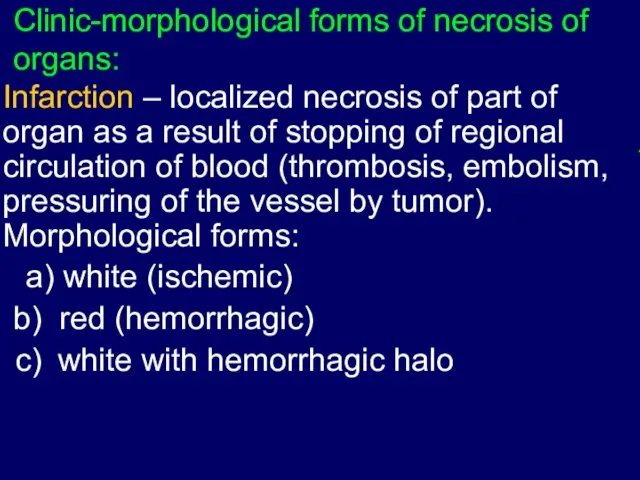
- 23. Clinic-morphological forms of necrosis of organs: Infarction – localized necrosis of part of organ as a
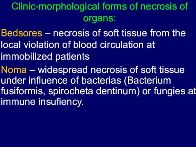
- 24. Clinic-morphological forms of necrosis of organs: Bedsores – necrosis of soft tissue from the local violation
- 25. Morphological forms of tissue necrosis: Coagulative necrosis Liquefactive necrosis Caseous necrosis Fat necrosis
- 26. Stages of development of necrosis: Before-necrotic changes – from the beginning of damage to stopping of
- 28. Скачать презентацию
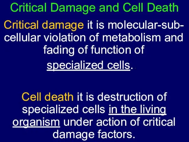
Critical Damage and Cell Death
Critical damage it is molecular-sub- cellular violation
Critical Damage and Cell Death
Critical damage it is molecular-sub- cellular violation
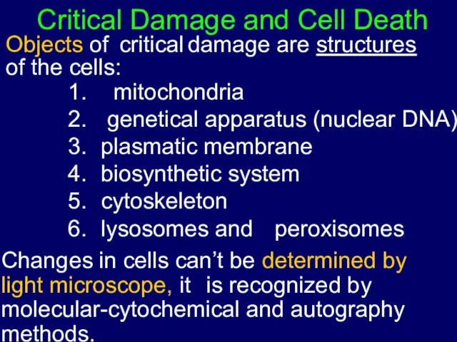
Critical Damage and Cell Death Objects of critical damage are structures of the
Critical Damage and Cell Death Objects of critical damage are structures of the
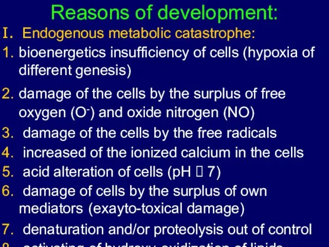
Reasons of development:
Endogenous metabolic catastrophe:
bioenergetics insufficiency of cells (hypoxia of
different genesis)
damage
Reasons of development:
Endogenous metabolic catastrophe:
bioenergetics insufficiency of cells (hypoxia of
different genesis)
damage
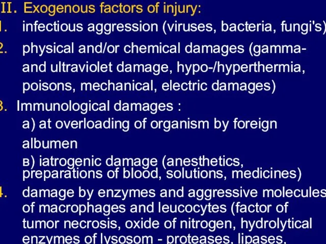
II. Exogenous factors of injury:
infectious aggression (viruses, bacteria, fungi's)
physical and/or chemical
II. Exogenous factors of injury:
infectious aggression (viruses, bacteria, fungi's)
physical and/or chemical
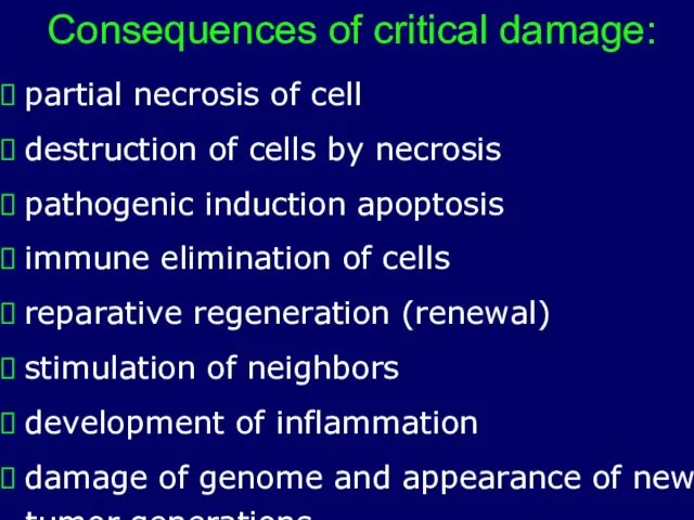
Consequences of critical damage:
⮚ partial necrosis of cell
⮚ destruction of cells
Consequences of critical damage:
⮚ partial necrosis of cell
⮚ destruction of cells
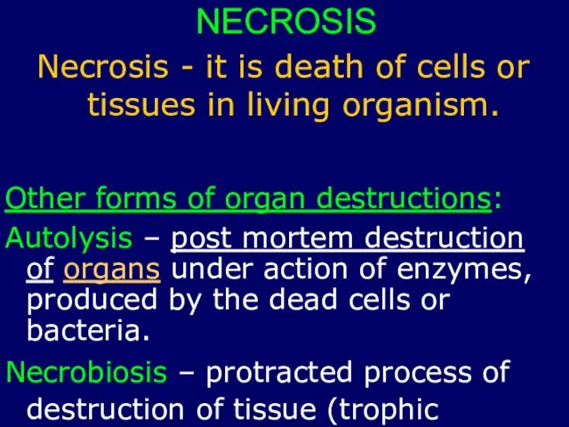
NECROSIS
Necrosis - it is death of cells or
tissues in living organism.
Other
NECROSIS
Necrosis - it is death of cells or
tissues in living organism.
Other
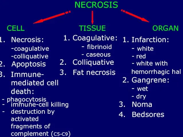
NECROSIS
CELL
Necrosis:
-coagulative
-colliquative
Apoptosis
Immune- mediated cell
death:
- phagocytosis
TISSUE
1. Coagulative:
- fibrinoid
- caseous
Colliquative
Fat necrosis
ORGAN
1. Infarction:
- white
red
white with
hemorrhagic
NECROSIS
CELL
Necrosis:
-coagulative
-colliquative
Apoptosis
Immune- mediated cell
death:
- phagocytosis
TISSUE
1. Coagulative:
- fibrinoid
- caseous
Colliquative
Fat necrosis
ORGAN
1. Infarction:
- white
red
white with
hemorrhagic
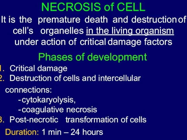
It is the premature death and destruction of cell’s organelles in the living organism under action of critical damage factors
Phases
It is the premature death and destruction of cell’s organelles in the living organism under action of critical damage factors
Phases
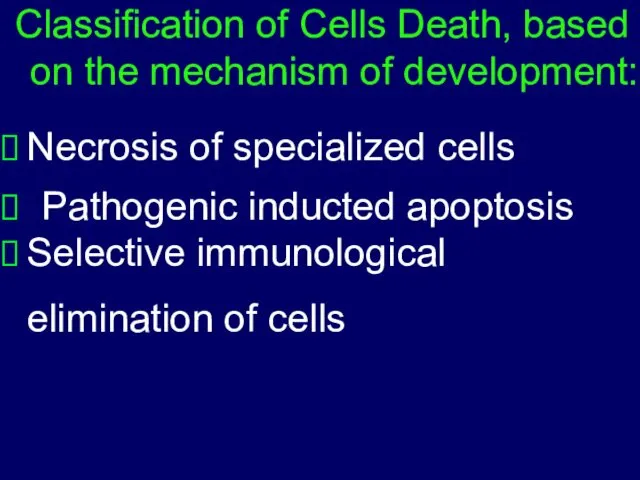
Classification of Cells Death, based
on the mechanism of development:
Necrosis of specialized
Classification of Cells Death, based
on the mechanism of development:
Necrosis of specialized
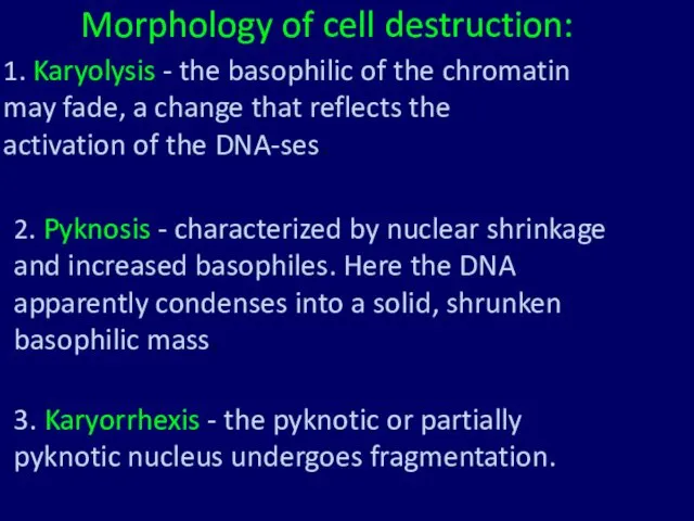
Morphology of cell destruction:
1. Karyolysis - the basophilic of the chromatin
Morphology of cell destruction:
1. Karyolysis - the basophilic of the chromatin
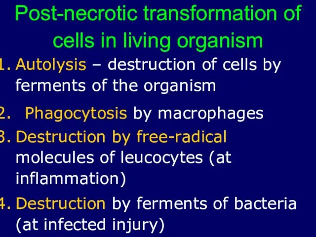
Post-necrotic transformation of
cells in living organism
Autolysis – destruction of cells by
Post-necrotic transformation of
cells in living organism
Autolysis – destruction of cells by
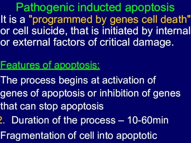
Pathogenic inducted apoptosis
It is a "programmed by genes cell death" or
Pathogenic inducted apoptosis
It is a "programmed by genes cell death" or
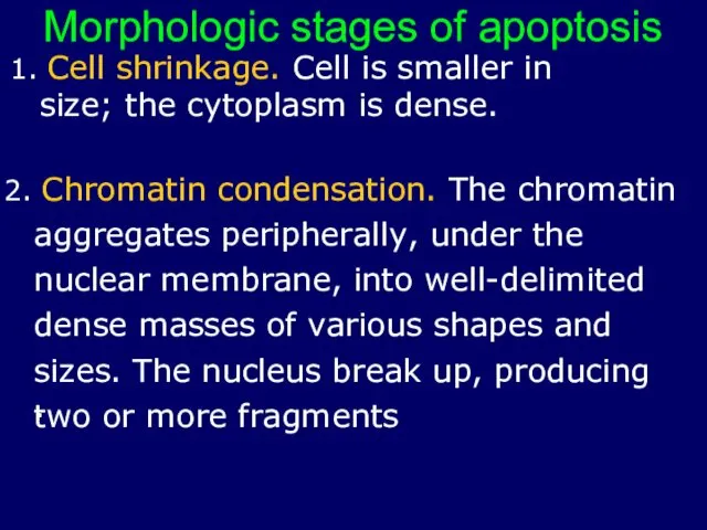
1. Cell shrinkage. Cell is smaller in size; the cytoplasm is
1. Cell shrinkage. Cell is smaller in size; the cytoplasm is
Morphologic stages of apoptosis
3. Formation of cytoplasmatical blebs and apoptotic bodies.
Morphologic stages of apoptosis
3. Formation of cytoplasmatical blebs and apoptotic bodies.
Immune-mediated cell death
It is damage of cells by immunocytes and auto-
Immune-mediated cell death
It is damage of cells by immunocytes and auto-
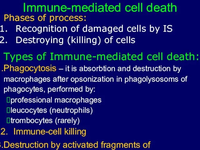
Phases of process:
Recognition of damaged cells by IS
Destroying (killing) of cells
Types
Phases of process:
Recognition of damaged cells by IS
Destroying (killing) of cells
Types
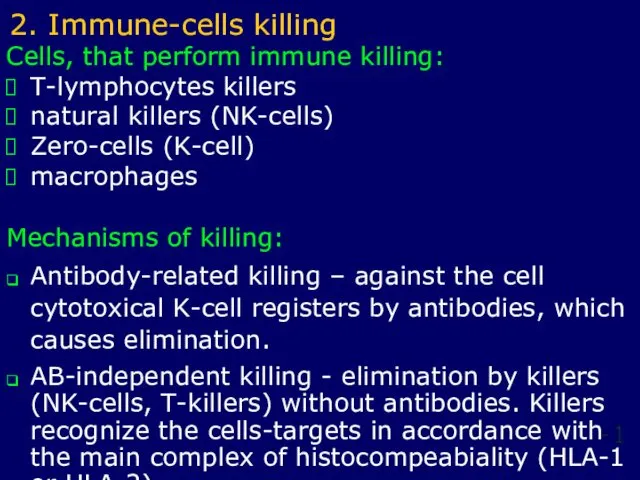
Cells, that perform immune killing:
T-lymphocytes killers
natural killers (NK-cells)
Zero-cells (K-cell)
macrophages
Mechanisms of killing:
❑ Antibody-related
Cells, that perform immune killing:
T-lymphocytes killers
natural killers (NK-cells)
Zero-cells (K-cell)
macrophages
Mechanisms of killing:
❑ Antibody-related
NECROSIS OF ORGANS
It is destruction of all
components of organs (specialized cells,
NECROSIS OF ORGANS
It is destruction of all
components of organs (specialized cells,
Reasons of development of necrosis:
⮚protracted ischemia
⮚stopping of arterial blood supplying
⮚damage by the
Reasons of development of necrosis:
⮚protracted ischemia
⮚stopping of arterial blood supplying
⮚damage by the
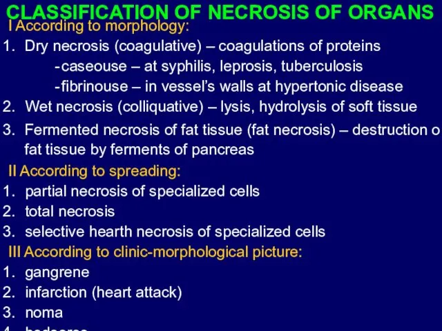
CLASSIFICATION OF NECROSIS OF ORGANS
I According to morphology:
Dry necrosis (coagulative) –
CLASSIFICATION OF NECROSIS OF ORGANS
I According to morphology:
Dry necrosis (coagulative) –
Clinic-morphological forms of necrosis of organs:
1. Gangrene – total necrosis of
Clinic-morphological forms of necrosis of organs:
1. Gangrene – total necrosis of
Clinic-morphological forms of necrosis of organs:
Infarction – localized necrosis of part
Clinic-morphological forms of necrosis of organs:
Infarction – localized necrosis of part
Clinic-morphological forms of necrosis of organs:
Bedsores – necrosis of soft tissue
Clinic-morphological forms of necrosis of organs:
Bedsores – necrosis of soft tissue
Morphological forms of tissue necrosis:
Coagulative necrosis
Liquefactive necrosis
Caseous necrosis
Fat necrosis
Morphological forms of tissue necrosis:
Coagulative necrosis
Liquefactive necrosis
Caseous necrosis
Fat necrosis
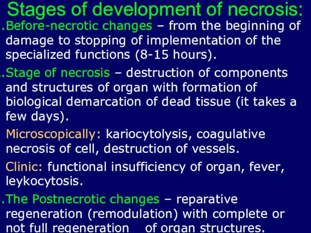
Stages of development of necrosis:
Before-necrotic changes – from the beginning of
Stages of development of necrosis:
Before-necrotic changes – from the beginning of
 Системная красная волчанка у детей
 Ньюкаслская болезнь (псевдочума)
 Чувствительная система. Анатомофизиологические особенности чувствительного анализатора. Виды нарушений чувствительности
 Международный день акушерки
 Spice (спайс)
 Династия врачей Грасмик
 Кардиогенный шок
 Маскүнемдік, нашақорлық және уытқорлықтың патофизиологиялық негіздері
 Производственная санитария, её задачи. Уход за производственным помещением
 Психические расстройства в результате употребления психодислептиков
 Влияние вредных факторов на плод
 Кишечная непроходимость
 Заболевания пищевода. Ахалазия кардии. Эзофагопатия при системной склеродермии. Варикозное расширение вен пищевода. (Часть 1)
 Методы исследования заболеваний органов дыхания (лекция 3)
 Патофизиология водно-солевого обмена
 Пульпит. Классификация пульпита
 Фармакокинетика лекарственных средств: пути введения, всасывание, превращение лекарств и выведение их из организма
 АиР Общая анестезия
 Ішек инфекциялары ауруларының асқынуы
 Лучевая диагностика заболеваний пищеварительного тракта
 Донорство крови
 Гинекологиялық ауруларды тексеру әдістері
 Лекция Лечебно-охранительный режим
 Воспалительные заболевания челюстно-лицевой области
 Шырғанақ өсімдігінің дәрілік мақсатта пайдаланылуы
 Анатомия и физиология спинного мозга. Спинномозговые нервы
 Групи крові. Переливання крові. Пересаджування кісткового мозку
 Сосудистая деменция