- Injury of genitourinary organs

Содержание
- 2. Emergency Diagnosis & Management About 10% of all injuries seen in the emergency room involve the
- 3. Many of them are subtle and difficult to define and require great diagnostic expertise. Early diagnosis
- 4. Emergency Diagnosis & Management Initial assessment should include control of hemorrhage and shock along with resuscitation
- 5. Emergency Diagnosis & Management The history should include a detailed description of the accident. In cases
- 6. Emergency Diagnosis & Management The abdomen and genitalia should be examined for evidence of contusions or
- 7. Fractures of the lower ribs are often associated with renal injuries, and pelvic fractures often accompany
- 8. Patients who do not have life-threatening injuries and whose blood pressure is stable can undergo more
- 9. Emergency Diagnosis & Management When genitourinary tract injury is suspected on the basis of the history
- 10. Assessment of Injury Assessment of the injury should be done in an orderly fashion so that
- 11. Catheterization Blood at the urethral meatus in men indicates urethral injury; catheterization should not be attempted
- 12. Catheterization If no blood is present at the meatus, a urethral catheter can be carefully passed
- 13. Catheterization If catheterization is traumatic despite the greatest care, the significance of hematuria cannot be determined,
- 14. Computed Tomography (CT) Abdominal CT with contrast media is the best imaging study to detect and
- 15. Computed Tomography (CT) It can define the size extent of the retroperitoneal hematoma
- 16. Computed Tomography (CT) Spiral CT scanning, now common, is very rapid, but it may not detect
- 17. Retrograde Cystography Filling of the bladder with contrast material is essential to establish whether bladder perforations
- 18. Retrograde Cystography A film should be obtained with the bladder filled and a second one after
- 19. Retrograde Cystography Cystography with CT is excellent for establishing bladder injury.
- 20. Urethrography A small (12F) catheter can be inserted into the urethral meatus and 3 mL of
- 21. Urethrography After retrograde injection of 20 mL of water-soluble contrast material, the urethra will be clearly
- 22. Arteriography Arteriography may help define renal parenchymal and renal vascular injuries.
- 23. Intravenous Urography Intravenous urography can be used to detect renal and ureteral injury.
- 24. Cystoscopy and Retrograde Urography Cystoscopy and retrograde urography may be useful to detect ureteral injury, but
- 25. Abdominal Sonography Abdominal sonography has not been shown to add substantial information during initial evaluation of
- 26. Injuries to the Kidney Renal injuries are the most common injuries of the urinary system.
- 27. Injuries to the Kidney Most injuries occur from automobile accidents or sporting mishaps, chiefly in men
- 28. Injuries to the Kidney Etiology Blunt trauma directly to the abdomen, flank, or back is the
- 29. Injuries to the Kidney Vehicle collisions at high speed may result in major renal trauma from
- 30. Injuries to the Kidney Associated abdominal visceral injuries are present in 80% of renal penetrating wounds.
- 31. Pathology & Classification Early Pathologic Findings Lacerations from blunt trauma usually occur in the transverse plane
- 32. Pathology & Classification Early Pathologic Findings In injuries from rapid deceleration, the kidney moves upward or
- 33. Pathology & Classification Early Pathologic Findings Acute thrombosis of the renal artery may be caused by
- 34. Pathology & Classification Hydronephrosis Follow-up excretory urography is indicated in all cases of major renal trauma.
- 35. Pathology & Classification Arteriovenous Fistula Arteriovenous fistulas may occur after penetrating injuries but are not common
- 36. Pathology & Classification Renal Vascular Hypertension The blood flow in tissue rendered nonviable by injury is
- 37. Clinical Findings & Indications for Studies Microscopic or gross hematuria following trauma to the abdomen indicates
- 38. Clinical Findings & Indications for Studies Some cases of renal vascular injury are not associated with
- 39. Clinical Findings & Indications for Studies The degree of renal injury does not correspond to the
- 40. Clinical Findings & Indications for Studies Miller and McAninch (1995) made the following recommendations based on
- 41. Clinical Findings & Indications for Studies However, should physical examination or associated injuries prompt reasonable suspicion
- 42. Clinical Findings & Indications for Studies Symptoms There is usually visible evidence of abdominal trauma. Pain
- 43. Clinical Findings & Indications for Studies Catheterization usually reveals hematuria.
- 44. Clinical Findings & Indications for Studies Signs Initially, shock or signs of a large loss of
- 45. Clinical Findings & Indications for Studies Signs Diffuse abdominal tenderness may be found on palpation; an
- 46. Clinical Findings & Indications for Studies Signs The abdomen may be distended and bowel sounds absent.
- 47. Clinical Findings & Indications for Studies Laboratory Findings Microscopic or gross hematuria is usually present.
- 48. Clinical Findings & Indications for Studies Staging and X-Ray Findings Staging of renal injuries allows a
- 49. Clinical Findings & Indications for Studies Staging and X-Ray Findings For example, blunt trauma to the
- 50. Clinical Findings & Indications for Studies Staging and X-Ray Findings Ultrasonography and retrograde urography are of
- 51. Clinical Findings & Indications for Studies Staging and X-Ray Findings Staging begins with an abdominal CT
- 52. Clinical Findings & Indications for Studies Staging and X-Ray Findings This noninvasive technique clearly defines parenchymal
- 53. Clinical Findings & Indications for Studies Staging and X-Ray Findings Arteriography defines major arterial and parenchymal
- 54. Clinical Findings & Indications for Studies Staging and X-Ray Findings The major causes of nonvisualization on
- 55. Clinical Findings & Indications for Studies Staging and X-Ray Findings Radionuclide renal scans have been used
- 56. Clinical Findings & Indications for Studies Differential Diagnosis Trauma to the abdomen and flank areas is
- 57. Clinical Findings & Indications for Studies Complications Early Complications Hemorrhage is perhaps the most important immediate
- 58. Clinical Findings & Indications for Studies Complications The size and expansion of palpable masses must be
- 59. Clinical Findings & Indications for Studies Complications Urinary extravasation from renal fracture may show as an
- 60. Clinical Findings & Indications for Studies Complications A resolving retroperitoneal hematoma may cause slight fever (38.3
- 61. Clinical Findings & Indications for Studies Complications Late Complications Hypertension, hydronephrosis, arteriovenous fistula, calculus formation, and
- 62. Clinical Findings & Indications for Studies Complications Heavy late bleeding may occur 4 weeks after injury.
- 63. Clinical Findings & Indications for Studies Treatment: Emergency Measures The objectives of early management are prompt
- 64. Clinical Findings & Indications for Studies Treatment: Surgical Measures Blunt Injuries Bleeding stops spontaneously with bed
- 65. Clinical Findings & Indications for Studies Treatment: Surgical Measures Cases in which operation is indicated include
- 66. Clinical Findings & Indications for Studies Treatment: Surgical Measures Penetrating Injuries Penetrating injuries should be surgically
- 67. Clinical Findings & Indications for Studies Treatment: Surgical Measures In 80% of cases of penetrating injury,
- 68. Clinical Findings & Indications for Studies Treatment: Surgical Measures Treatment of Complications Hydronephrosis may require surgical
- 69. Clinical Findings & Indications for Studies Treatment: Surgical Measures Prognosis With careful follow-up, most renal injuries
- 70. Clinical Findings & Indications for Studies Treatment: Surgical Measures Injuries to the Ureter Ureteral injury is
- 71. Clinical Findings & Indications for Studies Treatment: Surgical Measures Etiology Large pelvic masses (benign or malignant)
- 72. Clinical Findings & Indications for Studies Treatment: Surgical Measures Extensive carcinoma of the colon may invade
- 73. Clinical Findings & Indications for Studies Treatment: Surgical Measures Devascularization may occur with extensive pelvic lymph
- 74. Clinical Findings & Indications for Studies Treatment: Surgical Measures Endoscopic manipulation of a ureteral calculus with
- 75. Clinical Findings & Indications for Studies Treatment: Surgical Measures Pathogenesis & Pathology The ureter may be
- 76. Clinical Findings & Indications for Studies Treatment: Surgical Measures Intraperitoneal extravasation of urine can also occur,
- 77. Clinical Findings Symptoms If the ureter has been completely or partially ligated during operation, the postoperative
- 78. Clinical Findings Symptoms Ureteral injuries from external violence should be suspected in patients who have sustained
- 79. Clinical Findings Symptoms Signs The acute hydronephrosis of a totally ligated ureter results in severe flank
- 80. Clinical Findings Symptoms Watery discharge from the wound or vagina may be identified as urine by
- 81. Laboratory Findings Ureteral injury from external violence is manifested by microscopic hematuria in 90% of cases.
- 82. Imaging Findings Diagnosis is by excretory urography.
- 83. Imaging Findings Partial transection of the ureter results in more rapid excretion, but persistent hydronephrosis is
- 84. Imaging Findings In acute injury from external violence, the excretory urogram usually appears normal, with very
- 85. Ultrasonography Ultrasonography outlines hydroureter or urinary extravasation as it develops into a urinoma and is perhaps
- 86. Radionuclide Scanning Radionuclide scanning demonstrates delayed excretion on the injured side, with evidence of increasing counts
- 87. Differential Diagnosis Postoperative bowel obstruction and peritonitis may cause symptoms similar to those of acute ureteral
- 88. Differential Diagnosis Deep wound infection must be considered postoperatively in patients with fever, ileus, and localized
- 89. Differential Diagnosis Acute pyelonephritis in the early postoperative period may also result in findings similar to
- 90. Complications Ureteral injury may be complicated by stricture formation with resulting hydronephrosis in the area of
- 91. Treatment Prompt treatment of ureteral injuries is required. The best opportunity for successful repair is in
- 92. Treatment Proximal urinary drainage by percutaneous nephrostomy or formal nephrostomy should be considered if the injury
- 93. Treatment The goals of ureteral repair are to achieve complete debridement, a tension-free spatulated anastomosis, watertight
- 94. Lower Ureteral Injuries Injuries to the lower third of the ureter allow several options in management.
- 95. Lower Ureteral Injuries An antireflux procedure should be done when possible.
- 96. Lower Ureteral Injuries Transureteroureterostomy may be used in lower-third injuries if extensive urinoma and pelvic infection
- 97. Midureteral Injuries Midureteral injuries usually result from external violence and are best repaired by primary ureteroureterostomy
- 98. Upper Ureteral Injuries Injuries to the upper third of the ureter are best managed by primary
- 99. Stenting Most anastomoses after repair of ureteral injury should be stented.
- 100. Stenting After 3-4 weeks of healing, stents can be endoscopically removed from the bladder.
- 101. Prognosis The prognosis for ureteral injury is excellent if the diagnosis is made early and prompt
- 102. Injuries to the Bladder Bladder injuries occur most often from external force and are often associated
- 103. Injuries to the Bladder Iatrogenic injury may result from gynecologic and other extensive pelvic procedures as
- 104. Injuries to the Bladder Pathogenesis & Pathology The bony pelvis protects the urinary bladder very well.
- 105. Injuries to the Bladder Pathogenesis & Pathology When the bladder is filled to near capacity, a
- 106. Injuries to the Bladder Pathogenesis & Pathology If the diagnosis is not established immediately and if
- 107. Injuries to the Bladder Clinical Findings Pelvic fracture accompanies bladder rupture in 90% of cases.
- 108. Injuries to the Bladder Symptoms There is usually a history of lower abdominal trauma.
- 109. Injuries to the Bladder Signs Heavy bleeding associated with pelvic fracture may result in hemorrhagic shock,
- 110. Injuries to the Bladder Signs An acute abdomen may occur with intraperitoneal bladder rupture.
- 111. Injuries to the Bladder Laboratory Findings Catheterization usually is required in patients with pelvic trauma but
- 112. Injuries to the Bladder Laboratory Findings When catheterization is done, gross or, less commonly, microscopic hematuria
- 113. Injuries to the Bladder X-Ray Findings A plain abdominal film generally demonstrates pelvic fractures.
- 114. Injuries to the Bladder X-Ray Findings Bladder disruption is shown on cystography.
- 115. Injuries to the Bladder X-Ray Findings The drainage film is extremely important, because it demonstrates areas
- 116. Injuries to the Bladder X-Ray Findings CT cystography is an excellent method for detecting bladder rupture;
- 117. Injuries to the Bladder X-Ray Findings Incomplete distention with consequent missed diagnosis of bladder rupture often
- 118. Injuries to the Bladder Complications A pelvic abscess may develop from extraperitoneal bladder rupture; if the
- 119. Injuries to the Bladder Complications Partial incontinence may result from bladder injury when the laceration extends
- 120. Injuries to the Bladder Treatment Emergency Measures Shock and hemorrhage should be treated.
- 121. Injuries to the Bladder Treatment Surgical Measures A lower midline abdominal incision should be made.
- 122. Injuries to the Bladder Treatment The bladder should be opened in the midline and carefully inspected.
- 123. Injuries to the Bladder Treatment Extraperitoneal Bladder Rupture Extraperitoneal bladder rupture can be successfully managed with
- 124. Injuries to the Bladder Treatment As the bladder is opened in the midline, it should be
- 125. Injuries to the Bladder Treatment Extraperitoneal bladder lacerations occasionally extend into the bladder neck and should
- 126. Injuries to the Bladder Treatment Intraperitoneal Rupture Intraperitoneal bladder ruptures should be repaired via a transperitoneal
- 127. Injuries to the Bladder Treatment The bladder is then closed in separate layers by absorbable suture.
- 128. Injuries to the Bladder Treatment Pelvic Fracture Stable fracture of the pubic rami is usually present.
- 129. Injuries to the Bladder Treatment Pelvic Hematoma There may be heavy uncontrolled bleeding from rupture of
- 130. Injuries to the Bladder Treatment If bleeding persists, it may be necessary to leave the tapes
- 131. Injuries to the Bladder Treatment Prognosis With appropriate treatment, the prognosis is excellent.
- 132. Injuries to the Bladder Treatment Patients with lacerations extending into the bladder neck area may be
- 133. Injuries to the Urethra Urethral injuries are uncommon and occur most often in men, usually associated
- 134. Injuries to the Urethra Various parts of the urethra may be lacerated, transected, or contused.
- 135. Injuries to the Posterior Urethra Etiology The membranous urethra passes through the pelvic floor and voluntary
- 136. Injuries to the Posterior Urethra The urethra can be transected by the same mechanism at the
- 137. Injuries to the Posterior Urethra Clinical Findings Symptoms Patients usually complain of lower abdominal pain and
- 138. Injuries to the Posterior Urethra Clinical Findings Signs Blood at the urethral meatus is the single
- 139. Injuries to the Posterior Urethra Clinical Findings The presence of blood at the external urethral meatus
- 140. Injuries to the Posterior Urethra Clinical Findings Suprapubic tenderness and the presence of pelvic fracture are
- 141. Injuries to the Posterior Urethra Clinical Findings Rectal examination may reveal a large pelvic hematoma with
- 142. Injuries to the Posterior Urethra Clinical Findings Superior displacement of the prostate does not occur if
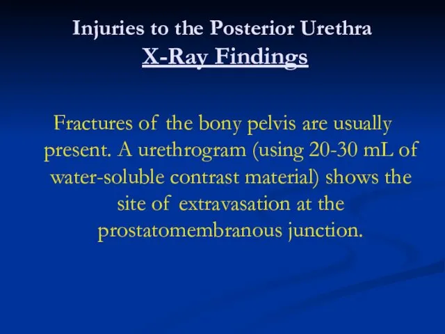
- 143. Injuries to the Posterior Urethra X-Ray Findings Fractures of the bony pelvis are usually present. A
- 144. Injuries to the Posterior Urethra X-Ray Findings Ordinarily, there is free extravasation of contrast material into
- 145. Injuries to the Posterior Urethra Instrumental Examination The only instrumentation involved should be for urethrography.
- 146. Injuries to the Posterior Urethra Differential Diagnosis Bladder rupture may be associated with posterior urethral injuries
- 147. Injuries to the Posterior Urethra Complications Stricture, impotence, and incontinence as complications of prostatomembranous disruption are
- 148. Injuries to the Posterior Urethra Complications Stricture following primary repair and anastomosis occurs in about 50%
- 149. Injuries to the Posterior Urethra Complications The incidence of impotence after primary repair is 30-80% (mean,
- 150. Injuries to the Posterior Urethra Treatment Emergency Measures Shock and hemorrhage should be treated.
- 151. Injuries to the Posterior Urethra Treatment Surgical Measures Urethral catheterization should be avoided.
- 152. Injuries to the Posterior Urethra Treatment Immediate Management Initial management should consist of suprapubic cystostomy to
- 153. Injuries to the Posterior Urethra Treatment The bladder often is distended by a large volume of
- 154. Injuries to the Posterior Urethra Treatment The bladder should be opened in the midline and carefully
- 155. Injuries to the Posterior Urethra Treatment This approach involves no urethral instrumentation or manipulation.
- 156. Injuries to the Posterior Urethra Treatment Incomplete laceration of the posterior urethra heals spontaneously, and the
- 157. Injuries to the Posterior Urethra Treatment Delayed Urethral Reconstruction Reconstruction of the urethra after prostatic disruption
- 158. Injuries to the Posterior Urethra Treatment This stricture usually is 1 -2 cm long and situated
- 159. Injuries to the Posterior Urethra Treatment A 16F silicone urethral catheter should be left in place
- 160. Injuries to the Posterior Urethra Treatment Immediate Urethral Realignment Some surgeons prefer to realign the urethra
- 161. Injuries to the Posterior Urethra Treatment General Measures After delayed reconstruction by a perineal approach, patients
- 162. Injuries to the Posterior Urethra Treatment Treatment of Complications Approximately 1 month after the delayed reconstruction,
- 163. Injuries to the Posterior Urethra Treatment If the cystogram shows a patent area of reconstruction free
- 164. Injuries to the Posterior Urethra Treatment Stricture, if present (
- 165. Injuries to the Posterior Urethra Treatment The patient may be impotent for several months after delayed
- 166. Injuries to the Posterior Urethra Treatment Incontinence after posterior urethral rupture and delayed repair is rare
- 167. Injuries to the Posterior Urethra Treatment Prognosis If complications can be avoided, the prognosis is excellent.
- 168. Injuries to the Anterior Urethra Etiology The anterior urethra is the portion distal to the urogenital
- 169. Injuries to the Anterior Urethra Pathogenesis & Pathology Contusion Contusion of the urethra is a sign
- 170. Injuries to the Anterior Urethra Pathogenesis & Pathology Laceration A severe straddle injury may result in
- 171. Injuries to the Anterior Urethra Clinical Findings Symptoms There is usually a history of a fall,
- 172. Injuries to the Anterior Urethra Clinical Findings If voiding has occurred and extravasation is noted, sudden
- 173. Injuries to the Anterior Urethra Clinical Findings Signs The perineum is very tender, and a mass
- 174. Injuries to the Anterior Urethra Clinical Findings No attempt should be made to pass a urethral
- 175. Injuries to the Anterior Urethra Clinical Findings When presentation of such injuries is delayed, there is
- 176. Injuries to the Anterior Urethra Laboratory Findings Blood loss is not usually excessive, particularly if secondary
- 177. Injuries to the Anterior Urethra X-Ray Findings A contused urethra shows no evidence of extravasation.
- 178. Injuries to the Anterior Urethra Complications Heavy bleeding from the corpus spongiosum injury may occur in
- 179. Injuries to the Anterior Urethra Complications The complications of urinary extravasation are chiefly sepsis and infection.
- 180. Injuries to the Anterior Urethra Complications Stricture at the site of injury is a common complication,
- 181. Injuries to the Anterior Urethra Treatment General Measures Major blood loss usually does not occur from
- 182. Injuries to the Anterior Urethra Treatment Specific Measures: Urethral Contusion The patient with urethral contusion shows
- 183. Injuries to the Anterior Urethra Treatment Urethral Lacerations Instrumentation of the urethra following urethrography should be
- 184. Injuries to the Anterior Urethra Treatment If only minor extravasation is noted on the urethrogram, a
- 185. Injuries to the Anterior Urethra Treatment Most of these strictures are not severe and do not
- 186. Injuries to the Anterior Urethra Treatment Urethral Laceration with Extensive Urinary Extravasation After major laceration, urinary
- 187. Injuries to the Anterior Urethra Treatment Immediate Repair Immediate repair of urethral lacerations can be performed,
- 188. Injuries to the Anterior Urethra Treatment Treatment of Complications Strictures at the site of injury may
- 189. Injuries to the Anterior Urethra Treatment Prognosis Urethral stricture is a major complication but in most
- 190. Injuries to the Penis Disruption of the tunica albuginea of the penis (penile fracture) can occur
- 191. Injuries to the Penis Gangrene and urethral injury may be caused by obstructing rings placed around
- 192. Injuries to the Penis Injuries to the penis should suggest possible urethral damage, which should be
- 193. Injuries to the Scrotum Superficial lacerations of the scrotum may be debrided and closed primarily. Blunt
- 194. Injuries to the Scrotum Total avulsion of the scrotal skin may be caused by machinery accidents
- 195. Injuries to the Scrotum Later reconstruction of the scrotum can be done with a skin graft
- 197. Скачать презентацию

Emergency Diagnosis & Management
About 10% of all injuries seen in the
Emergency Diagnosis & Management
About 10% of all injuries seen in the

Many of them are subtle and difficult to define and require
Many of them are subtle and difficult to define and require
Emergency Diagnosis & Management
Initial assessment should include control of hemorrhage and
Emergency Diagnosis & Management
Initial assessment should include control of hemorrhage and
Emergency Diagnosis & Management
The history should include a detailed description of
Emergency Diagnosis & Management
The history should include a detailed description of
Emergency Diagnosis & Management
The abdomen and genitalia should be examined for
Emergency Diagnosis & Management
The abdomen and genitalia should be examined for
Fractures of the lower ribs are often associated with renal injuries,
Fractures of the lower ribs are often associated with renal injuries,

Patients who do not have life-threatening injuries and whose blood pressure
Patients who do not have life-threatening injuries and whose blood pressure
Emergency Diagnosis & Management
When genitourinary tract injury is suspected on the
Emergency Diagnosis & Management
When genitourinary tract injury is suspected on the
Assessment of Injury
Assessment of the injury should be done in
Assessment of Injury
Assessment of the injury should be done in
Catheterization
Blood at the urethral meatus in men indicates urethral injury;
Catheterization
Blood at the urethral meatus in men indicates urethral injury;
Catheterization
If no blood is present at the meatus, a urethral catheter
Catheterization
If no blood is present at the meatus, a urethral catheter

Catheterization
If catheterization is traumatic despite the greatest care, the significance of
Catheterization
If catheterization is traumatic despite the greatest care, the significance of

Computed Tomography (CT)
Abdominal CT with contrast media is the best imaging
Computed Tomography (CT)
Abdominal CT with contrast media is the best imaging

Computed Tomography (CT)
It can define
the size
extent of the retroperitoneal
Computed Tomography (CT)
It can define
the size
extent of the retroperitoneal
Computed Tomography (CT)
Spiral CT scanning, now common, is very rapid, but
Computed Tomography (CT)
Spiral CT scanning, now common, is very rapid, but
Retrograde Cystography
Filling of the bladder with contrast material is essential to
Retrograde Cystography
Filling of the bladder with contrast material is essential to
Retrograde Cystography
A film should be obtained with the bladder filled and
Retrograde Cystography
A film should be obtained with the bladder filled and

Retrograde Cystography
Cystography with CT is excellent for establishing bladder injury.
Retrograde Cystography
Cystography with CT is excellent for establishing bladder injury.
Urethrography
A small (12F) catheter can be inserted into the urethral meatus
Urethrography
A small (12F) catheter can be inserted into the urethral meatus

Urethrography
After retrograde injection of 20 mL of water-soluble contrast material,
Urethrography
After retrograde injection of 20 mL of water-soluble contrast material,

Arteriography
Arteriography may help define renal parenchymal and renal vascular injuries.
Arteriography
Arteriography may help define renal parenchymal and renal vascular injuries.
Intravenous Urography
Intravenous urography can be used to detect renal and ureteral
Intravenous Urography
Intravenous urography can be used to detect renal and ureteral
Cystoscopy and Retrograde Urography
Cystoscopy and retrograde urography may be useful to
Cystoscopy and Retrograde Urography
Cystoscopy and retrograde urography may be useful to

Abdominal Sonography
Abdominal sonography has not been shown to add substantial information
Abdominal Sonography
Abdominal sonography has not been shown to add substantial information
Injuries to the Kidney
Renal injuries are the most common injuries of
Injuries to the Kidney
Renal injuries are the most common injuries of

Injuries to the Kidney
Most injuries occur from automobile accidents or sporting
Injuries to the Kidney
Most injuries occur from automobile accidents or sporting

Injuries to the Kidney
Etiology
Blunt trauma directly to the abdomen, flank, or
Injuries to the Kidney
Etiology
Blunt trauma directly to the abdomen, flank, or
Injuries to the Kidney
Vehicle collisions at high speed may result in
Injuries to the Kidney
Vehicle collisions at high speed may result in
Injuries to the Kidney
Associated abdominal visceral injuries are present in 80%
Injuries to the Kidney
Associated abdominal visceral injuries are present in 80%
Pathology & Classification
Early Pathologic Findings
Lacerations from blunt trauma usually occur
Pathology & Classification
Early Pathologic Findings
Lacerations from blunt trauma usually occur
Pathology & Classification
Early Pathologic Findings
In injuries from rapid deceleration, the
Pathology & Classification
Early Pathologic Findings
In injuries from rapid deceleration, the
Pathology & Classification
Early Pathologic Findings
Acute thrombosis of the renal artery
Pathology & Classification
Early Pathologic Findings
Acute thrombosis of the renal artery
Pathology & Classification Hydronephrosis
Follow-up excretory urography is indicated in all cases
Pathology & Classification Hydronephrosis
Follow-up excretory urography is indicated in all cases

Pathology & Classification Arteriovenous Fistula
Arteriovenous fistulas may occur after penetrating injuries
Pathology & Classification Arteriovenous Fistula
Arteriovenous fistulas may occur after penetrating injuries
Pathology & Classification Renal Vascular Hypertension
The blood flow in tissue rendered
Pathology & Classification Renal Vascular Hypertension
The blood flow in tissue rendered
Clinical Findings & Indications for Studies
Microscopic or gross hematuria following trauma
Clinical Findings & Indications for Studies
Microscopic or gross hematuria following trauma
Clinical Findings & Indications for Studies
Some cases of renal vascular injury
Clinical Findings & Indications for Studies
Some cases of renal vascular injury
Clinical Findings & Indications for Studies
The degree of renal injury does
Clinical Findings & Indications for Studies
The degree of renal injury does

Clinical Findings & Indications for Studies
Miller and McAninch (1995) made the
Clinical Findings & Indications for Studies
Miller and McAninch (1995) made the
Clinical Findings & Indications for Studies
However, should physical examination or associated
Clinical Findings & Indications for Studies
However, should physical examination or associated
Clinical Findings & Indications for Studies
Symptoms
There is usually visible evidence of
Clinical Findings & Indications for Studies
Symptoms
There is usually visible evidence of

Clinical Findings & Indications for Studies
Catheterization usually reveals hematuria.
Clinical Findings & Indications for Studies
Catheterization usually reveals hematuria.

Clinical Findings & Indications for Studies
Signs
Initially, shock or signs of
Clinical Findings & Indications for Studies
Signs
Initially, shock or signs of

Clinical Findings & Indications for Studies
Signs
Diffuse abdominal tenderness may be
Clinical Findings & Indications for Studies
Signs
Diffuse abdominal tenderness may be

Clinical Findings & Indications for Studies
Signs
The abdomen may be distended
Clinical Findings & Indications for Studies
Signs
The abdomen may be distended

Clinical Findings & Indications for Studies
Laboratory Findings
Microscopic or gross hematuria
Clinical Findings & Indications for Studies
Laboratory Findings
Microscopic or gross hematuria
Clinical Findings & Indications for Studies
Staging and X-Ray Findings
Staging of
Clinical Findings & Indications for Studies
Staging and X-Ray Findings
Staging of
Clinical Findings & Indications for Studies
Staging and X-Ray Findings
For example,
Clinical Findings & Indications for Studies
Staging and X-Ray Findings
For example,
Clinical Findings & Indications for Studies
Staging and X-Ray Findings
Ultrasonography and
Clinical Findings & Indications for Studies
Staging and X-Ray Findings
Ultrasonography and
Clinical Findings & Indications for Studies
Staging and X-Ray Findings
Staging begins
Clinical Findings & Indications for Studies
Staging and X-Ray Findings
Staging begins
Clinical Findings & Indications for Studies
Staging and X-Ray Findings
This noninvasive
Clinical Findings & Indications for Studies
Staging and X-Ray Findings
This noninvasive

Clinical Findings & Indications for Studies
Staging and X-Ray Findings
Arteriography defines
Clinical Findings & Indications for Studies
Staging and X-Ray Findings
Arteriography defines
Clinical Findings & Indications for Studies
Staging and X-Ray Findings
The major
Clinical Findings & Indications for Studies
Staging and X-Ray Findings
The major
Clinical Findings & Indications for Studies
Staging and X-Ray Findings
Radionuclide renal
Clinical Findings & Indications for Studies
Staging and X-Ray Findings
Radionuclide renal

Clinical Findings & Indications for Studies
Differential Diagnosis
Trauma to the abdomen
Clinical Findings & Indications for Studies
Differential Diagnosis
Trauma to the abdomen
Clinical Findings & Indications for Studies
Complications
Early Complications
Hemorrhage is perhaps the
Clinical Findings & Indications for Studies
Complications
Early Complications
Hemorrhage is perhaps the
Clinical Findings & Indications for Studies
Complications
The size and expansion of
Clinical Findings & Indications for Studies
Complications
The size and expansion of
Clinical Findings & Indications for Studies
Complications
Urinary extravasation from renal fracture
Clinical Findings & Indications for Studies
Complications
Urinary extravasation from renal fracture

Clinical Findings & Indications for Studies
Complications
A resolving retroperitoneal hematoma may
Clinical Findings & Indications for Studies
Complications
A resolving retroperitoneal hematoma may
Clinical Findings & Indications for Studies
Complications
Late Complications
Hypertension, hydronephrosis, arteriovenous fistula,
Clinical Findings & Indications for Studies
Complications
Late Complications
Hypertension, hydronephrosis, arteriovenous fistula,
Clinical Findings & Indications for Studies
Complications
Heavy late bleeding may occur
Clinical Findings & Indications for Studies
Complications
Heavy late bleeding may occur

Clinical Findings & Indications for Studies
Treatment: Emergency Measures
The objectives of
Clinical Findings & Indications for Studies
Treatment: Emergency Measures
The objectives of
Clinical Findings & Indications for Studies
Treatment: Surgical Measures
Blunt Injuries
Bleeding stops
Clinical Findings & Indications for Studies
Treatment: Surgical Measures
Blunt Injuries
Bleeding stops
Clinical Findings & Indications for Studies
Treatment: Surgical Measures
Cases in which
Clinical Findings & Indications for Studies
Treatment: Surgical Measures
Cases in which

Clinical Findings & Indications for Studies
Treatment: Surgical Measures
Penetrating Injuries
Penetrating injuries
Clinical Findings & Indications for Studies
Treatment: Surgical Measures
Penetrating Injuries
Penetrating injuries
Clinical Findings & Indications for Studies
Treatment: Surgical Measures
In 80% of
Clinical Findings & Indications for Studies
Treatment: Surgical Measures
In 80% of
Clinical Findings & Indications for Studies
Treatment: Surgical Measures
Treatment of Complications
Hydronephrosis
Clinical Findings & Indications for Studies
Treatment: Surgical Measures
Treatment of Complications
Hydronephrosis
Clinical Findings & Indications for Studies
Treatment: Surgical Measures
Prognosis
With careful follow-up,
Clinical Findings & Indications for Studies
Treatment: Surgical Measures
Prognosis
With careful follow-up,
Clinical Findings & Indications for Studies
Treatment: Surgical Measures
Injuries to the
Clinical Findings & Indications for Studies
Treatment: Surgical Measures
Injuries to the

Clinical Findings & Indications for Studies
Treatment: Surgical Measures
Etiology
Large pelvic masses
Clinical Findings & Indications for Studies
Treatment: Surgical Measures
Etiology
Large pelvic masses
Clinical Findings & Indications for Studies
Treatment: Surgical Measures
Extensive carcinoma of
Clinical Findings & Indications for Studies
Treatment: Surgical Measures
Extensive carcinoma of
Clinical Findings & Indications for Studies
Treatment: Surgical Measures
Devascularization may occur
Clinical Findings & Indications for Studies
Treatment: Surgical Measures
Devascularization may occur
Clinical Findings & Indications for Studies
Treatment: Surgical Measures
Endoscopic manipulation of
Clinical Findings & Indications for Studies
Treatment: Surgical Measures
Endoscopic manipulation of

Clinical Findings & Indications for Studies
Treatment: Surgical Measures
Pathogenesis & Pathology
The
Clinical Findings & Indications for Studies
Treatment: Surgical Measures
Pathogenesis & Pathology
The
Clinical Findings & Indications for Studies
Treatment: Surgical Measures
Intraperitoneal extravasation of
Clinical Findings & Indications for Studies
Treatment: Surgical Measures
Intraperitoneal extravasation of

Clinical Findings
Symptoms
If the ureter has been completely or partially ligated during
Clinical Findings
Symptoms
If the ureter has been completely or partially ligated during
Clinical Findings
Symptoms
Ureteral injuries from external violence should be suspected in patients
Clinical Findings
Symptoms
Ureteral injuries from external violence should be suspected in patients
Clinical Findings
Symptoms
Signs
The acute hydronephrosis of a totally ligated ureter results in
Clinical Findings
Symptoms
Signs
The acute hydronephrosis of a totally ligated ureter results in
Clinical Findings
Symptoms
Watery discharge from the wound or vagina may be identified
Clinical Findings
Symptoms
Watery discharge from the wound or vagina may be identified
Laboratory Findings
Ureteral injury from external violence is manifested by microscopic hematuria
Laboratory Findings
Ureteral injury from external violence is manifested by microscopic hematuria

Imaging Findings
Diagnosis is by excretory urography.
Imaging Findings
Diagnosis is by excretory urography.
Imaging Findings
Partial transection of the ureter results in more rapid excretion,
Imaging Findings
Partial transection of the ureter results in more rapid excretion,
Imaging Findings
In acute injury from external violence, the excretory urogram usually
Imaging Findings
In acute injury from external violence, the excretory urogram usually
Ultrasonography
Ultrasonography outlines hydroureter or urinary extravasation as it develops into a
Ultrasonography
Ultrasonography outlines hydroureter or urinary extravasation as it develops into a
Radionuclide Scanning
Radionuclide scanning demonstrates delayed excretion on the injured side, with
Radionuclide Scanning
Radionuclide scanning demonstrates delayed excretion on the injured side, with
Differential Diagnosis
Postoperative bowel obstruction and peritonitis may cause symptoms similar to
Differential Diagnosis
Postoperative bowel obstruction and peritonitis may cause symptoms similar to

Differential Diagnosis
Deep wound infection must be considered postoperatively in patients with
Differential Diagnosis
Deep wound infection must be considered postoperatively in patients with
Differential Diagnosis
Acute pyelonephritis in the early postoperative period may also result
Differential Diagnosis
Acute pyelonephritis in the early postoperative period may also result
Complications
Ureteral injury may be complicated by stricture formation with resulting hydronephrosis
Complications
Ureteral injury may be complicated by stricture formation with resulting hydronephrosis
Treatment
Prompt treatment of ureteral injuries is required. The best opportunity for
Treatment
Prompt treatment of ureteral injuries is required. The best opportunity for
Treatment
Proximal urinary drainage by percutaneous nephrostomy or formal nephrostomy should be
Treatment
Proximal urinary drainage by percutaneous nephrostomy or formal nephrostomy should be
Treatment
The goals of ureteral repair are to achieve complete debridement, a
Treatment
The goals of ureteral repair are to achieve complete debridement, a
Lower Ureteral Injuries
Injuries to the lower third of the ureter allow
Lower Ureteral Injuries
Injuries to the lower third of the ureter allow
Lower Ureteral Injuries
An antireflux procedure should be done when possible.
Lower Ureteral Injuries
An antireflux procedure should be done when possible.
Lower Ureteral Injuries
Transureteroureterostomy may be used in lower-third injuries if extensive
Lower Ureteral Injuries
Transureteroureterostomy may be used in lower-third injuries if extensive

Midureteral Injuries
Midureteral injuries usually result from external violence and are best
Midureteral Injuries
Midureteral injuries usually result from external violence and are best
Upper Ureteral Injuries
Injuries to the upper third of the ureter are
Upper Ureteral Injuries
Injuries to the upper third of the ureter are
Stenting
Most anastomoses after repair of ureteral injury should be stented.
Stenting
Most anastomoses after repair of ureteral injury should be stented.
Stenting
After 3-4 weeks of healing, stents can be endoscopically removed from
Stenting
After 3-4 weeks of healing, stents can be endoscopically removed from

Prognosis
The prognosis for ureteral injury is excellent if the diagnosis is
Prognosis
The prognosis for ureteral injury is excellent if the diagnosis is
Injuries to the Bladder
Bladder injuries occur most often from external force
Injuries to the Bladder
Bladder injuries occur most often from external force
Injuries to the Bladder
Iatrogenic injury may result from gynecologic and other
Injuries to the Bladder
Iatrogenic injury may result from gynecologic and other
Injuries to the Bladder
Pathogenesis & Pathology
The bony pelvis protects the urinary
Injuries to the Bladder
Pathogenesis & Pathology
The bony pelvis protects the urinary
Injuries to the Bladder
Pathogenesis & Pathology
When the bladder is filled to
Injuries to the Bladder
Pathogenesis & Pathology
When the bladder is filled to
Injuries to the Bladder
Pathogenesis & Pathology
If the diagnosis is not established
Injuries to the Bladder
Pathogenesis & Pathology
If the diagnosis is not established
Injuries to the Bladder
Clinical Findings
Pelvic fracture accompanies bladder rupture in
Injuries to the Bladder
Clinical Findings
Pelvic fracture accompanies bladder rupture in
Injuries to the Bladder
Symptoms
There is usually a history of lower
Injuries to the Bladder
Symptoms
There is usually a history of lower
Injuries to the Bladder
Signs
Heavy bleeding associated with pelvic fracture may
Injuries to the Bladder
Signs
Heavy bleeding associated with pelvic fracture may
Injuries to the Bladder
Signs
An acute abdomen may occur with intraperitoneal
Injuries to the Bladder
Signs
An acute abdomen may occur with intraperitoneal
Injuries to the Bladder
Laboratory Findings
Catheterization usually is required in patients
Injuries to the Bladder
Laboratory Findings
Catheterization usually is required in patients

Injuries to the Bladder
Laboratory Findings
When catheterization is done, gross or,
Injuries to the Bladder
Laboratory Findings
When catheterization is done, gross or,

Injuries to the Bladder
X-Ray Findings
A plain abdominal film generally demonstrates
Injuries to the Bladder
X-Ray Findings
A plain abdominal film generally demonstrates
Injuries to the Bladder
X-Ray Findings
Bladder disruption is shown on cystography.
Injuries to the Bladder
X-Ray Findings
Bladder disruption is shown on cystography.
Injuries to the Bladder
X-Ray Findings
The drainage film is extremely important,
Injuries to the Bladder
X-Ray Findings
The drainage film is extremely important,
Injuries to the Bladder
X-Ray Findings
CT cystography is an excellent method
Injuries to the Bladder
X-Ray Findings
CT cystography is an excellent method
Injuries to the Bladder
X-Ray Findings
Incomplete distention with consequent missed diagnosis
Injuries to the Bladder
X-Ray Findings
Incomplete distention with consequent missed diagnosis
Injuries to the Bladder
Complications
A pelvic abscess may develop from extraperitoneal
Injuries to the Bladder
Complications
A pelvic abscess may develop from extraperitoneal
Injuries to the Bladder
Complications
Partial incontinence may result from bladder injury
Injuries to the Bladder
Complications
Partial incontinence may result from bladder injury
Injuries to the Bladder
Treatment
Emergency Measures
Shock and hemorrhage should be treated.
Injuries to the Bladder
Treatment
Emergency Measures
Shock and hemorrhage should be treated.
Injuries to the Bladder
Treatment
Surgical Measures
A lower midline abdominal incision should
Injuries to the Bladder
Treatment
Surgical Measures
A lower midline abdominal incision should
Injuries to the Bladder
Treatment
The bladder should be opened in the
Injuries to the Bladder
Treatment
The bladder should be opened in the
Injuries to the Bladder
Treatment
Extraperitoneal Bladder Rupture
Extraperitoneal bladder rupture can be
Injuries to the Bladder
Treatment
Extraperitoneal Bladder Rupture
Extraperitoneal bladder rupture can be
Injuries to the Bladder
Treatment
As the bladder is opened in the
Injuries to the Bladder
Treatment
As the bladder is opened in the
Injuries to the Bladder
Treatment
Extraperitoneal bladder lacerations occasionally extend into the
Injuries to the Bladder
Treatment
Extraperitoneal bladder lacerations occasionally extend into the
Injuries to the Bladder
Treatment
Intraperitoneal Rupture
Intraperitoneal bladder ruptures should be repaired
Injuries to the Bladder
Treatment
Intraperitoneal Rupture
Intraperitoneal bladder ruptures should be repaired
Injuries to the Bladder
Treatment
The bladder is then closed in separate
Injuries to the Bladder
Treatment
The bladder is then closed in separate
Injuries to the Bladder
Treatment
Pelvic Fracture
Stable fracture of the pubic rami
Injuries to the Bladder
Treatment
Pelvic Fracture
Stable fracture of the pubic rami
Injuries to the Bladder
Treatment
Pelvic Hematoma
There may be heavy uncontrolled bleeding
Injuries to the Bladder
Treatment
Pelvic Hematoma
There may be heavy uncontrolled bleeding
Injuries to the Bladder
Treatment
If bleeding persists, it may be necessary
Injuries to the Bladder
Treatment
If bleeding persists, it may be necessary
Injuries to the Bladder
Treatment
Prognosis
With appropriate treatment, the prognosis is excellent.
Injuries to the Bladder
Treatment
Prognosis
With appropriate treatment, the prognosis is excellent.
Injuries to the Bladder
Treatment
Patients with lacerations extending into the bladder
Injuries to the Bladder
Treatment
Patients with lacerations extending into the bladder
Injuries to the Urethra
Urethral injuries are uncommon and occur most often
Injuries to the Urethra
Urethral injuries are uncommon and occur most often
Injuries to the Urethra
Various parts of the urethra may be lacerated,
Injuries to the Urethra
Various parts of the urethra may be lacerated,
Injuries to the Posterior Urethra
Etiology
The membranous urethra passes through the pelvic
Injuries to the Posterior Urethra
Etiology
The membranous urethra passes through the pelvic
Injuries to the Posterior Urethra
The urethra can be transected by the
Injuries to the Posterior Urethra
The urethra can be transected by the
Injuries to the Posterior Urethra
Clinical Findings
Symptoms
Patients usually complain of lower
Injuries to the Posterior Urethra
Clinical Findings
Symptoms
Patients usually complain of lower
Injuries to the Posterior Urethra
Clinical Findings
Signs
Blood at the urethral meatus
Injuries to the Posterior Urethra
Clinical Findings
Signs
Blood at the urethral meatus
Injuries to the Posterior Urethra
Clinical Findings
The presence of blood at
Injuries to the Posterior Urethra
Clinical Findings
The presence of blood at
Injuries to the Posterior Urethra
Clinical Findings
Suprapubic tenderness and the presence
Injuries to the Posterior Urethra
Clinical Findings
Suprapubic tenderness and the presence
Injuries to the Posterior Urethra
Clinical Findings
Rectal examination may reveal a
Injuries to the Posterior Urethra
Clinical Findings
Rectal examination may reveal a
Injuries to the Posterior Urethra
Clinical Findings
Superior displacement of the prostate
Injuries to the Posterior Urethra
Clinical Findings
Superior displacement of the prostate
Injuries to the Posterior Urethra
X-Ray Findings
Fractures of the bony pelvis
Injuries to the Posterior Urethra
X-Ray Findings
Fractures of the bony pelvis

Injuries to the Posterior Urethra
X-Ray Findings
Ordinarily, there is free extravasation
Injuries to the Posterior Urethra
X-Ray Findings
Ordinarily, there is free extravasation
Injuries to the Posterior Urethra
Instrumental Examination
The only instrumentation involved should
Injuries to the Posterior Urethra
Instrumental Examination
The only instrumentation involved should
Injuries to the Posterior Urethra
Differential Diagnosis
Bladder rupture may be associated
Injuries to the Posterior Urethra
Differential Diagnosis
Bladder rupture may be associated
Injuries to the Posterior Urethra
Complications
Stricture, impotence, and incontinence as complications
Injuries to the Posterior Urethra
Complications
Stricture, impotence, and incontinence as complications
Injuries to the Posterior Urethra
Complications
Stricture following primary repair and anastomosis
Injuries to the Posterior Urethra
Complications
Stricture following primary repair and anastomosis

Injuries to the Posterior Urethra
Complications
The incidence of impotence after primary
Injuries to the Posterior Urethra
Complications
The incidence of impotence after primary
Injuries to the Posterior Urethra
Treatment
Emergency Measures
Shock and hemorrhage should be
Injuries to the Posterior Urethra
Treatment
Emergency Measures
Shock and hemorrhage should be
Injuries to the Posterior Urethra
Treatment
Surgical Measures
Urethral catheterization should be avoided.
Injuries to the Posterior Urethra
Treatment
Surgical Measures
Urethral catheterization should be avoided.
Injuries to the Posterior Urethra
Treatment
Immediate Management
Initial management should consist of
Injuries to the Posterior Urethra
Treatment
Immediate Management
Initial management should consist of
Injuries to the Posterior Urethra
Treatment
The bladder often is distended by
Injuries to the Posterior Urethra
Treatment
The bladder often is distended by
Injuries to the Posterior Urethra
Treatment
The bladder should be opened in
Injuries to the Posterior Urethra
Treatment
The bladder should be opened in
Injuries to the Posterior Urethra
Treatment
This approach involves no urethral instrumentation
Injuries to the Posterior Urethra
Treatment
This approach involves no urethral instrumentation
Injuries to the Posterior Urethra
Treatment
Incomplete laceration of the posterior urethra
Injuries to the Posterior Urethra
Treatment
Incomplete laceration of the posterior urethra
Injuries to the Posterior Urethra
Treatment
Delayed Urethral Reconstruction
Reconstruction of the urethra
Injuries to the Posterior Urethra
Treatment
Delayed Urethral Reconstruction
Reconstruction of the urethra
Injuries to the Posterior Urethra
Treatment
This stricture usually is 1 -2
Injuries to the Posterior Urethra
Treatment
This stricture usually is 1 -2
Injuries to the Posterior Urethra
Treatment
A 16F silicone urethral catheter should
Injuries to the Posterior Urethra
Treatment
A 16F silicone urethral catheter should
Injuries to the Posterior Urethra
Treatment
Immediate Urethral Realignment
Some surgeons prefer to
Injuries to the Posterior Urethra
Treatment
Immediate Urethral Realignment
Some surgeons prefer to
Injuries to the Posterior Urethra
Treatment
General Measures
After delayed reconstruction by a
Injuries to the Posterior Urethra
Treatment
General Measures
After delayed reconstruction by a
Injuries to the Posterior Urethra
Treatment
Treatment of Complications
Approximately 1 month
Injuries to the Posterior Urethra
Treatment
Treatment of Complications
Approximately 1 month
Injuries to the Posterior Urethra
Treatment
If the cystogram shows a patent
Injuries to the Posterior Urethra
Treatment
If the cystogram shows a patent
Injuries to the Posterior Urethra
Treatment
Stricture, if present (< 5%), is
Injuries to the Posterior Urethra
Treatment
Stricture, if present (< 5%), is
Injuries to the Posterior Urethra
Treatment
The patient may be impotent for
Injuries to the Posterior Urethra
Treatment
The patient may be impotent for
Injuries to the Posterior Urethra
Treatment
Incontinence after posterior urethral rupture and
Injuries to the Posterior Urethra
Treatment
Incontinence after posterior urethral rupture and
Injuries to the Posterior Urethra
Treatment
Prognosis
If complications can be avoided, the
Injuries to the Posterior Urethra
Treatment
Prognosis
If complications can be avoided, the
Injuries to the Anterior Urethra
Etiology
The anterior urethra is the portion distal
Injuries to the Anterior Urethra
Etiology
The anterior urethra is the portion distal
Injuries to the Anterior Urethra
Pathogenesis & Pathology
Contusion
Contusion of the
Injuries to the Anterior Urethra
Pathogenesis & Pathology
Contusion
Contusion of the
Injuries to the Anterior Urethra
Pathogenesis & Pathology
Laceration
A severe straddle
Injuries to the Anterior Urethra
Pathogenesis & Pathology
Laceration
A severe straddle
Injuries to the Anterior Urethra
Clinical Findings
Symptoms
There is usually a history
Injuries to the Anterior Urethra
Clinical Findings
Symptoms
There is usually a history
Injuries to the Anterior Urethra
Clinical Findings
If voiding has occurred and
Injuries to the Anterior Urethra
Clinical Findings
If voiding has occurred and
Injuries to the Anterior Urethra
Clinical Findings
Signs
The perineum is very tender,
Injuries to the Anterior Urethra
Clinical Findings
Signs
The perineum is very tender,
Injuries to the Anterior Urethra
Clinical Findings
No attempt should be made
Injuries to the Anterior Urethra
Clinical Findings
No attempt should be made
Injuries to the Anterior Urethra
Clinical Findings
When presentation of such injuries
Injuries to the Anterior Urethra
Clinical Findings
When presentation of such injuries
Injuries to the Anterior Urethra
Laboratory Findings
Blood loss is not usually
Injuries to the Anterior Urethra
Laboratory Findings
Blood loss is not usually
Injuries to the Anterior Urethra
X-Ray Findings
A contused urethra shows no
Injuries to the Anterior Urethra
X-Ray Findings
A contused urethra shows no
Injuries to the Anterior Urethra
Complications
Heavy bleeding from the corpus spongiosum
Injuries to the Anterior Urethra
Complications
Heavy bleeding from the corpus spongiosum
Injuries to the Anterior Urethra
Complications
The complications of urinary extravasation are
Injuries to the Anterior Urethra
Complications
The complications of urinary extravasation are
Injuries to the Anterior Urethra
Complications
Stricture at the site of injury
Injuries to the Anterior Urethra
Complications
Stricture at the site of injury
Injuries to the Anterior Urethra
Treatment
General Measures
Major blood loss usually does
Injuries to the Anterior Urethra
Treatment
General Measures
Major blood loss usually does
Injuries to the Anterior Urethra
Treatment
Specific Measures: Urethral Contusion
The patient with
Injuries to the Anterior Urethra
Treatment
Specific Measures: Urethral Contusion
The patient with
Injuries to the Anterior Urethra
Treatment
Urethral Lacerations
Instrumentation of the urethra following
Injuries to the Anterior Urethra
Treatment
Urethral Lacerations
Instrumentation of the urethra following
Injuries to the Anterior Urethra
Treatment
If only minor extravasation is noted
Injuries to the Anterior Urethra
Treatment
If only minor extravasation is noted
Injuries to the Anterior Urethra
Treatment
Most of these strictures are not
Injuries to the Anterior Urethra
Treatment
Most of these strictures are not
Injuries to the Anterior Urethra
Treatment
Urethral Laceration with Extensive Urinary Extravasation
After
Injuries to the Anterior Urethra
Treatment
Urethral Laceration with Extensive Urinary Extravasation
After
Injuries to the Anterior Urethra
Treatment
Immediate Repair
Immediate repair of urethral lacerations
Injuries to the Anterior Urethra
Treatment
Immediate Repair
Immediate repair of urethral lacerations
Injuries to the Anterior Urethra
Treatment
Treatment of Complications
Strictures at the site
Injuries to the Anterior Urethra
Treatment
Treatment of Complications
Strictures at the site
Injuries to the Anterior Urethra
Treatment
Prognosis
Urethral stricture is a major complication
Injuries to the Anterior Urethra
Treatment
Prognosis
Urethral stricture is a major complication
Injuries to the Penis
Disruption of the tunica albuginea of the penis
Injuries to the Penis
Disruption of the tunica albuginea of the penis
Injuries to the Penis
Gangrene and urethral injury may be caused by
Injuries to the Penis
Gangrene and urethral injury may be caused by
Injuries to the Penis
Injuries to the penis should suggest possible urethral
Injuries to the Penis
Injuries to the penis should suggest possible urethral
Injuries to the Scrotum
Superficial lacerations of the scrotum may be debrided
Injuries to the Scrotum
Superficial lacerations of the scrotum may be debrided
Injuries to the Scrotum
Total avulsion of the scrotal skin may be
Injuries to the Scrotum
Total avulsion of the scrotal skin may be

Injuries to the Scrotum
Later reconstruction of the scrotum can be done
Injuries to the Scrotum
Later reconstruction of the scrotum can be done
 Протезирование на имплантатах
 Здоровое питание - активное долголетие
 Общая характеристика группы инфекционных болезней с воздушнокапельным механизмом передачи. Грипп
 Болезни, передаваемые половым путем
 Шығармашылық модельдеу және рестоврациялау
 Ішкі сәулеленуден медициналық қорғану
 Вакцины и сыворотки
 Вирусты гепатит В
 Гормональні зміни впродовж статевого циклу у коров
 Естественное вскармливание. Преимущества. Питание, режим и гигиена кормящей матери
 Лабораторная диагностика нарушений гемостаза. Коагуляционный гемостаз
 Современные алгоритмы лечения СД и его осложнений
 Лекарственная терапия диссеминированного колоректального рака
 Фармакоэпидемиология және фармакоэканомика: анықтамасы, даму кезеңдері, негізгі қағидалары. 2 лекция
 Техникалық электр токтың әсерінен болған өлімнің сот медициналық сараптамасы
 Теоретичні основи фізичної рекреації
 ОКИ. Клиника, диагностика, лечение
 Нарушения периферического кровообращения
 Кесарево сечение в современном акушерстве
 Гигиенические принципы здорового образа жизни
 Лечебная гимнастика и массаж при вибрационной болезни
 Нефролгия и урология
 Кишечные инфекции. Эшерихиозы
 Алкогольная болезнь печени
 Инсомния: проблема надуманная или актуальная
 Поняття про онкогенні фактори та онкологічні захворювання
 Самай – төменгі жақсүйек буындарының аурулары мен зақымданулары. Жіктелуі. Ортопедиялық емі
 Техника и методика инъекций