- Кровоснабжение проводящей системы сердца

Содержание
- 2. A. Right & Left Coronary Arteries B. Branches of Right Coronary Artery 1. Main 2. Posterior
- 3. Diagram showing normal aortic origin and initial distribution of four major coronary arteries: left anterior descending
- 4. Drawing illustrating the normal blood supply of the human ventricular septum.
- 5. Diagram illustrating the principal arteries and veins on (A) the anterior surface of the heart and
- 6. Diagram showing myocardial perfusion patterns of major epicardial coronary arteries as viewed from three tomographic cuts:
- 7. Diagram showing arterial blood supply of the cardiac conduction system. The nodal artery (NA) arises from
- 10. Schematic diagram of blood supply to cardiac conduction system. The first septal branch of the left
- 12. Скачать презентацию
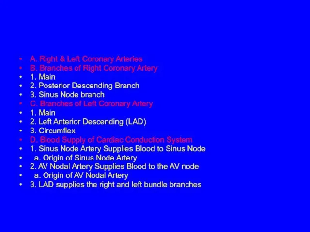
A. Right & Left Coronary Arteries
B. Branches of Right Coronary
A. Right & Left Coronary Arteries
B. Branches of Right Coronary
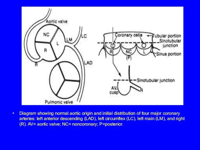
Diagram showing normal aortic origin and initial distribution of four major
Diagram showing normal aortic origin and initial distribution of four major
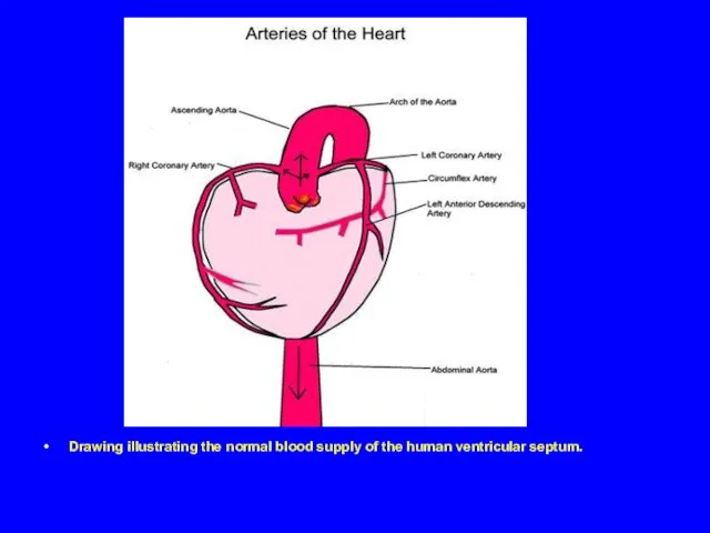
Drawing illustrating the normal blood supply of the human ventricular septum.
Drawing illustrating the normal blood supply of the human ventricular septum.
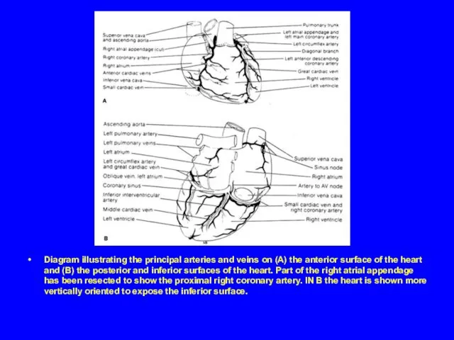
Diagram illustrating the principal arteries and veins on (A) the anterior
Diagram illustrating the principal arteries and veins on (A) the anterior
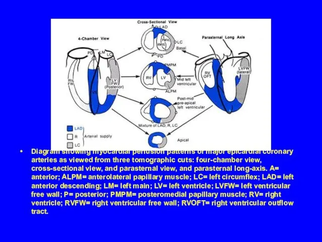
Diagram showing myocardial perfusion patterns of major epicardial coronary arteries as
Diagram showing myocardial perfusion patterns of major epicardial coronary arteries as
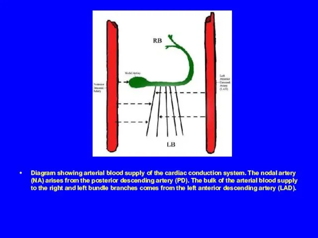
Diagram showing arterial blood supply of the cardiac conduction system. The
Diagram showing arterial blood supply of the cardiac conduction system. The


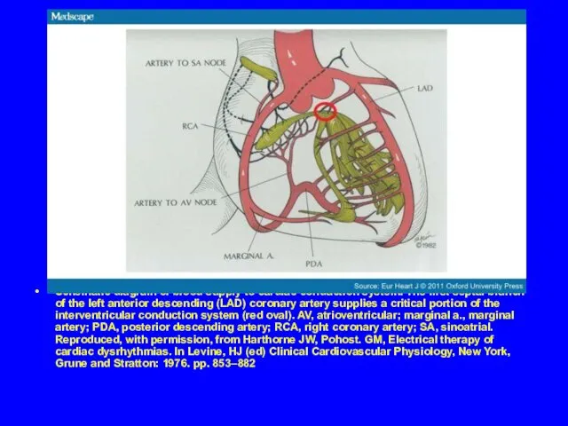
Schematic diagram of blood supply to cardiac conduction system. The first
Schematic diagram of blood supply to cardiac conduction system. The first
 Инфекционный мононуклеоз
 Эпидемиология и дерматовенерология
 Витамины. Классификация витаминов
 Избирательное прошлифовывание зубов
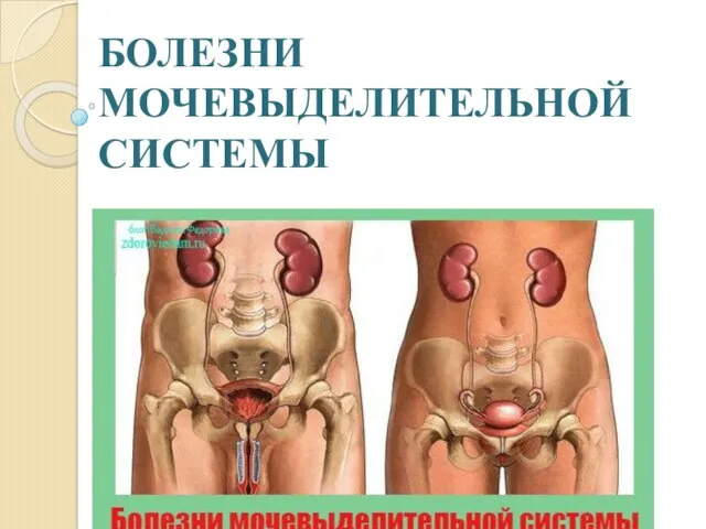 Болезни мочевыделительной системы
 Мочекаменная болезнь
 Острый тяжелый панкреатит. Современные принципы диагностики и лечения
 Гигиенические требования к качеству и безопасности пищевых продуктов
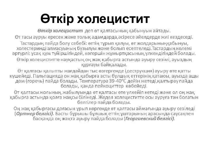 Өткір холецистит
 Дәрігердің кәсіби деформациясы
 Нейролептики (антипсихотики)
 Использование УЗИ при катетеризации центральных вен
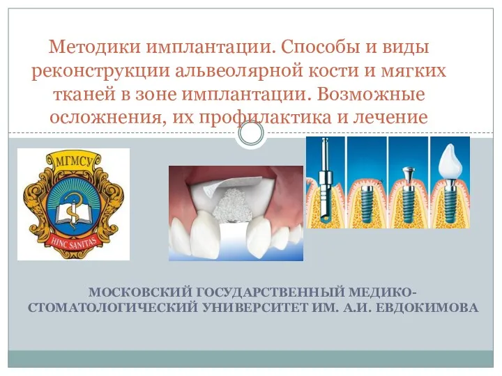 Методики имплантации. Способы и виды реконструкции альвеолярной кости и мягких тканей в зоне имплантации
 ]Противовоспалительные средства, применяемые в стоматологии
 Синдром дефицита внимания и гиперактивности
 Туберкулез эпидемиологиясы. Қазақстандағы туберкулез эпидемиологиясының ерекшеліктері
 Медико-санитарное обеспечение при ликвидации последствий чрезвычайных ситуаций природного характера
 Информация по исполнению Дорожной карты по внедрению интегрированной модели управления острыми инсультами по г. Алматы
 Правила чистки зубов
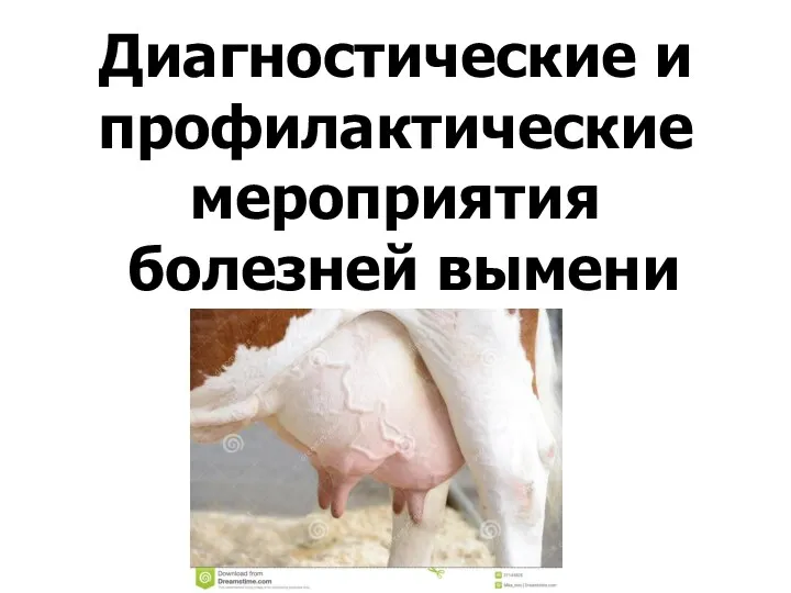 Диагностические и профилактические мероприятия болезней вымени
 Хронический гепатит
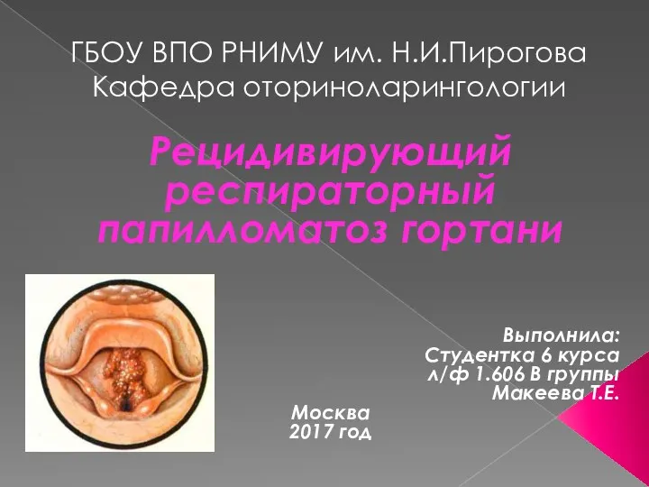 Рецидивирующий респираторный папилломатоз гортани
 Приобретенные структуры полости рта
 Классификация токсичных веществ и отравлений. Факторы, влияющие на токсичность химических веществ
 Пропедевтика внутренних болезней с профессиональными болезнями и сестринское дело. Аллергические состояния, бронхиальная астма
 Профілактика анемії у дітей
 Гострі алергози. Невідкладна допомога
 Лабораторная диагностика воздушно-капельных инфекций