- Malignant Lymphomas

Содержание
- 2. Definition Lymphoma means a malignant tumor of the lymphatic system. There are two main types of
- 3. The lymphatic system The lymphatic systemThe lymphatic system is part of the immune systemThe lymphatic system
- 4. Epidemiology
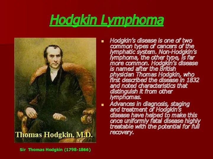
- 6. Hodgkin Lymphoma Hodgkin’s disease is one of two common types of cancers of the lymphatic system.
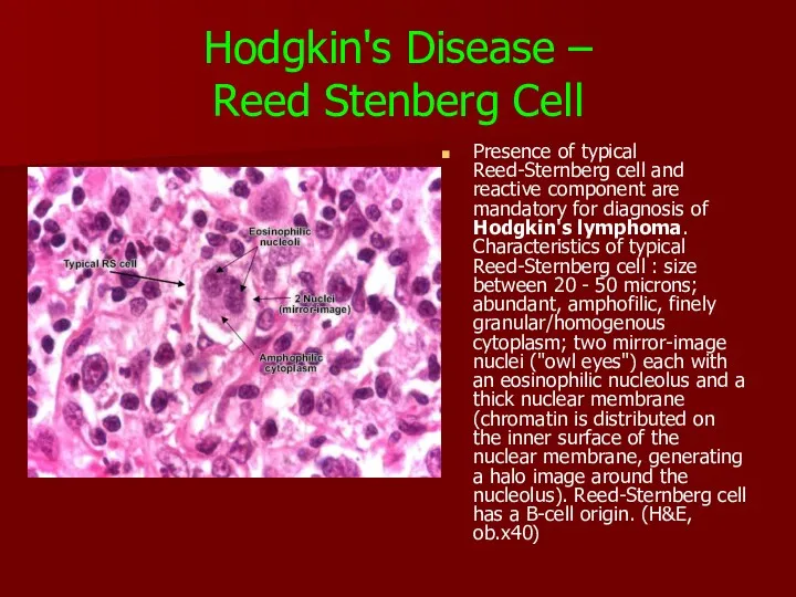
- 7. Hodgkin's Disease – Reed Stenberg Cell Presence of typical Reed-Sternberg cell and reactive component are mandatory
- 8. Within the latter, four subtypes have been distinguished: Hodgkin Lymphoma nodular sclerosis, mixed cellularity, lymphocyte-rich lymphocyte-depleted.
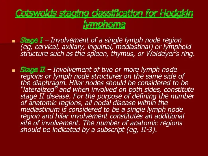
- 9. Cotswolds staging classification for Hodgkin lymphoma Stage I – Involvement of a single lymph node region
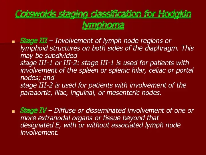
- 10. Cotswolds staging classification for Hodgkin lymphoma Stage III – Involvement of lymph node regions or lymphoid
- 11. All cases are subclassified to indicate the absence (A) or presence (B) of the systemic symptoms
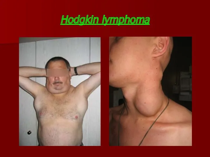
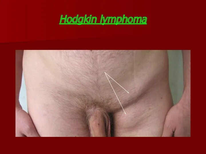
- 12. Hodgkin lymphoma
- 13. Hodgkin lymphoma
- 14. Hodgkin Lymphoma Findings: Subtle soft tissue swelling is present along the left side of the patient's
- 15. Hodgkin disease Massive involvement of paratracheal, hilar and subcarinal lymph nodes as well as two vertebral
- 16. Nonenhanced CT scan through the mediastinum shows multiple enlarged lymph nodes in the prevascular space, in
- 17. CT scan Can demonstrate the relationship of the mass to vessels and other structures. Can help
- 18. Comments: 78 y/o man with hepatosplenomegaly. Splenectomy specimen showed scattered gray-white nodules. The tumor cells were
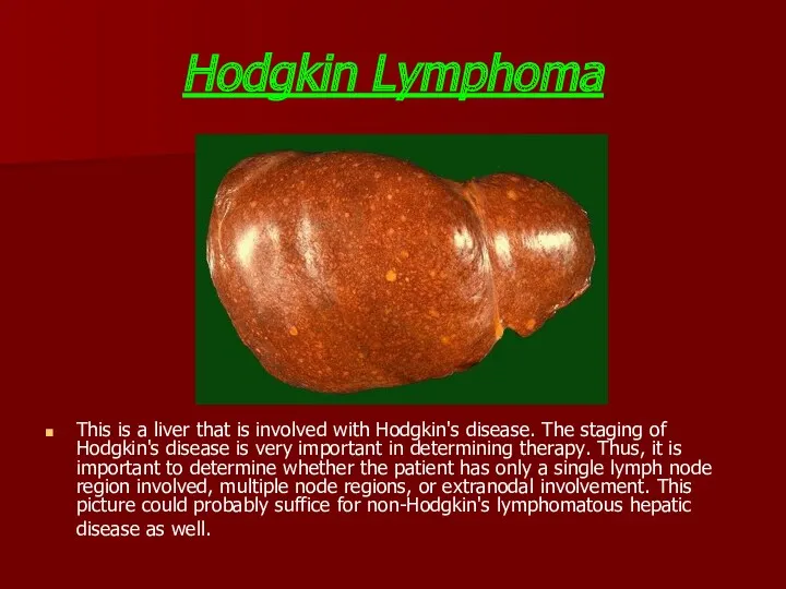
- 19. Hodgkin Lymphoma This is a liver that is involved with Hodgkin's disease. The staging of Hodgkin's
- 20. Staging of Hodgkin's lymphoma using FDG-PET Maximum intensity projection image of a staging FDG-PET scan of
- 22. Positron emission tomography/computed tomography images of Hodgkin’s disease staging Clinical stage I left axillary disease was
- 24. The 301,000 cases of non-Hodgkin Lymphoma (NHL) that occurred in 2002 (2.8% of all cancers) comprise
- 25. NHLs are slightly more common in developed countries (50.5% of cases worldwide), with rates highest in
- 26. There have been marked increases in the incidence of NHL in many parts of the world.
- 27. REAL: Classification for Non-Hodgkin’s Lymphoma
- 28. BURKITT LYMPHOMA
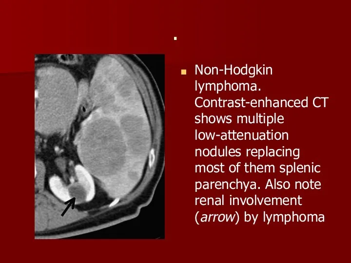
- 29. . Non-Hodgkin lymphoma. Contrast-enhanced CT shows multiple low-attenuation nodules replacing most of them splenic parenchya. Also
- 31. Скачать презентацию
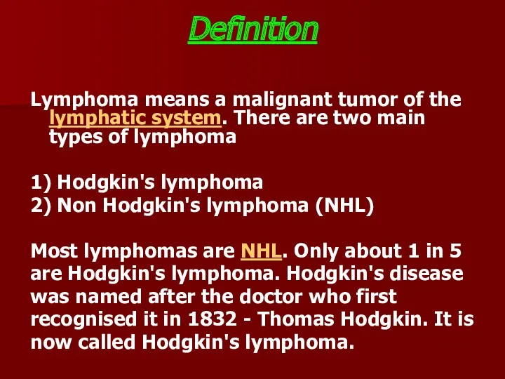
Definition
Lymphoma means a malignant tumor of the lymphatic system. There are
Definition
Lymphoma means a malignant tumor of the lymphatic system. There are
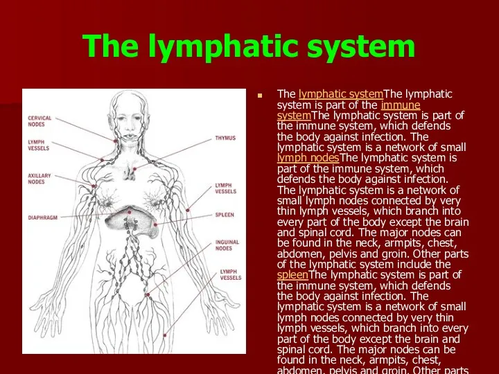
The lymphatic system
The lymphatic systemThe lymphatic system is part of
The lymphatic system
The lymphatic systemThe lymphatic system is part of
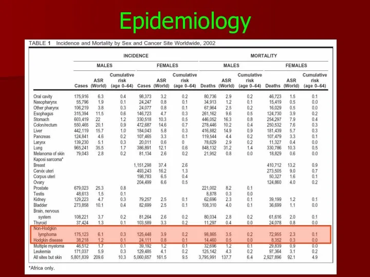
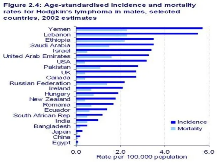
Epidemiology
Epidemiology
Hodgkin Lymphoma
Hodgkin’s disease is one of two common types of
Hodgkin Lymphoma
Hodgkin’s disease is one of two common types of
Hodgkin's Disease –
Reed Stenberg Cell
Presence of typical Reed-Sternberg cell and
Hodgkin's Disease –
Reed Stenberg Cell
Presence of typical Reed-Sternberg cell and
Within the latter, four subtypes have been distinguished:
Hodgkin Lymphoma
nodular
Within the latter, four subtypes have been distinguished:
Hodgkin Lymphoma
nodular
Cotswolds staging classification for Hodgkin lymphoma
Stage I – Involvement of a
Cotswolds staging classification for Hodgkin lymphoma
Stage I – Involvement of a
Cotswolds staging classification for Hodgkin lymphoma
Stage III – Involvement of lymph
Cotswolds staging classification for Hodgkin lymphoma
Stage III – Involvement of lymph
All cases are subclassified to indicate the absence (A) or presence
All cases are subclassified to indicate the absence (A) or presence
Hodgkin lymphoma
Hodgkin lymphoma
Hodgkin lymphoma
Hodgkin lymphoma
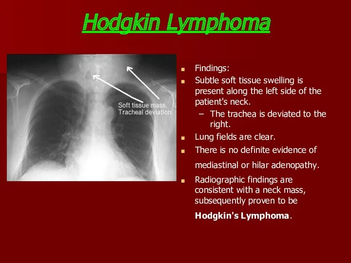
Hodgkin Lymphoma
Findings:
Subtle soft tissue swelling is present along the
Hodgkin Lymphoma
Findings:
Subtle soft tissue swelling is present along the
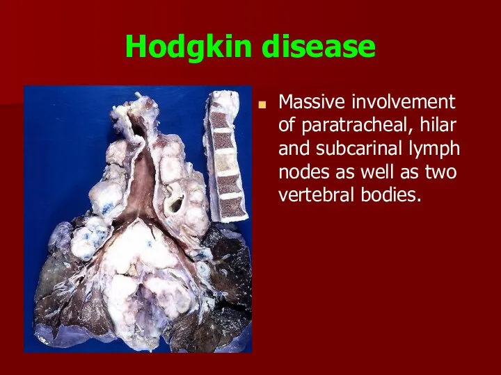
Hodgkin disease
Massive involvement of paratracheal, hilar and subcarinal lymph nodes
Hodgkin disease
Massive involvement of paratracheal, hilar and subcarinal lymph nodes
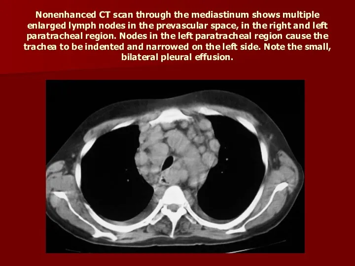
Nonenhanced CT scan through the mediastinum shows multiple enlarged lymph nodes
Nonenhanced CT scan through the mediastinum shows multiple enlarged lymph nodes
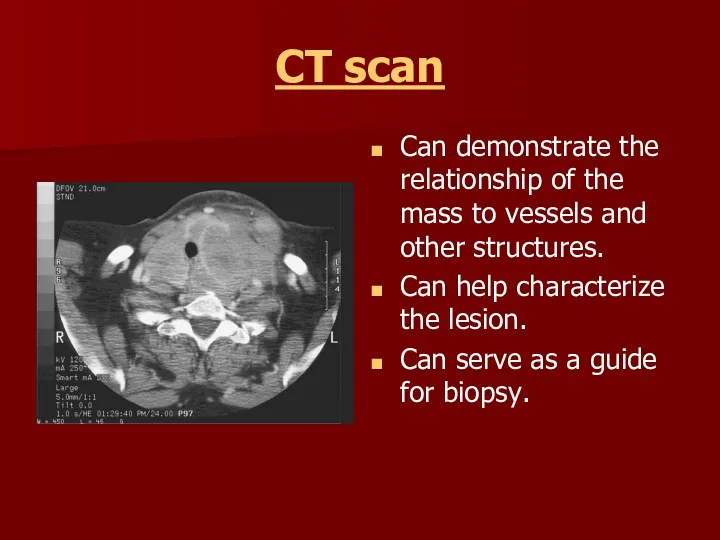
CT scan
Can demonstrate the relationship of the mass to vessels and
CT scan
Can demonstrate the relationship of the mass to vessels and
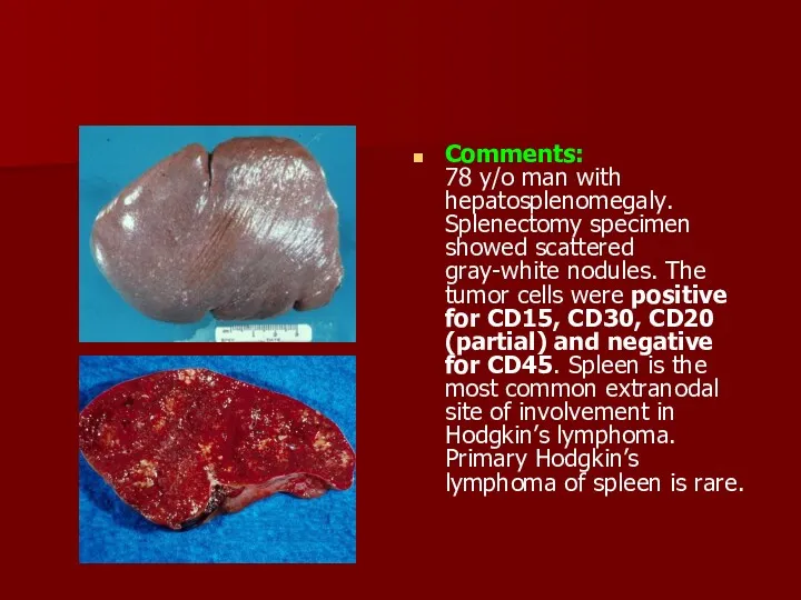
Comments:
78 y/o man with hepatosplenomegaly. Splenectomy specimen showed scattered gray-white nodules.
Comments: 78 y/o man with hepatosplenomegaly. Splenectomy specimen showed scattered gray-white nodules.
Hodgkin Lymphoma
This is a liver that is involved with Hodgkin's disease.
Hodgkin Lymphoma
This is a liver that is involved with Hodgkin's disease.
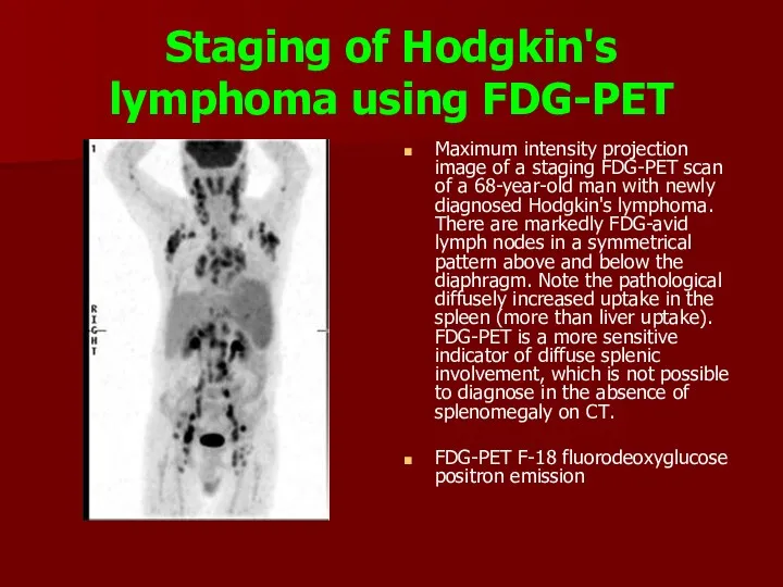
Staging of Hodgkin's lymphoma using FDG-PET
Maximum intensity projection image of a
Staging of Hodgkin's lymphoma using FDG-PET
Maximum intensity projection image of a
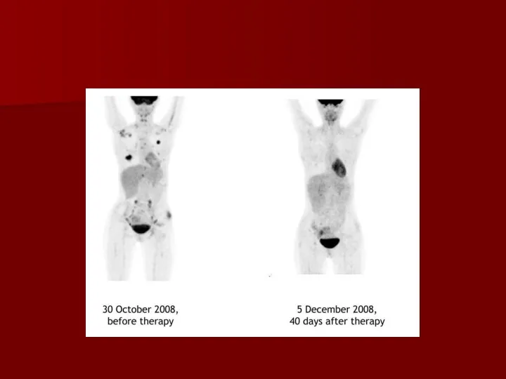
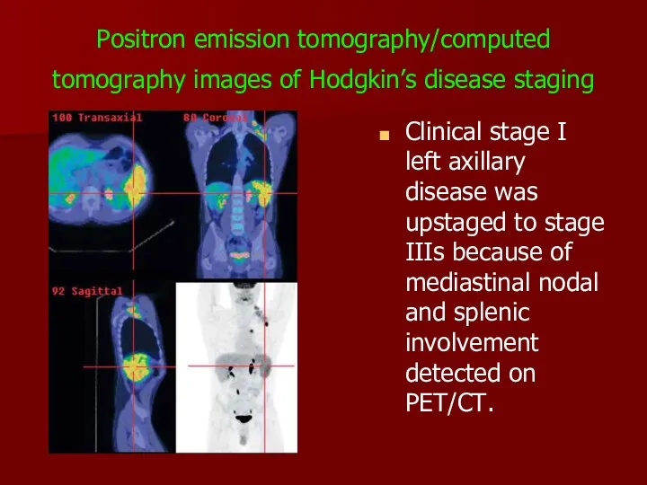
Positron emission tomography/computed tomography images of Hodgkin’s disease staging
Clinical stage
Positron emission tomography/computed tomography images of Hodgkin’s disease staging
Clinical stage
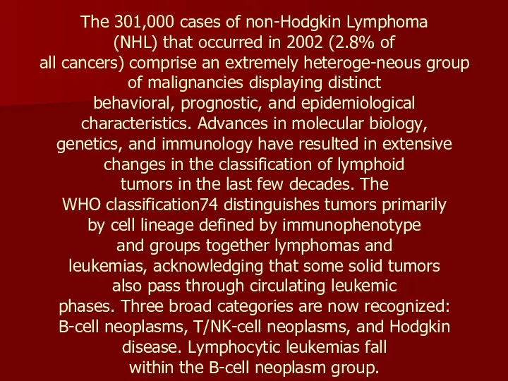
The 301,000 cases of non-Hodgkin Lymphoma
(NHL) that occurred in 2002 (2.8%
The 301,000 cases of non-Hodgkin Lymphoma (NHL) that occurred in 2002 (2.8%
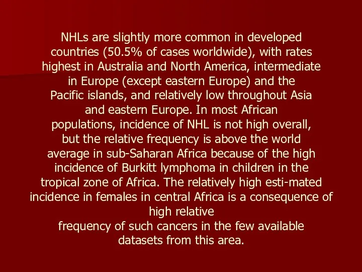
NHLs are slightly more common in developed
countries (50.5% of cases worldwide),
NHLs are slightly more common in developed countries (50.5% of cases worldwide),
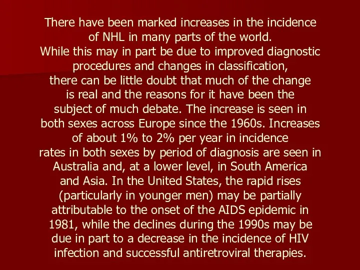
There have been marked increases in the incidence
of NHL in many
There have been marked increases in the incidence of NHL in many
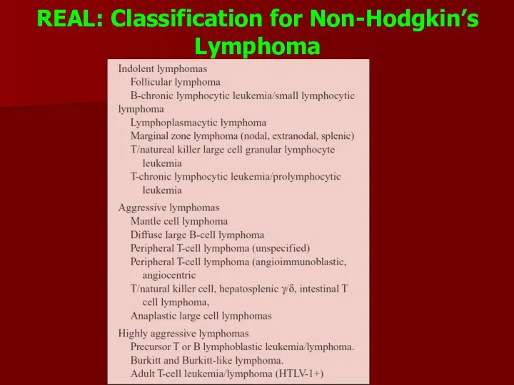
REAL: Classification for Non-Hodgkin’s
Lymphoma
REAL: Classification for Non-Hodgkin’s
Lymphoma
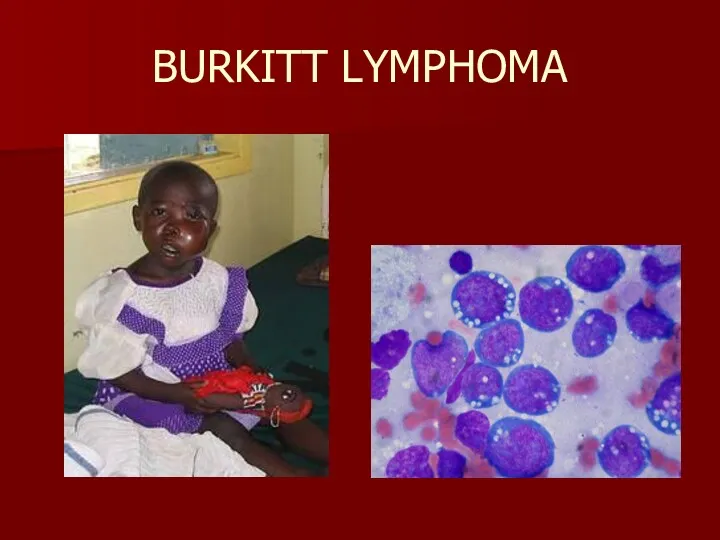
BURKITT LYMPHOMA
BURKITT LYMPHOMA
.
Non-Hodgkin lymphoma. Contrast-enhanced CT shows multiple low-attenuation nodules replacing most
.
Non-Hodgkin lymphoma. Contrast-enhanced CT shows multiple low-attenuation nodules replacing most
 Профилактическая медицина
 Боль и обезболивание
 Егде жастағы және қарт адамдардың тамақтануы
 Улучшение качества медицинской помощи на основе информационных технологий
 Жапонияның денсаулық сақтау жүйесі
 Налоги и налогообложение в здравоохранении
 Гнойно-воспалительные заболевания мягких тканей у детей
 Кариес зубов. Причины и механизм образования. Основные теории и гипотезы кариеса (Миллер, Лукомский, Энтин, Шарпенак, Боровский)
 Дифференциальная диагностика при легочных инфильтратах. Принципы лечения
 Коронавирус 2019-nCoV
 Металло керамикалық сауыт дайындаудың бірінші лабораториялық кезеңінде қолданылатын құрал саймандар мен апараттар
 Доброкачественные органоспецифические опухоли челюстей, лица, шеи
 Fizika kafedrasi
 Невроз. Психотравма высшей нервной деятельности с конфликтом в ценостеях, ориентации, отношениях
 Қазіргі кездегі өнеркәсіптік қала тұрғындарының денсаулығы
 Ішек өтімсіздігі (инвагинация)
 СПИД – что это такое?
 Течение и ведение послеродового периода. Становление лактации. Послеродовая контрацепция
 Oral cavity
 Malignant Melanoma
 Вещества, влияющие на эфферентную иннервацию. Адренергические средства
 Основы здорового образа жизни студентов
 Болезни желчного пузыря и желчевыводящих путей. Желчно-каменная болезнь. Хронический панкреатит
 Неоадъювантное лечение II-III стадии HER2-положительного рака молочной железы
 Туберкулинодиагностика
 Тромболитики. Механизм действия
 Стахиботриотоксикоз
 Защита медицинского персонала от ВБИ