- Medical helmintology. Cestodes

Содержание
- 2. CESTODES (TAPEWORMS) The tapeworms are hermaphroditic and require an intermediate host. The adult tapeworms found in
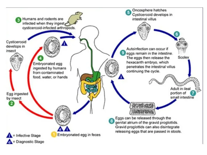
- 3. Hymenolepis nana (dwarf tapeworm) Morphology Adult worm measures 1-3 cm in length. It is made up
- 5. Infection takes place by: 1. Ingestion of egg with contaminated raw vegetables. 2. Direct infection from
- 6. Hymenolepis diminuta (rat tapeworm) Hymenolepis diminuta differs from Hymenolepis nana in that: ♦ The adult worm
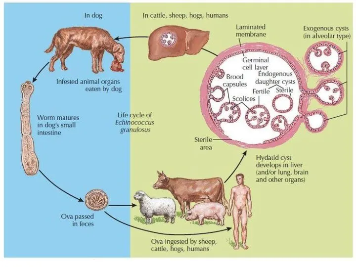
- 8. Echinococcus granulosus (dog tape worm) Responsible for most cases of echinococcosis. Echinococcosis is caused by larval
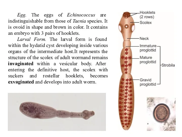
- 9. Egg. The eggs of Echinococcus are indistinguishable from those of Taenia species. It is ovoid in
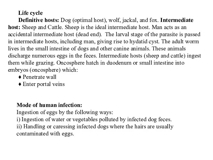
- 10. Life cycle Definitive hosts: Dog (optimal host), wolf, jackal, and fox. Intermediate host: Sheep and Cattle.
- 12. Migrate via portal blood supply to organs: eg: lungs, liver, brain etc., thus, causing extra intestinal
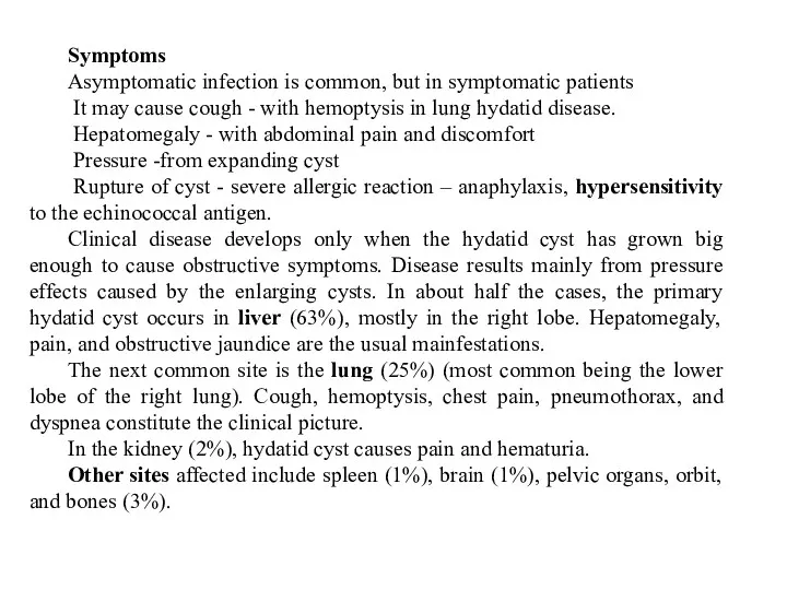
- 13. Symptoms Asymptomatic infection is common, but in symptomatic patients It may cause cough - with hemoptysis
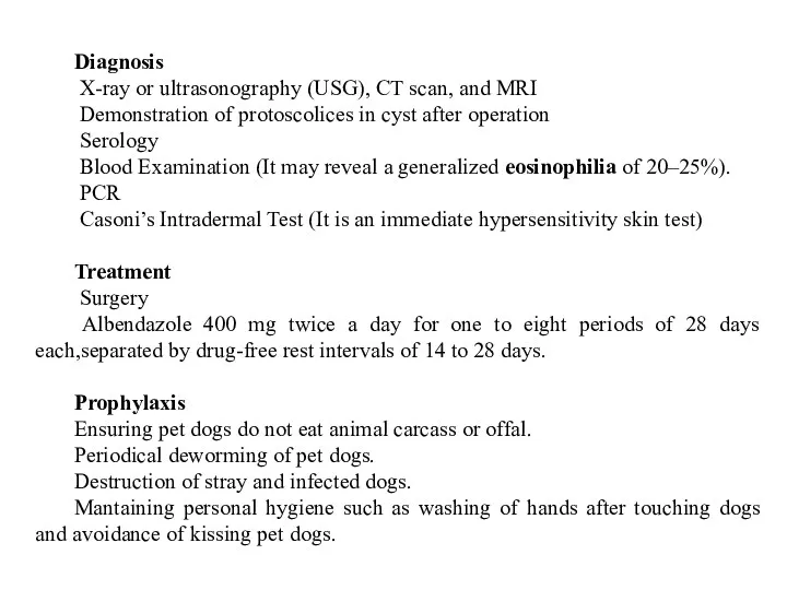
- 14. Diagnosis X-ray or ultrasonography (USG), CT scan, and MRI Demonstration of protoscolices in cyst after operation
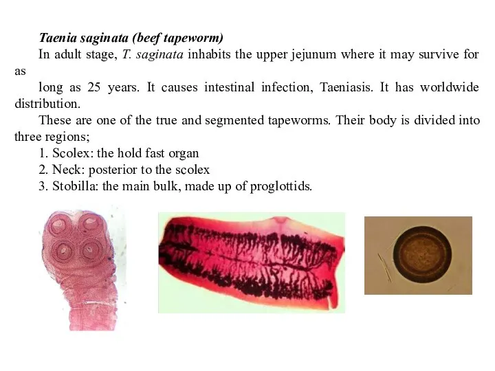
- 15. Taenia saginata (beef tapeworm) In adult stage, T. saginata inhabits the upper jejunum where it may
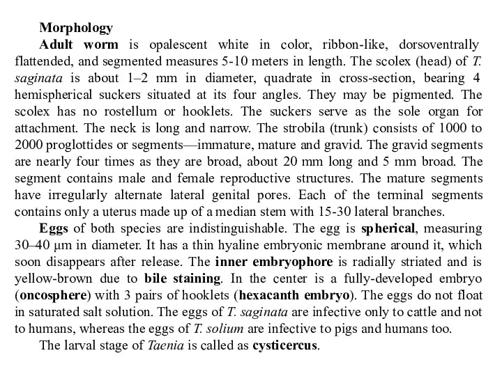
- 16. Morphology Adult worm is opalescent white in color, ribbon-like, dorsoventrally flattended, and segmented measures 5-10 meters
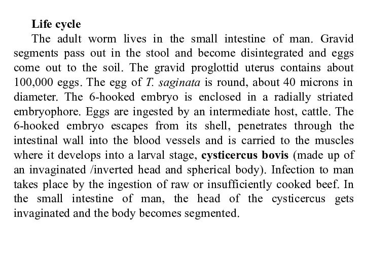
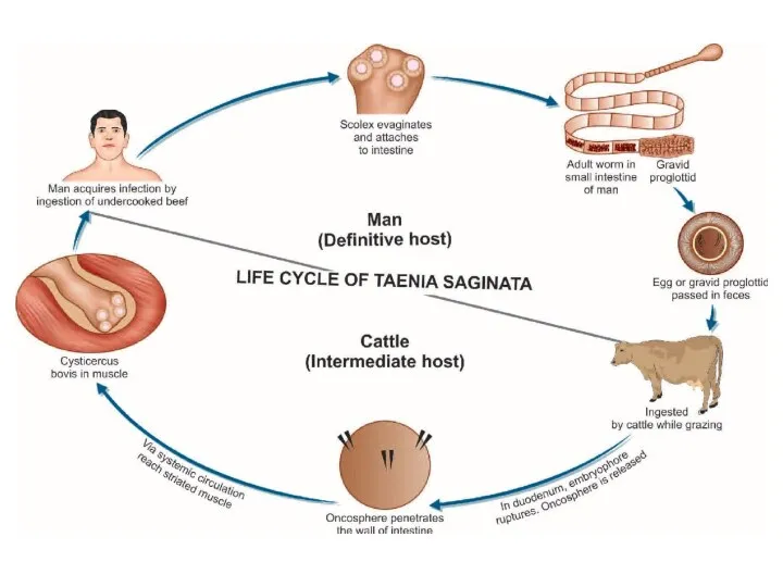
- 17. Life cycle The adult worm lives in the small intestine of man. Gravid segments pass out
- 19. Symptoms Infected persons may complain of epigastric pain, abdominal discomfort, indigestion, diarrhea, nausea, weight loss, hunger
- 20. Taenia solium (pork tapeworm) The adult worms of T. solium reside or inhabit the upper jejunum.
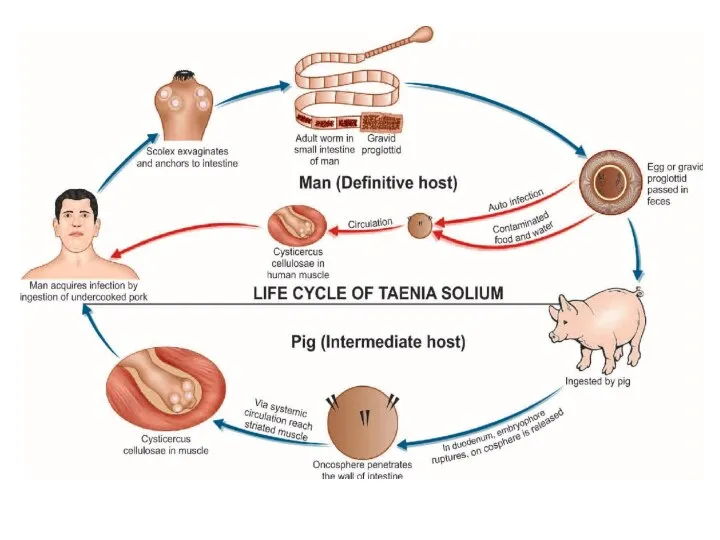
- 21. Life cycle Definitive host: Man Intermediate host: Pig, occasionally man (in case of cysticercosis). Mode of
- 23. Symptoms Infected persons may complain of epigastric pain, abdominal discomfort, indigestion, diarrhea, nausea, weight loss, hunger
- 24. Diagnosis Stool Examination: Microscopy of stool specimen, cellophane swab method (NIH Swab), Detection of Taenia Antigen
- 25. Diphylobotrium latum (fish tapeworm or broad tapeworm) The broad tapeworm infecting man has worldwide distribution, occurring
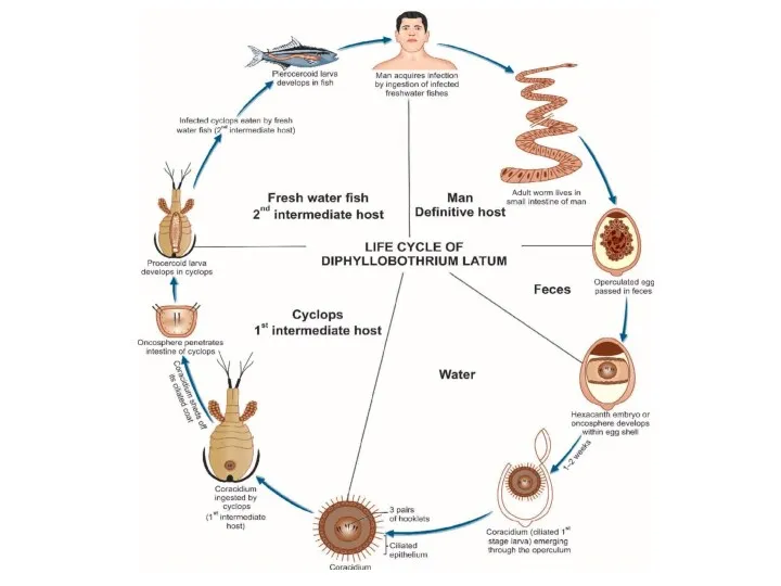
- 26. Life cycle Definitive hosts: Man, dog, and cat. Man is the optimal host. First intermediate host:
- 28. Symptoms Most infections are asymptomatic. Rarely, it causes severe cramping, mechanical Obstruction, abdominal discomfort, vomiting, diarrhea,
- 30. Скачать презентацию
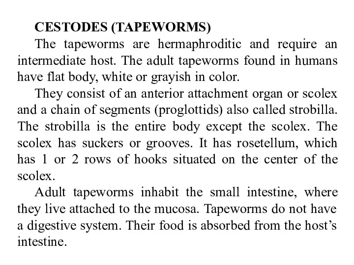
CESTODES (TAPEWORMS)
The tapeworms are hermaphroditic and require an intermediate host. The
CESTODES (TAPEWORMS)
The tapeworms are hermaphroditic and require an intermediate host. The
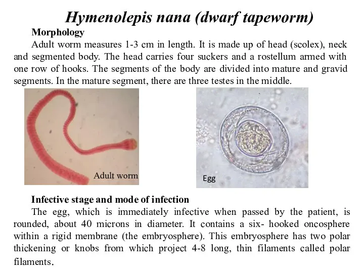
Hymenolepis nana (dwarf tapeworm)
Morphology
Adult worm measures 1-3 cm in length. It
Hymenolepis nana (dwarf tapeworm)
Morphology
Adult worm measures 1-3 cm in length. It
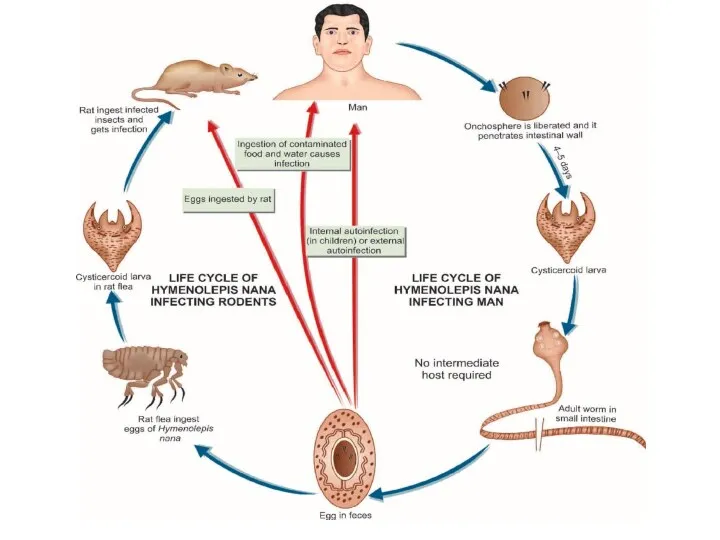

Infection takes place by:
1. Ingestion of egg with contaminated raw vegetables.
2.
Infection takes place by:
1. Ingestion of egg with contaminated raw vegetables.
2.
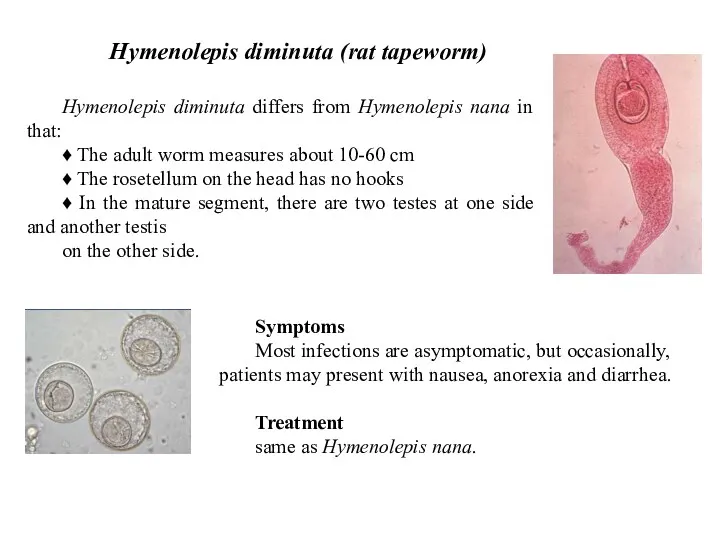
Hymenolepis diminuta (rat tapeworm)
Hymenolepis diminuta differs from Hymenolepis nana in that:
♦
Hymenolepis diminuta (rat tapeworm)
Hymenolepis diminuta differs from Hymenolepis nana in that:
♦
Echinococcus granulosus (dog tape worm)
Responsible for most cases of echinococcosis. Echinococcosis
Echinococcus granulosus (dog tape worm)
Responsible for most cases of echinococcosis. Echinococcosis
Egg. The eggs of Echinococcus are indistinguishable from those of Taenia
Egg. The eggs of Echinococcus are indistinguishable from those of Taenia
Life cycle
Definitive hosts: Dog (optimal host), wolf, jackal, and fox.
Life cycle
Definitive hosts: Dog (optimal host), wolf, jackal, and fox.
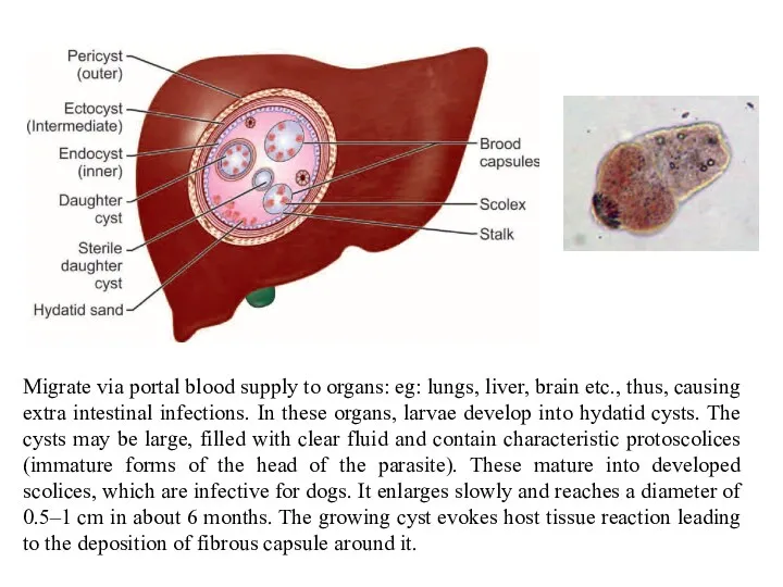
Migrate via portal blood supply to organs: eg: lungs, liver, brain
Migrate via portal blood supply to organs: eg: lungs, liver, brain
Symptoms
Asymptomatic infection is common, but in symptomatic patients
It may cause
Symptoms
Asymptomatic infection is common, but in symptomatic patients
It may cause
Diagnosis
X-ray or ultrasonography (USG), CT scan, and MRI
Demonstration of
Diagnosis
X-ray or ultrasonography (USG), CT scan, and MRI
Demonstration of
Taenia saginata (beef tapeworm)
In adult stage, T. saginata inhabits the upper
Taenia saginata (beef tapeworm)
In adult stage, T. saginata inhabits the upper
Morphology
Adult worm is opalescent white in color, ribbon-like, dorsoventrally flattended, and
Morphology
Adult worm is opalescent white in color, ribbon-like, dorsoventrally flattended, and
Life cycle
The adult worm lives in the small intestine of man.
Life cycle
The adult worm lives in the small intestine of man.

Symptoms
Infected persons may complain of epigastric pain, abdominal discomfort, indigestion, diarrhea,
Symptoms
Infected persons may complain of epigastric pain, abdominal discomfort, indigestion, diarrhea,
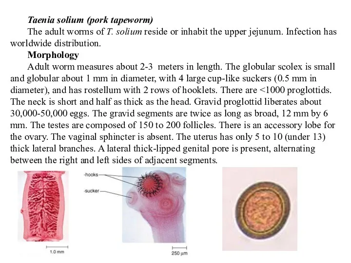
Taenia solium (pork tapeworm)
The adult worms of T. solium reside or
Taenia solium (pork tapeworm)
The adult worms of T. solium reside or

Life cycle
Definitive host: Man
Intermediate host: Pig, occasionally man (in case of
Life cycle
Definitive host: Man
Intermediate host: Pig, occasionally man (in case of
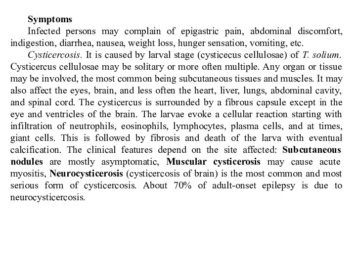
Symptoms
Infected persons may complain of epigastric pain, abdominal discomfort, indigestion, diarrhea,
Symptoms
Infected persons may complain of epigastric pain, abdominal discomfort, indigestion, diarrhea,
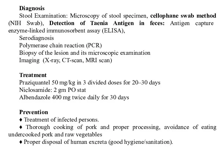
Diagnosis
Stool Examination: Microscopy of stool specimen, cellophane swab method (NIH Swab),
Diagnosis
Stool Examination: Microscopy of stool specimen, cellophane swab method (NIH Swab),
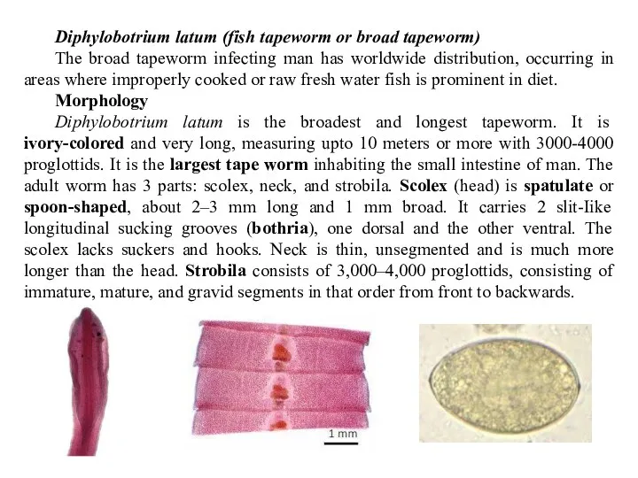
Diphylobotrium latum (fish tapeworm or broad tapeworm)
The broad tapeworm infecting man
Diphylobotrium latum (fish tapeworm or broad tapeworm)
The broad tapeworm infecting man
Life cycle
Definitive hosts: Man, dog, and cat. Man is the optimal
Life cycle
Definitive hosts: Man, dog, and cat. Man is the optimal
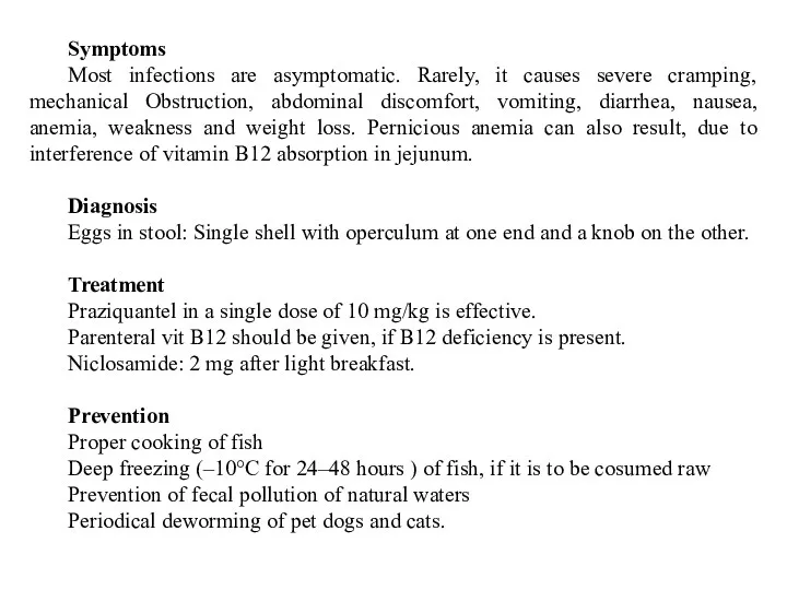
Symptoms
Most infections are asymptomatic. Rarely, it causes severe cramping, mechanical Obstruction,
Symptoms
Most infections are asymptomatic. Rarely, it causes severe cramping, mechanical Obstruction,
 Гемофилия В
 ЛФК при нарушениях осанки детей младшего школьного возраста
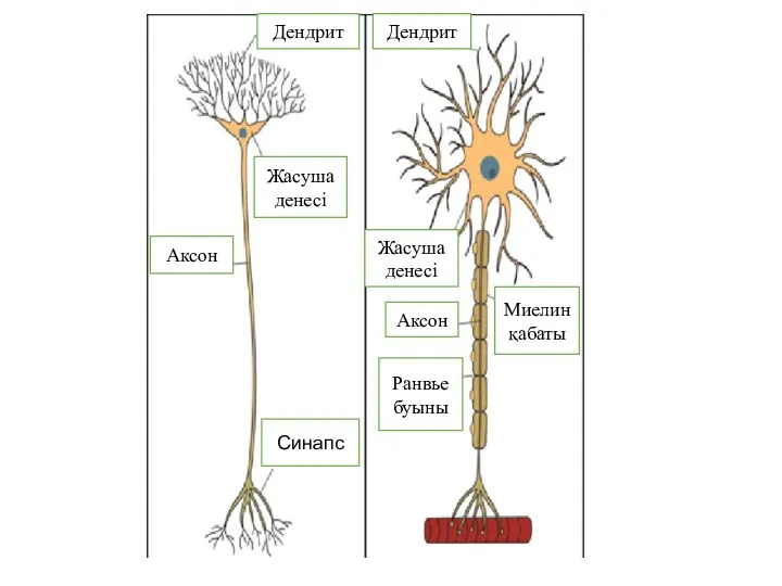 Миеленді, миеленсіз аксондарда жүйке импульстарының туындауы және өткізілуі. Өткізу жылдамдығы
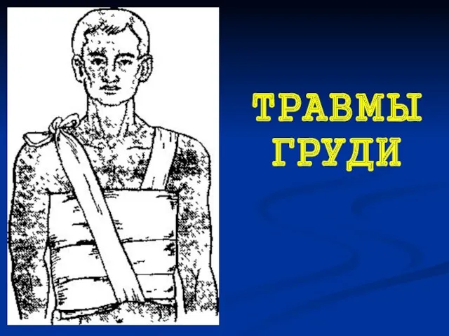 Травмы груди
 USMLE Exams and Post Graduate Training in US
 Электродные процессы, их биологическая роль и применение в медицине
 Острый инфаркт миокарда: современные подходы к диагностике и лечению
 Акушерские кровотечения во время беременности. Классификация
 Наследственные и генетические заболевания человека
 Дизартрия. Дефекты при дизартрии
 Буын— бір сүйекпен екінші сүйекті жалғастыратын аралық. Сүйектер бір-бірімен кірігіп немесе қозғалмалы болып жалғасады
 Сердечно-сосудистые заболевания. Атеросклероз
 Техника удаления временных и постоянных зубов у детей
 Гигиенические основы профилактики ВИЧ, Спид,туберкулеза и других опасных заболеваний при оказании стоматологической помощи
 Потребности человека
 Ішкі аурулар пропедевтикасы
 Асептика и антисептика
 Патология белой крови
 Поражения перикарда после лучевой терапии
 Балық өнімдерінің тағамдық нутритивтік кұндылығы
 Пороки развития ЦНС
 John Hunter
 Сестринская помощь при хронической сердечной недостаточности. Тема 4.3
 Кроветворение, органы кроветворения и иммунной защиты
 Экзема. Факторы, вызывающие экзему
 Рак печени
 Острый аппендицит
 Медсестринский процесс