- Methods of examination in gynecology

Содержание
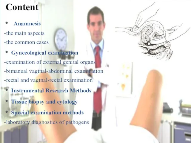
- 2. Anamnesis -the main aspects -the common cases Gynecological examination -examination of external genital organs -bimanual vaginal-abdominal
- 3. • age; • complaints; • family history; • lifestyle, nutrition, bad habits, working and living conditions;
- 4. Amenorrhea - absence of menstruation; Hypomencastral syndrome is expressed in reduction (hypomenorrhoea), shortening (oligomenorrhea), and decreasing
- 5. *is carried out on the gynecological chair. In a healthy, mature, nonpregnant and unkempt woman, the
- 6. The normal position of female genital organs is provided by: • own tone of genital organs;
- 7. Examination of external genitalia: the condition and magnitude of small and greater the labia; condition of
- 8. The index and middle fingers of one hand (usually right), dressed in a glove, are inserted
- 9. is mandatory in postmenopause to clarify the status of the uterine appendages should be given to
- 10. !!!To women, leading sex life. Timely recognition of diseases of the cervix, erosions, polyps and other
- 11. Endoscopic methods. Colposcopy - examination of the vaginal part of the cervix with an increase in
- 12. examination of the vaginal part of the cervix with an optical system (contrast luminescent colpomicroscope or
- 13. Hysterocervicaloscopy - examination with the help of optical systems of the internal surface of the uterus
- 14. an invasive diagnostic method used to establish position and direction of the uterine cavity, its length
- 15. This puncture is performed when it is necessary to ascertain the presence or lack of free
- 16. The ultrasound technique assumes an assessment of the location of the uterus, its size, outer contour
- 17. Hysterosalpingography (at present - rarely) X-ray examination of the skull Computed tomography (CT) Magnetic resonance imaging
- 18. - examination of the abdominal cavity organs by an endoscope inserted through the anterior abdominal wall
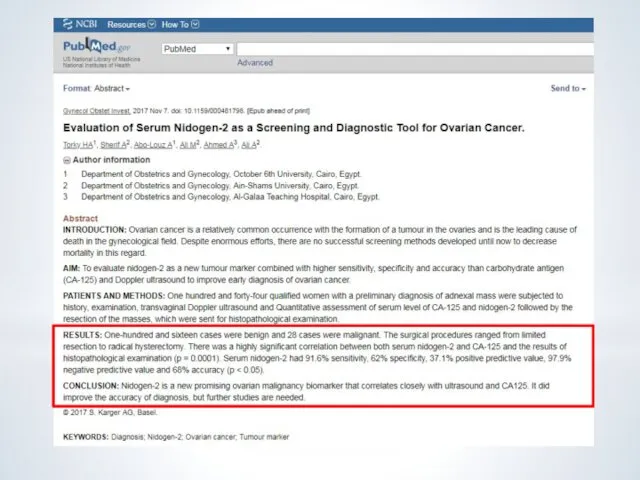
- 19. Instead laparascopy Source: https://www.ncbi.nlm.nih.gov/pubmed/29033397
- 20. In gynecology used excisional biopsy (excision of a piece of tissue), targeted biopsy - under the
- 21. The brush together with the dilator Cusco is injected into the vagina. The brush is brought
- 22. The symptom of “pupil” The symptom of the extension of the cervical mucus KPI Basal temperature
- 23. Bactrioscopic (microscopic) The cultural method Molecular biological methods Bacteriological diagnosis Bacteria Source: The vaginal microbiome: new
- 29. Скачать презентацию
Anamnesis
-the main aspects
-the common cases
Gynecological examination
-examination of external genital organs
-bimanual vaginal-abdominal
Anamnesis
-the main aspects
-the common cases
Gynecological examination
-examination of external genital organs
-bimanual vaginal-abdominal
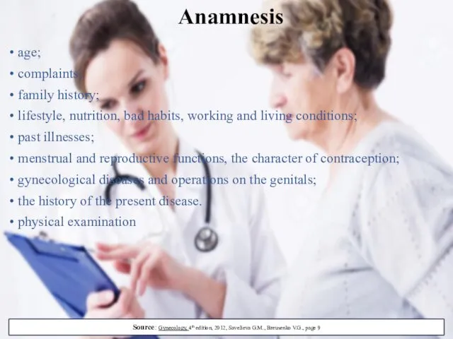
• age;
• complaints;
• family history;
• lifestyle, nutrition, bad habits, working and
• age;
• complaints;
• family history;
• lifestyle, nutrition, bad habits, working and
Amenorrhea - absence of menstruation;
Hypomencastral syndrome is expressed in reduction
Amenorrhea - absence of menstruation;
Hypomencastral syndrome is expressed in reduction
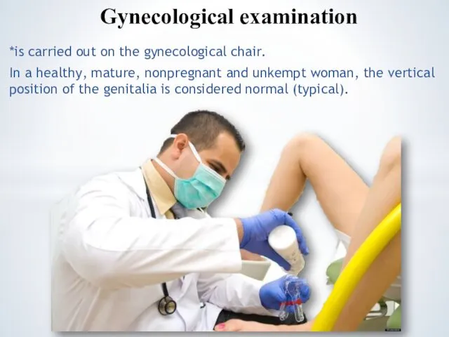
*is carried out on the gynecological chair.
In a healthy, mature,
*is carried out on the gynecological chair.
In a healthy, mature,
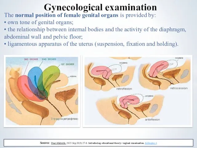
The normal position of female genital organs is provided by:
• own
The normal position of female genital organs is provided by:
• own
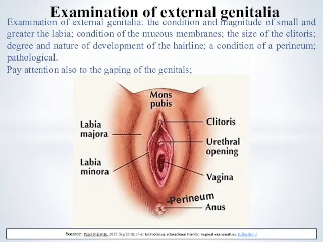
Examination of external genitalia: the condition and magnitude of small and
Examination of external genitalia: the condition and magnitude of small and
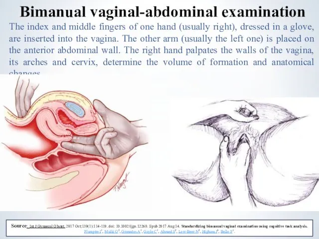
The index and middle fingers of one hand (usually right), dressed
The index and middle fingers of one hand (usually right), dressed
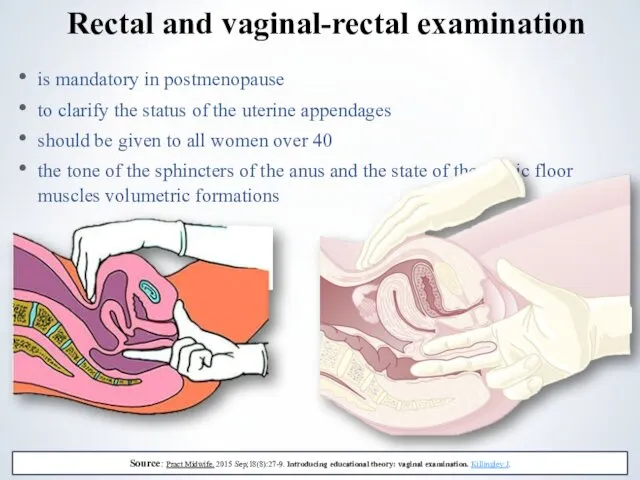
is mandatory in postmenopause
to clarify the status of the uterine appendages
should
is mandatory in postmenopause
to clarify the status of the uterine appendages
should
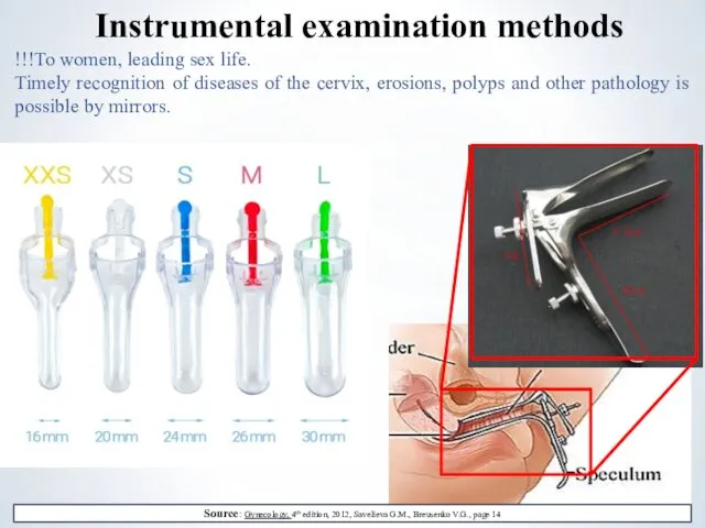
!!!To women, leading sex life.
Timely recognition of diseases of the
!!!To women, leading sex life.
Timely recognition of diseases of the
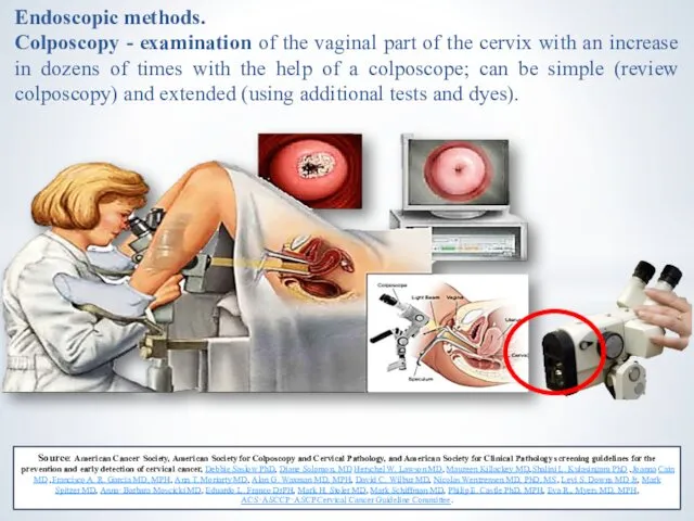
Endoscopic methods.
Colposcopy - examination of the vaginal part of the
Endoscopic methods.
Colposcopy - examination of the vaginal part of the
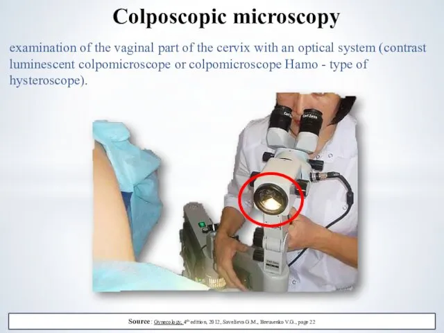
examination of the vaginal part of the cervix with an optical
examination of the vaginal part of the cervix with an optical
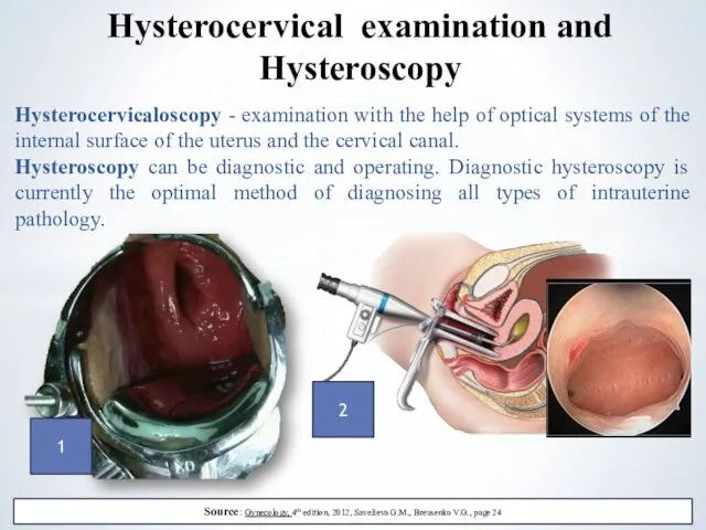
Hysterocervicaloscopy - examination with the help of optical systems of the
Hysterocervicaloscopy - examination with the help of optical systems of the
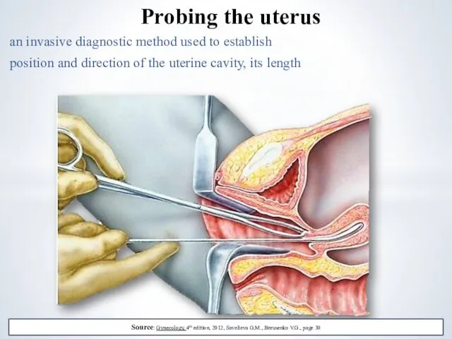
an invasive diagnostic method used to establish
position and direction of the
an invasive diagnostic method used to establish
position and direction of the
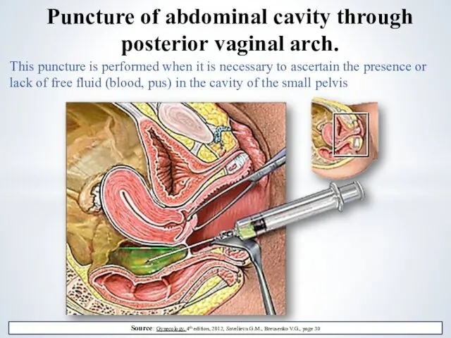
This puncture is performed when it is necessary to ascertain the
This puncture is performed when it is necessary to ascertain the
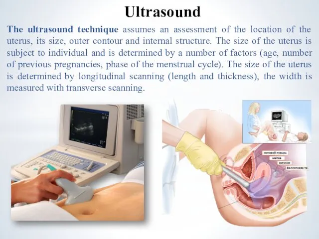
The ultrasound technique assumes an assessment of the location of the
The ultrasound technique assumes an assessment of the location of the
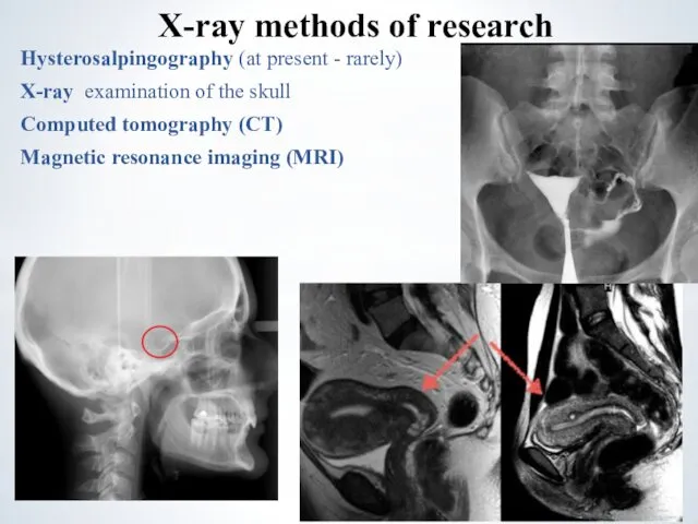
Hysterosalpingography (at present - rarely)
X-ray examination of the skull
Computed tomography
Hysterosalpingography (at present - rarely)
X-ray examination of the skull
Computed tomography
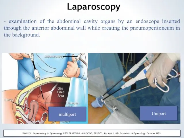
- examination of the abdominal cavity organs by an endoscope inserted
- examination of the abdominal cavity organs by an endoscope inserted
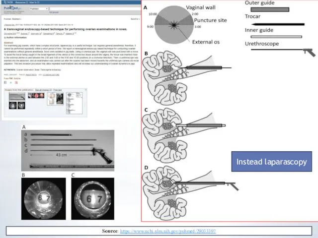
Instead laparascopy
Source: https://www.ncbi.nlm.nih.gov/pubmed/29033397
Instead laparascopy
Source: https://www.ncbi.nlm.nih.gov/pubmed/29033397
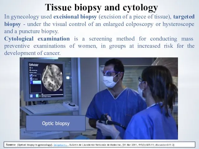
In gynecology used excisional biopsy (excision of a piece of tissue),
In gynecology used excisional biopsy (excision of a piece of tissue),
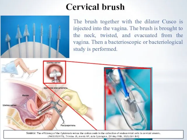
The brush together with the dilator Cusco is injected into the
The brush together with the dilator Cusco is injected into the
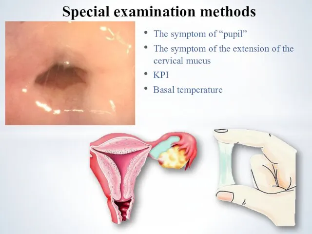
The symptom of “pupil”
The symptom of the extension of the cervical
The symptom of “pupil”
The symptom of the extension of the cervical
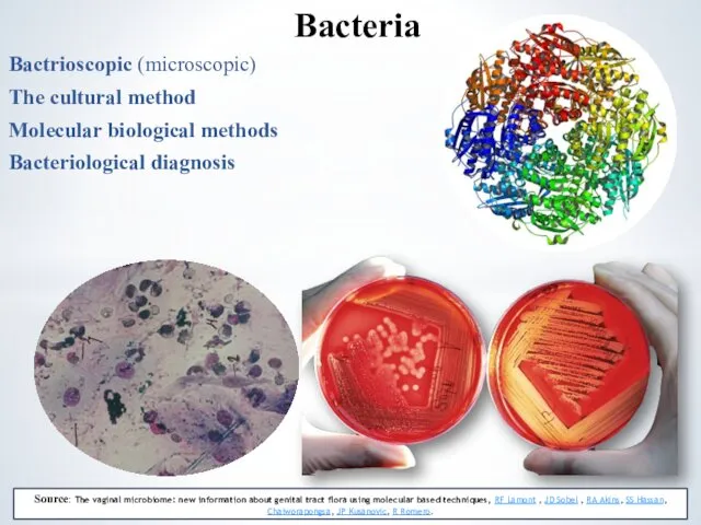
Bactrioscopic (microscopic)
The cultural method
Molecular biological methods
Bacteriological diagnosis
Bacteria
Source: The vaginal microbiome:
Bactrioscopic (microscopic)
The cultural method
Molecular biological methods
Bacteriological diagnosis
Bacteria
Source: The vaginal microbiome:


 Эпидемиология и профилактика ВИЧ-инфекции
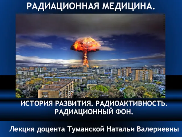 Радиационная медицина. История развития. Радиоактивность. Радиационный фон
 Епідеміологія як наука. Основи епідеміологічного методу дослідження
 Диспансерное наблюдение детей
 Анестезия в России. Опыт применения наркоза
 Заболевания органов дыхания и их профилактика
 Современные методы изготовления бюгельных протезов на огнеупорной модели
 Үлкен жартышар қызметінің асимметриясы
 Рентгеновская семиотика
 Школы больных и центр здоровья
 Основні переваги грудного вигодовування малят
 Синдромы и методы функциональной диагностики при патологии ЖВП и печени
 ВИЧ инфекция. Клиника, лечение, профилактика
 Черепно-мозговая травма. Классификация
 Рак полового члена
 Загальні принципи діагностики і лікування онкологічних хворих
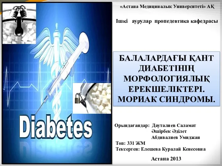 Балалардағы қант диабетінің морфологиялық ерекшеліктері. Мориак синдромы
 Науқасқа, оның отбасына және туыстарына каралы хабарды жеткiзу
 Черный мор (чума)
 Фехтование. Рациональное питание спортсменов
 Лихорадка. Типы лихорадок в зависимости от величины температуры тела
 Gastroenterology. Exam preparation
 Esophageal Cancer
 Лабораторное оборудование
 Иммундық жауаптың нейро эндокринді реттелуі
 zapor_Smagulova_342
 Паратуберкулез. Індеттік ерекшеліктері
 Анемиялық синдром кезінде клиникалық лабараториялық сипаттама