- Radiation methods for studying the organs of the cardiovascular system in childhood

Содержание
- 2. USM The method of choice in the diagnosis of pathological changes in the cardiovascular system in
- 3. The level of the capabilities of modern ultrasonic devices allows to assess the anatomical and functional
- 4. It is possible to perform a non-invasive assessment of the size of the valvular apparatus and
- 5. The main indications for Echocardiography are: - auscultative picture (noise); - complaints of the child for
- 6. The main scanning modes for echocardiography are: - B-mode: visualization of the heart in real time
- 7. EchoCG in B-mode: 1 - cavity of the right ventricle; 2 - pulmonary artery trunk: a
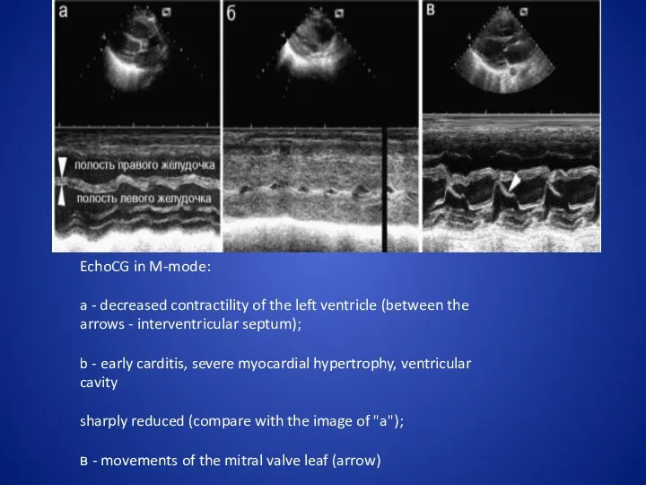
- 8. EchoCG in M-mode: a - decreased contractility of the left ventricle (between the arrows - interventricular
- 9. Echocardiography in the color doppler mode: 1 - the cavity of the left ventricle; 2 -
- 10. Echocardiography in the regime of constant wave doppler (arrows show the components of Doppler curves characterizing
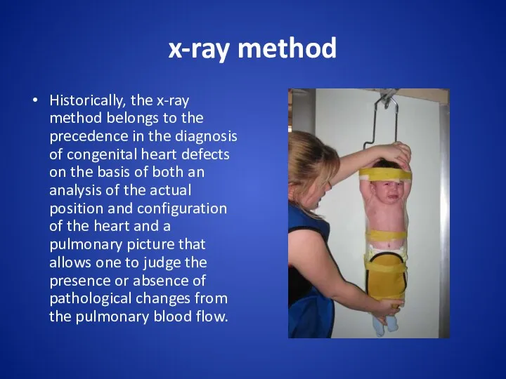
- 11. x-ray method Historically, the x-ray method belongs to the precedence in the diagnosis of congenital heart
- 12. With the review of radiography can be identified only such views that lead to a change
- 13. Normal radiographic anatomy of the heart in a direct projection: a - arches of the heart
- 14. On the right contour, 2 arcs are distinguished: the upper one is formed by the ascending
- 15. To determine the size of the heart cardiothoracic index is used - the ratio of the
- 16. X-ray signs of pulmonary edema in children: a - newborn, arrows show "butterfly wings"; b -
- 17. X-ray signs of heart defects with normovolemia of the small circle of blood circulation: a -
- 18. Angiocardiography Congenital abnormalities of the aortic arch and pulmonary bifurcation may occur in isolation or in
- 19. Левая дуга аорты с аберрантной правой подключичной артерией: а - субтракционная аортограмма с нормальным отхождением правой
- 21. Скачать презентацию

USM
The method of choice in the diagnosis of pathological changes in
USM
The method of choice in the diagnosis of pathological changes in
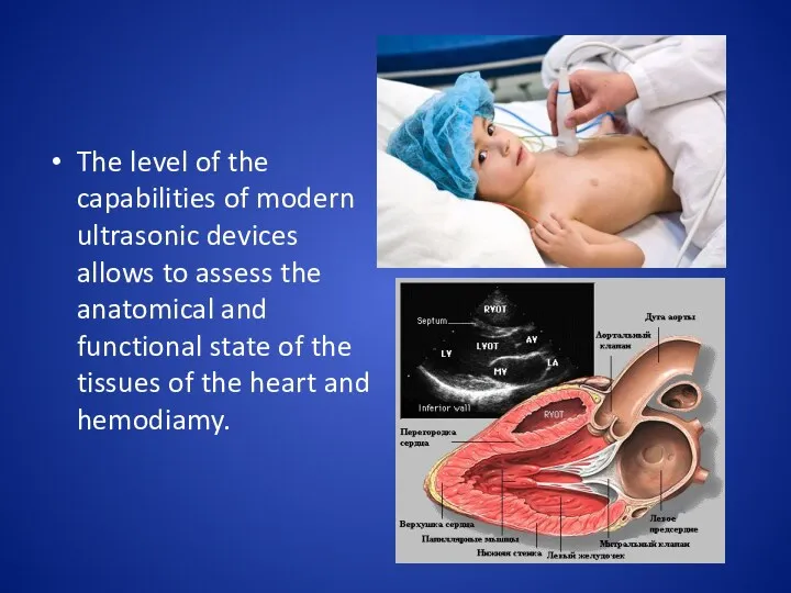
The level of the capabilities of modern ultrasonic devices allows to
The level of the capabilities of modern ultrasonic devices allows to
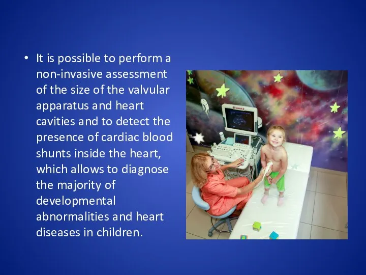
It is possible to perform a non-invasive assessment of the size
It is possible to perform a non-invasive assessment of the size
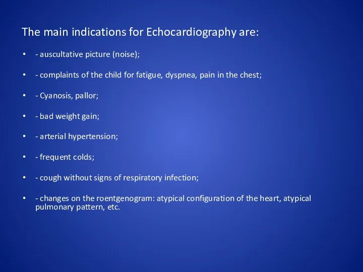
The main indications for Echocardiography are:
- auscultative picture (noise);
- complaints of
The main indications for Echocardiography are:
- auscultative picture (noise);
- complaints of
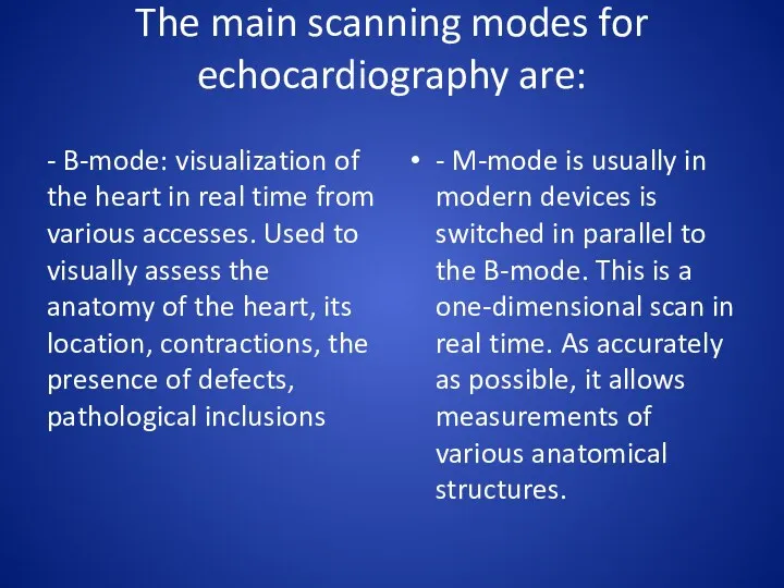
The main scanning modes for echocardiography are:
- B-mode: visualization of the
The main scanning modes for echocardiography are:
- B-mode: visualization of the
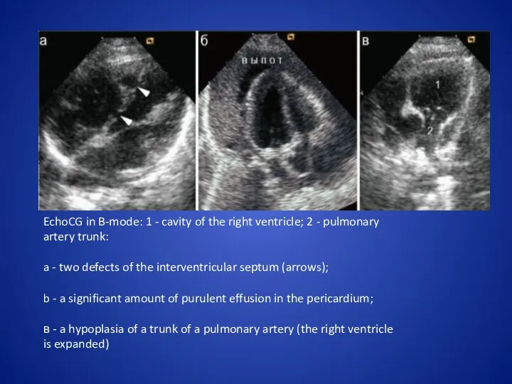
EchoCG in B-mode: 1 - cavity of the right ventricle; 2
EchoCG in B-mode: 1 - cavity of the right ventricle; 2
EchoCG in M-mode:
a - decreased contractility of the left ventricle (between
EchoCG in M-mode:
a - decreased contractility of the left ventricle (between
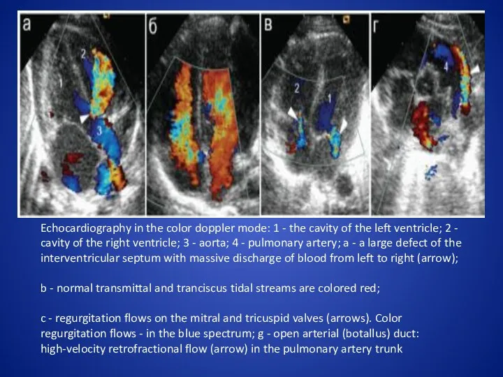
Echocardiography in the color doppler mode: 1 - the cavity of
Echocardiography in the color doppler mode: 1 - the cavity of
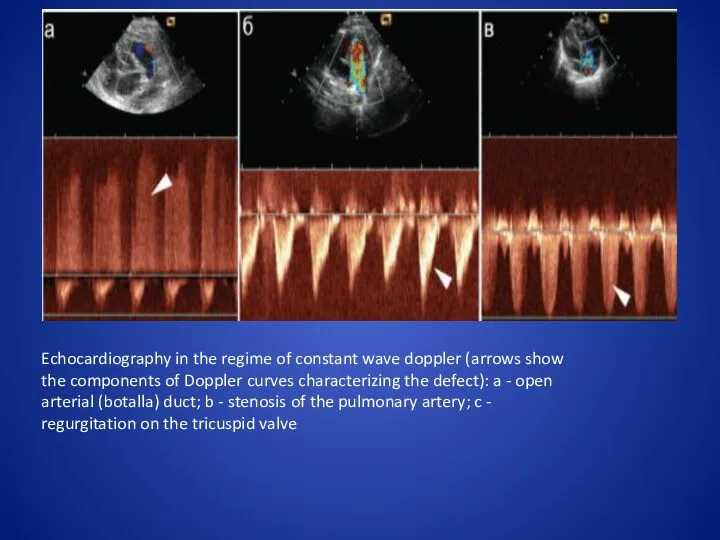
Echocardiography in the regime of constant wave doppler (arrows show the
Echocardiography in the regime of constant wave doppler (arrows show the
x-ray method
Historically, the x-ray method belongs to the precedence in the
x-ray method
Historically, the x-ray method belongs to the precedence in the
With the review of radiography can be identified only such views
With the review of radiography can be identified only such views
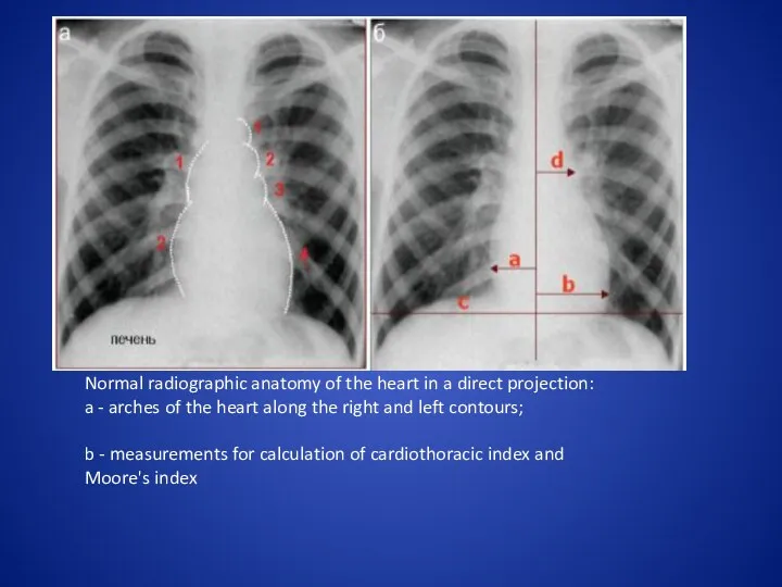
Normal radiographic anatomy of the heart in a direct projection:
a
Normal radiographic anatomy of the heart in a direct projection:
a
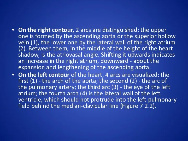
On the right contour, 2 arcs are distinguished: the upper one
On the right contour, 2 arcs are distinguished: the upper one
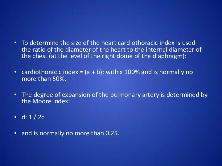
To determine the size of the heart cardiothoracic index is used
To determine the size of the heart cardiothoracic index is used
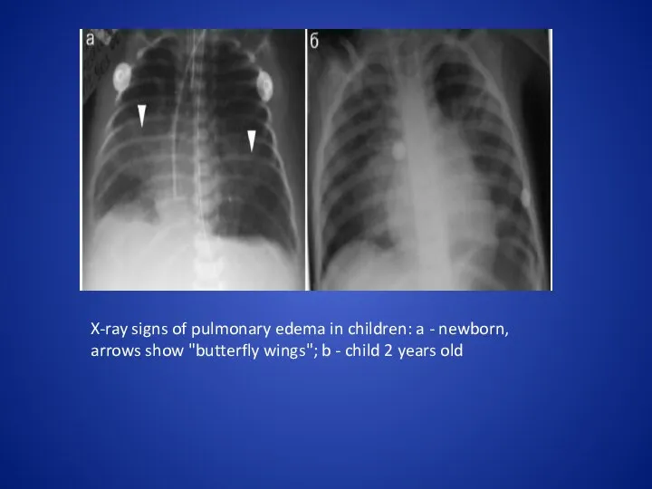
X-ray signs of pulmonary edema in children: a - newborn, arrows
X-ray signs of pulmonary edema in children: a - newborn, arrows
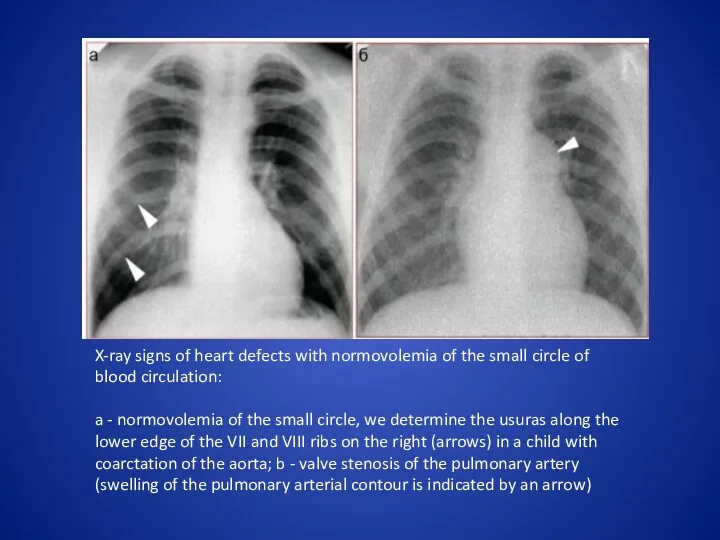
X-ray signs of heart defects with normovolemia of the small circle
X-ray signs of heart defects with normovolemia of the small circle
Angiocardiography
Congenital abnormalities of the aortic arch and pulmonary bifurcation may occur
Angiocardiography
Congenital abnormalities of the aortic arch and pulmonary bifurcation may occur
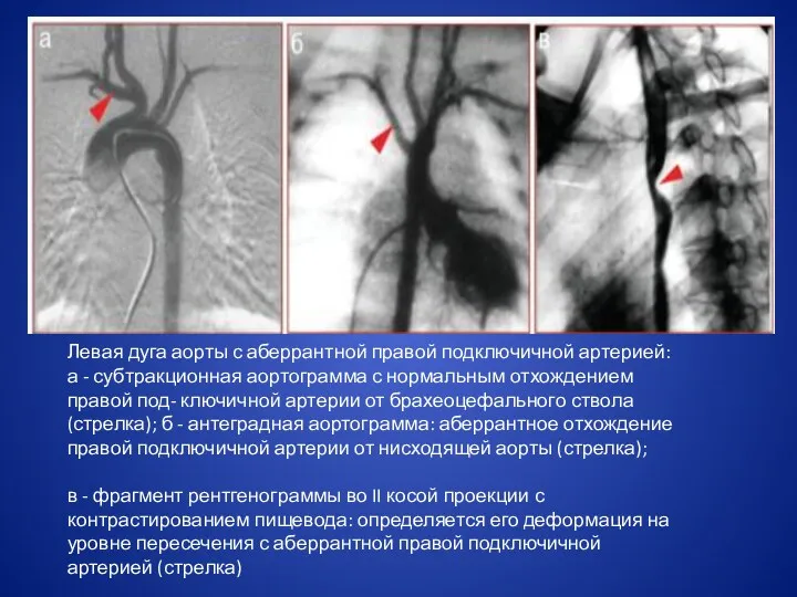
Левая дуга аорты с аберрантной правой подключичной артерией: а - субтракционная
Левая дуга аорты с аберрантной правой подключичной артерией: а - субтракционная
 Рак желудка
 Технология гомеопатических таблеток. Викторина
 Художественное творчество больных шизофренией и эпилепсией
 Эпителиальные злокачественные опухоли поджелудочной железы
 Вибрационная болезнь
 Государственная система управления здравоохранением. Современные формы управления в системе здравоохранения
 Анатомія нервової системи
 Health and safety legislation
 Алынбайтын протездер
 Заболевания периферической нервной системы
 Здоровое питание
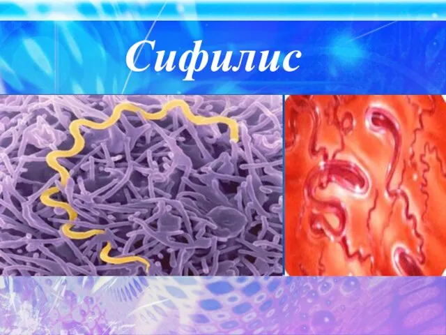 Сифилис
 Атипичное удаление зубов
 Кровотечение
 Витамин Д зависимый рахит, витамин Д резистентный рахит
 Концепция охраны репродуктивного здоровья и активного социального долголетия
 ЭКГ при электролитных нарушениях в организме
 Ожирение среди подростков. Ожирение - хроническое заболевание
 Patologik anatomiya mavzu: distrofiya
 Ауески ауруы
 Заболевания вен, нижних конечностей
 Преимущество естественного вскармливания перед искусственным
 Физика в моей будущей профессии
 Анемия. Классификация анемий
 Фармакогенетика негіздері. Дәрілік препараттағы ағзаның тұқым қуалау негізделген полимарфизм реакциясы
 Эритроциттегі глюкоза-6-фосфатдегидрогеназа ферменті тапшылығына байланысты туындайтын гемолиздік анемия
 Острая почечная недостaточность
 Коленный сустав. Биомеханика, физиология