- RBC’s count by haemocytometer

Содержание
- 2. 1- RBC’s count by haemocytometer Aim: The number of RBC’s is counted by haemocytometer in a
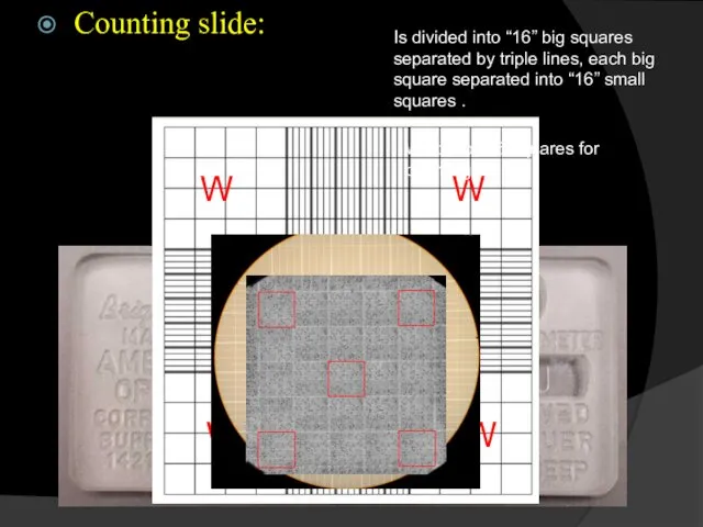
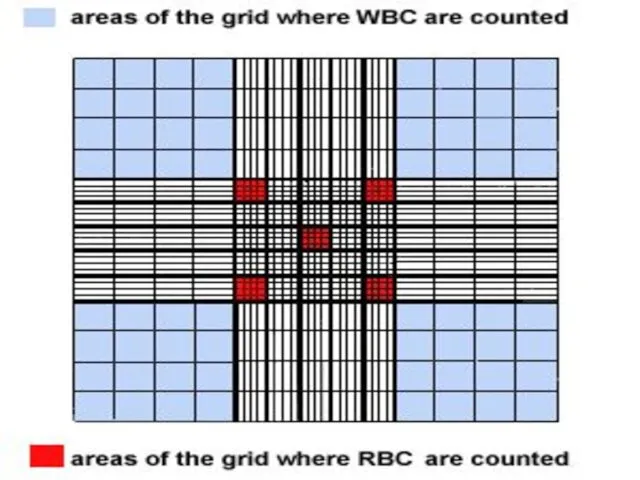
- 3. Counting slide: Is divided into “16” big squares separated by triple lines, each big square separated
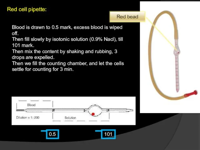
- 4. Red cell pipette: 0.5 101 Blood is drawn to 0.5 mark, excess blood is wiped off.
- 5. Shake well to mix with the hose end sealed with your finger.
- 6. Unmixed cell free fluid in the capillary portion of the pipette Empty 2-3 drops off pipette
- 8. Carefully adjust the haemocytometer on the microscope and cover
- 9. Add a small amount of the diluted RBCs to just fill the first chamber of the
- 10. It should flow in to fill the chamber by capillary action. Do not over fill. Notice
- 11. To improve your skill, repeat the dilution a second time and fill the second chamber. The
- 14. Counting chamber It is the space formed between the cover placed on the counting slide and
- 15. Calculations
- 16. Calculations: No, of RBC’s / small square = Volume of small square = No, of RBC’s
- 17. Normal value: ♂ = 4.8 -5.6 million cell/ mm3 ♀ = 4.6 – 5.2 million cell
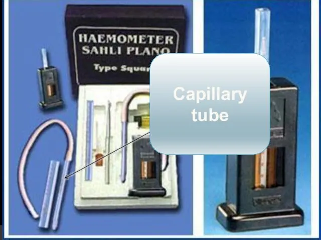
- 18. 2- colorimetric determination of “Hb” by haemometer Aim: Determination of amount of “Hb” by change in
- 19. Principle: The haemolysis of RBC’s by using the acid “HCl” to get a free “Hb” in
- 20. Haemometer: Consists of: “2” standard colored tubes Graduated tube Capillary tube
- 21. Standard coloured tubes
- 22. Graduated tube
- 23. Capillary tube
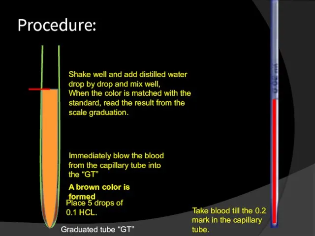
- 24. Procedure: Graduated tube “GT” Place 5 drops of 0.1 HCL. Take blood till the 0.2 mark
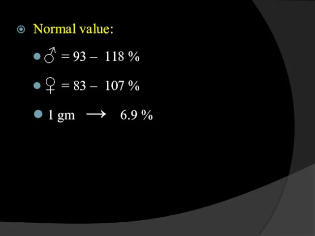
- 25. Normal value: ♂ = 93 – 118 % ♀ = 83 – 107 % 1 gm
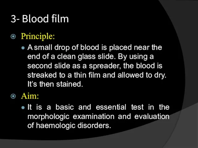
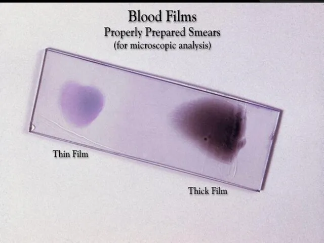
- 26. 3- Blood film Principle: A small drop of blood is placed near the end of a
- 27. Method: A finger puncture in made, and a small drop of blood is placed on the
- 28. The thickness of the film can be varied by:- the spread with which the slide is
- 29. Speed Angle Thinner blood film
- 31. Stains used: Leishman Stain : and it consists of Methylene blue : It stains nuclear DNA
- 32. Examination of the Blood film: Evaluation of RBC’s Evaluation of platelets Differentail leucocytic count
- 33. GRANULOCYTES
- 34. AGRANULOCYTES
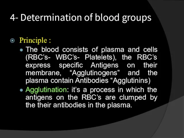
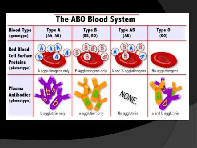
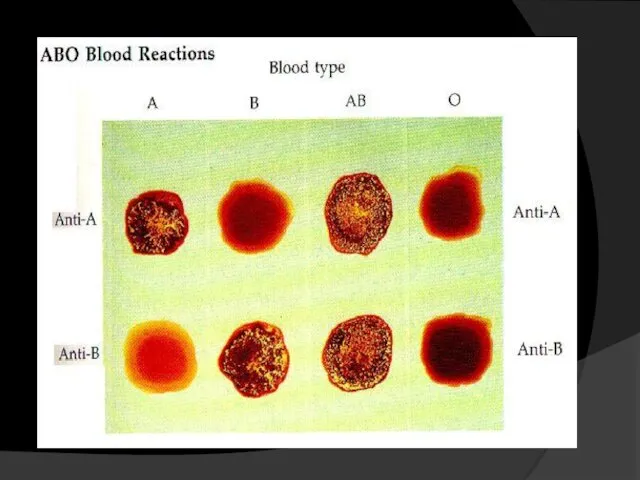
- 35. 4- Determination of blood groups Principle : The blood consists of plasma and cells (RBC’s- WBC’s-
- 37. This diagram shows the possible ways of blood transfusion without causing agglutination to the blood: O:
- 38. Preparation of the slide: Add a drop of antibody A “blue” Add a drop of antibody
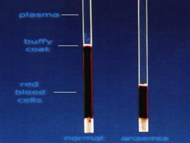
- 40. Haematocrite value
- 41. Define:- hematocrit (Ht ), also known as packed cell volume (PCV) or erythrocyte volume fraction (EVF),
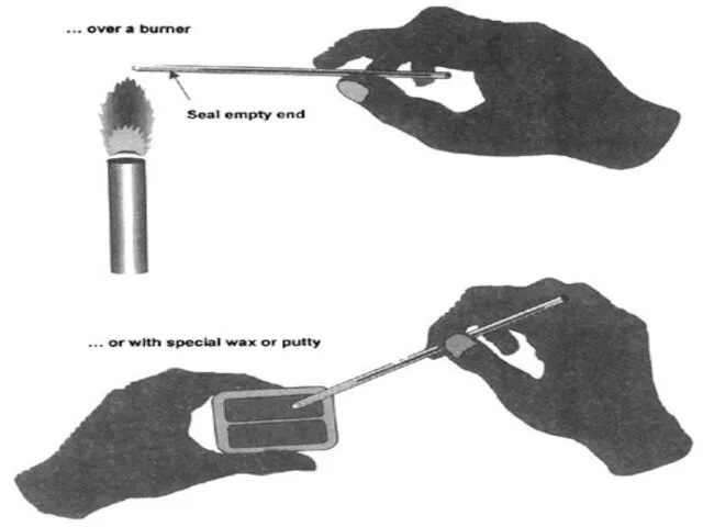
- 42. How to calculate Ht/PCV? The packed cell volume (PCV) can be determined by centrifugingThe packed cell
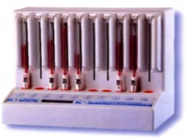
- 49. Rate of sedimentation (ESR=Erythrocyte sedimentation rate)
- 50. Define:- Rate of sedimentation is the rate at which red blood cells sediment in a period
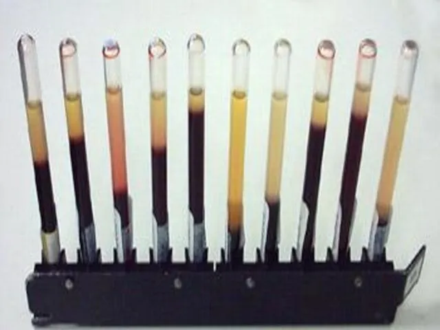
- 51. How To perform the test? Anticoagulated blood is placed in an upright tube, known as a
- 54. Depend on gravitational force Depend on centrifugal force ESR Ht
- 56. Скачать презентацию
1- RBC’s count by haemocytometer
Aim:
The number of RBC’s is counted by
1- RBC’s count by haemocytometer
Aim:
The number of RBC’s is counted by
Counting slide:
Is divided into “16” big squares separated by triple lines,
Counting slide:
Is divided into “16” big squares separated by triple lines,
Red cell pipette:
0.5
101
Blood is drawn to 0.5 mark, excess blood is
Red cell pipette:
0.5
101
Blood is drawn to 0.5 mark, excess blood is
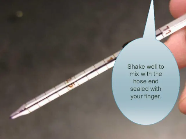
Shake well to mix with the hose end sealed with your
Shake well to mix with the hose end sealed with your
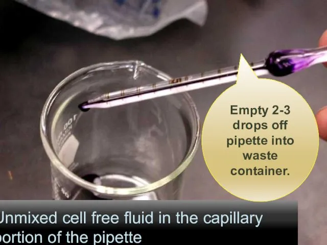
Unmixed cell free fluid in the capillary portion of the pipette
Unmixed cell free fluid in the capillary portion of the pipette
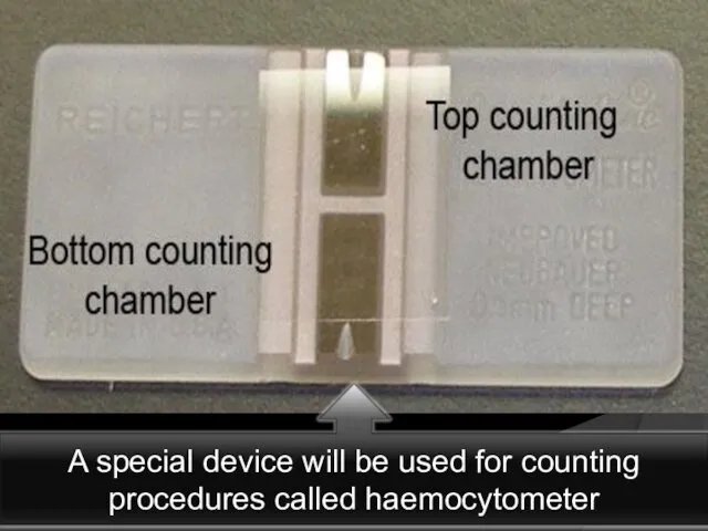
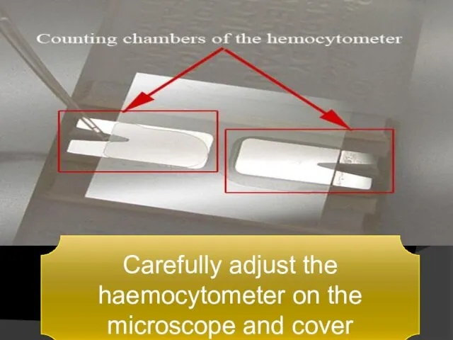
Carefully adjust the haemocytometer on the microscope and cover
Carefully adjust the haemocytometer on the microscope and cover
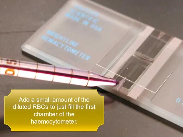
Add a small amount of the diluted RBCs to just fill
Add a small amount of the diluted RBCs to just fill
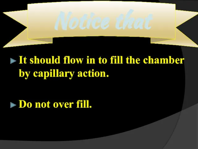
It should flow in to fill the chamber by capillary action.
Do
It should flow in to fill the chamber by capillary action.
Do
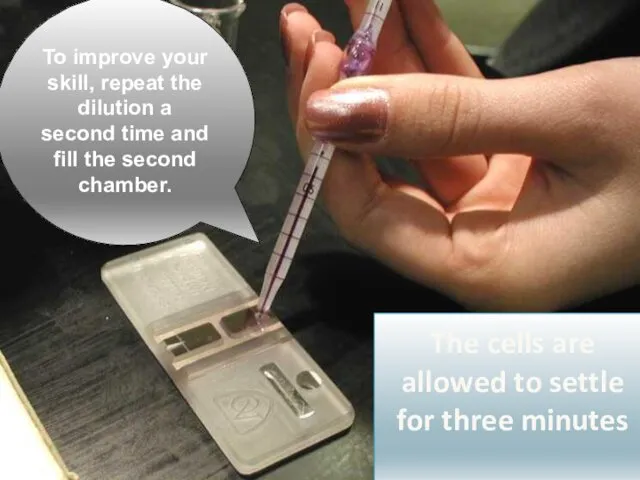
To improve your skill, repeat the dilution a second time and
To improve your skill, repeat the dilution a second time and
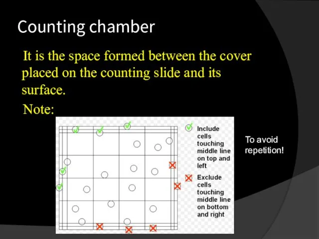
Counting chamber
It is the space formed between the cover placed on
Counting chamber
It is the space formed between the cover placed on

Calculations
Calculations
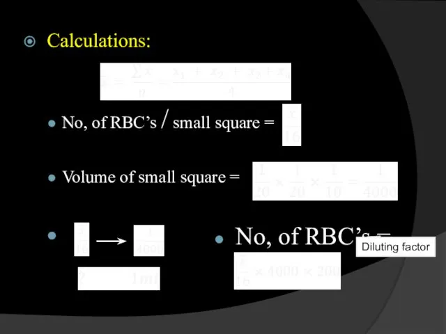
Calculations:
No, of RBC’s / small square =
Volume of small square =
Calculations:
No, of RBC’s / small square =
Volume of small square =
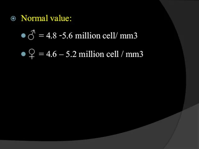
Normal value:
♂ = 4.8 -5.6 million cell/ mm3
♀ = 4.6 –
Normal value:
♂ = 4.8 -5.6 million cell/ mm3
♀ = 4.6 –
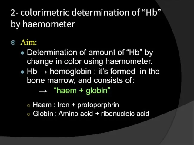
2- colorimetric determination of “Hb” by haemometer
Aim:
Determination of amount of
2- colorimetric determination of “Hb” by haemometer
Aim:
Determination of amount of
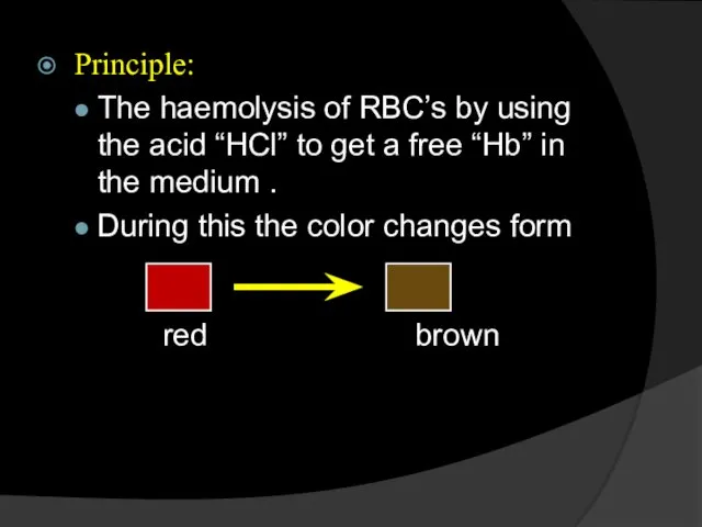
Principle:
The haemolysis of RBC’s by using the acid “HCl” to get
Principle:
The haemolysis of RBC’s by using the acid “HCl” to get
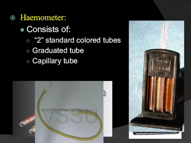
Haemometer:
Consists of:
“2” standard colored tubes
Graduated tube
Capillary tube
Haemometer:
Consists of:
“2” standard colored tubes
Graduated tube
Capillary tube
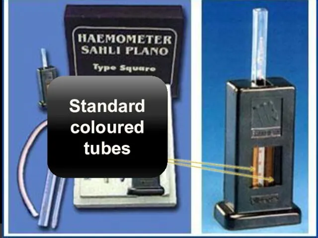
Standard coloured tubes
Standard coloured tubes
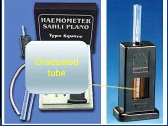
Graduated tube
Graduated tube
Capillary tube
Capillary tube
Procedure:
Graduated tube “GT”
Place 5 drops of 0.1 HCL.
Take blood till
Procedure:
Graduated tube “GT”
Place 5 drops of 0.1 HCL.
Take blood till
Normal value:
♂ = 93 – 118 %
♀ = 83 – 107
Normal value:
♂ = 93 – 118 %
♀ = 83 – 107
3- Blood film
Principle:
A small drop of blood is placed near
3- Blood film
Principle:
A small drop of blood is placed near
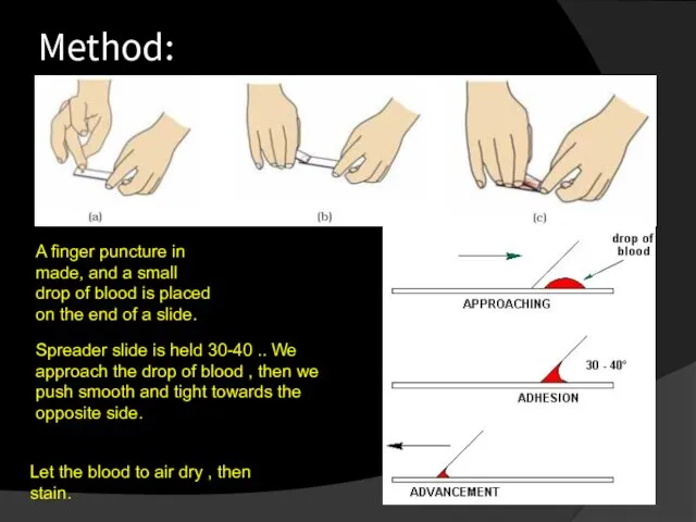
Method:
A finger puncture in made, and a small drop of blood
Method:
A finger puncture in made, and a small drop of blood
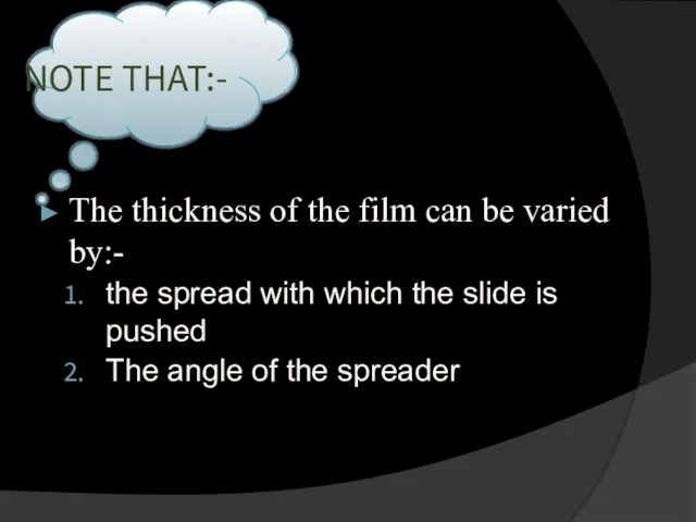
The thickness of the film can be varied by:-
the spread
The thickness of the film can be varied by:-
the spread
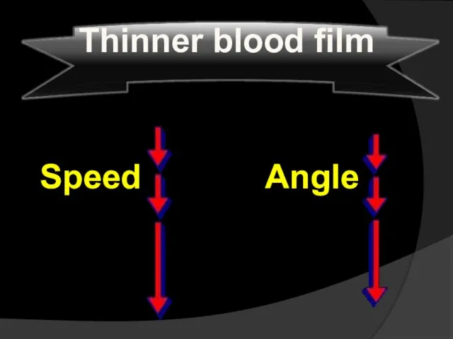
Speed
Angle
Thinner blood film
Speed
Angle
Thinner blood film
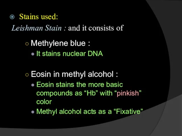
Stains used:
Leishman Stain : and it consists of
Methylene blue :
It
Stains used:
Leishman Stain : and it consists of
Methylene blue :
It
Examination of the Blood film:
Evaluation of RBC’s
Evaluation of platelets
Differentail leucocytic count
Examination of the Blood film:
Evaluation of RBC’s
Evaluation of platelets
Differentail leucocytic count
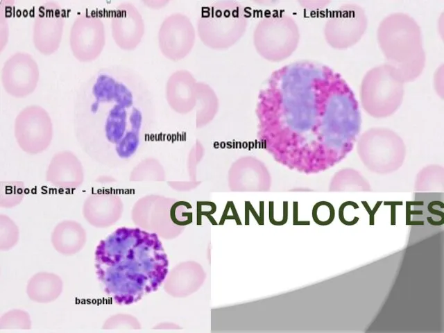
GRANULOCYTES
GRANULOCYTES
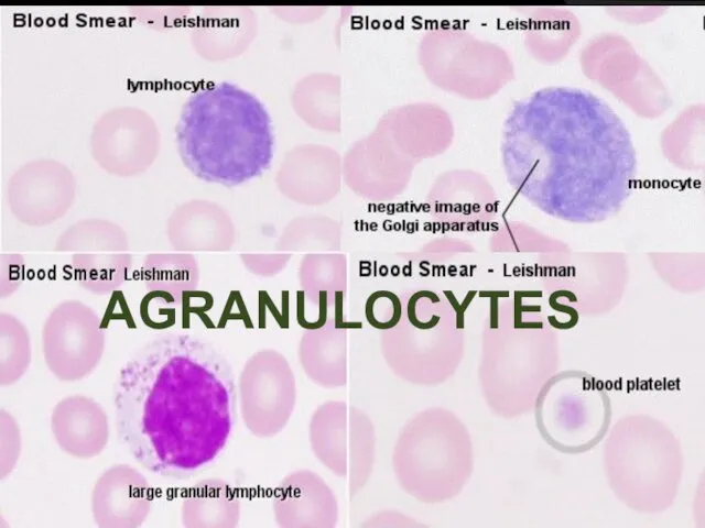
AGRANULOCYTES
AGRANULOCYTES
4- Determination of blood groups
Principle :
The blood consists of plasma and
4- Determination of blood groups
Principle :
The blood consists of plasma and
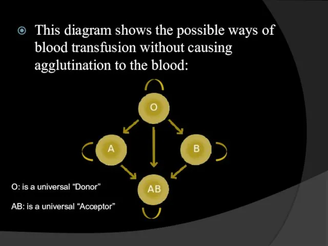
This diagram shows the possible ways of blood transfusion without causing
This diagram shows the possible ways of blood transfusion without causing
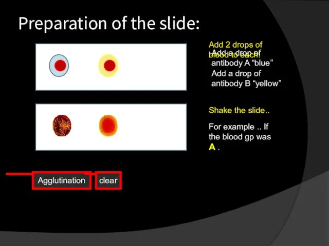
Preparation of the slide:
Add a drop of antibody A “blue”
Add a
Preparation of the slide:
Add a drop of antibody A “blue”
Add a

Haematocrite value
Haematocrite value
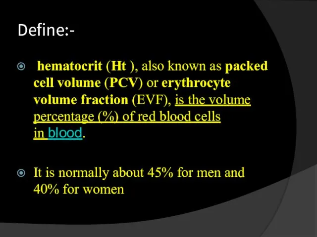
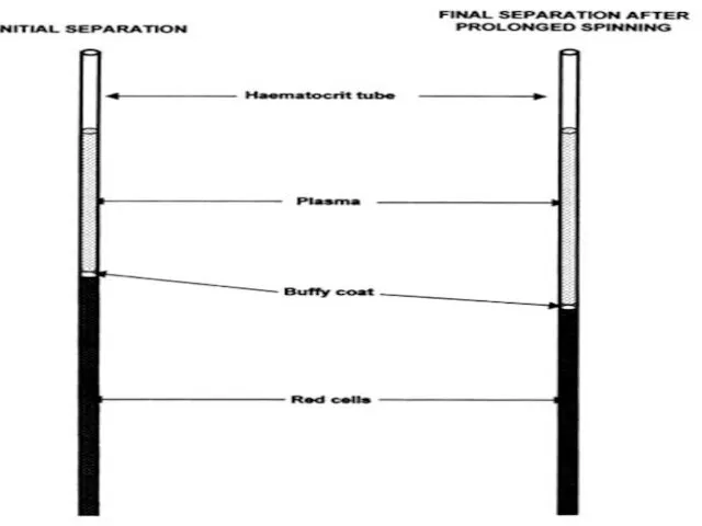
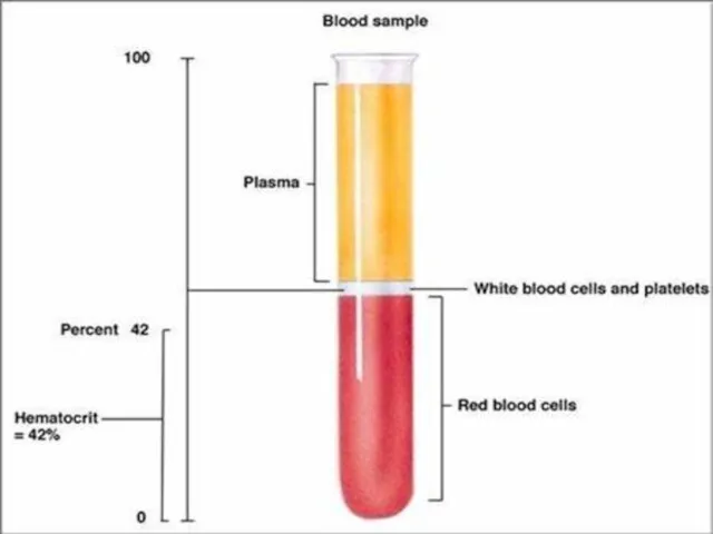
Define:-
hematocrit (Ht ), also known as packed cell volume (PCV) or erythrocyte volume fraction (EVF), is the
Define:-
hematocrit (Ht ), also known as packed cell volume (PCV) or erythrocyte volume fraction (EVF), is the
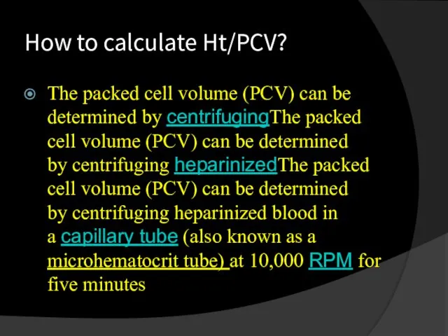
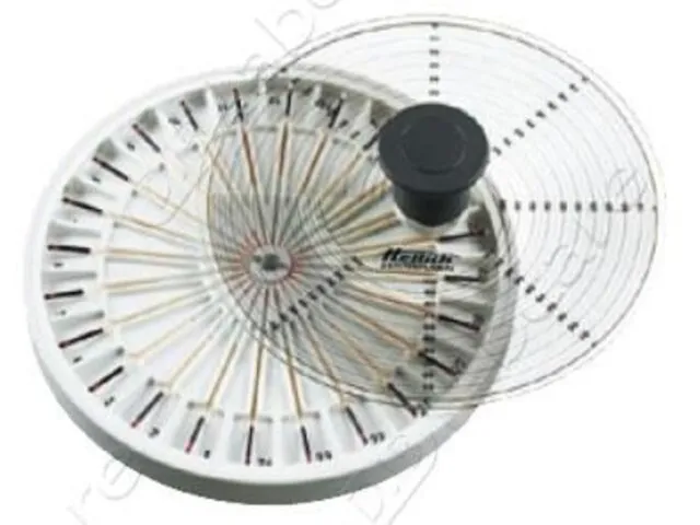
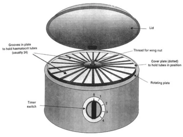
How to calculate Ht/PCV?
The packed cell volume (PCV) can be determined
How to calculate Ht/PCV?
The packed cell volume (PCV) can be determined

Rate of sedimentation
(ESR=Erythrocyte sedimentation rate)
Rate of sedimentation
(ESR=Erythrocyte sedimentation rate)
Define:-
Rate of sedimentation is the rate at which red blood cells sediment in
Define:-
Rate of sedimentation is the rate at which red blood cells sediment in
How To perform the test?
Anticoagulated blood is placed in an upright tube, known
How To perform the test?
Anticoagulated blood is placed in an upright tube, known
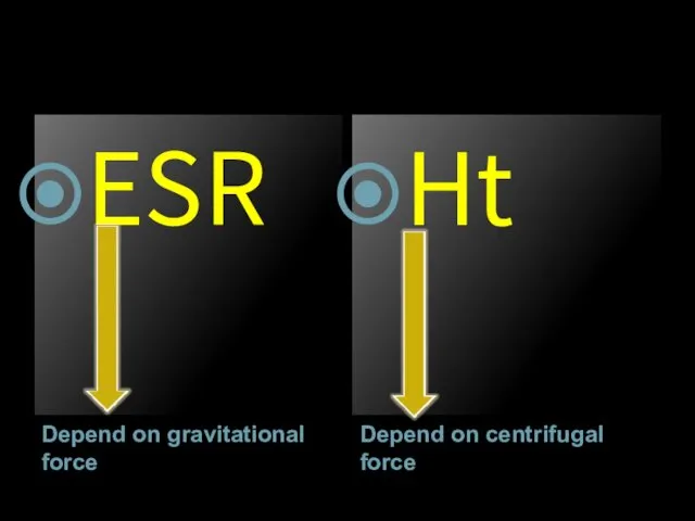
Depend on gravitational force
Depend on centrifugal force
ESR
Ht
Depend on gravitational force
Depend on centrifugal force
ESR
Ht
Похожие презентации
 Кардиогенный шок. Дефиниции: термины и понятия
 Жанұя денсаулығы. Бала денсаулығы
 Подходы к организации логопедической работы с детьми при нарушении зрения
 Пищевая аллергия
 Балалардағы ішек дисбактериозы
 Профилактика внутрибольничной инфекции
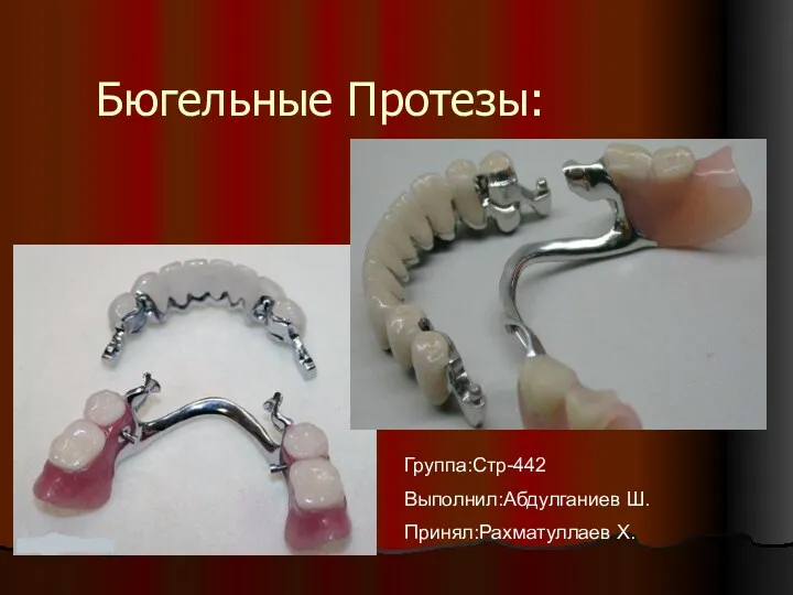 Бюгельные протезы
 Нурофен для детей
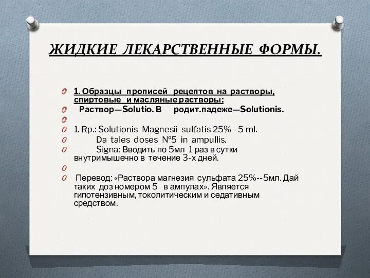 Жидкие лекарственные формы
 Стероид резистентті нефротикалық синдром
 Становление естественнонаучных знаний в эпоху Возрождения
 Перикардит. Перикардиттің даму себептері мен жіктелісі
 Аборт – экстренная контрацепция или убийство?
 СУ при кровотечениях
 Ақшаның мәні және атқаратын қызметтері
 Адам ағзасындағы фотохимиялық түрленулер
 Хронические гепатиты у детей
 Қан құюдан кейінгі асқынулар. Уақытында диагностикалау. Емдеу тактикасы
 Рак шейки матки (клиника, диагностика, лечение)
 Инфекциялық үрдістің патофизиологиясы
 Психология пациента
 Вирус ГРИППа
 Pacienta komforta nodrošinājums un pacienta drošība
 История, предмет и задачи гигиены и экологии человека
 Чистые руки
 Вторичный период сифилиса. Скрытый сифилис. Серологическая диагностика сифилиса
 Хроническая сердечная недостаточность