- Respiratory system power point

Содержание
- 2. Moving air to the exchange surface of the lungs Gas exchange between air and circulating blood
- 3. The Components of the Respiratory System
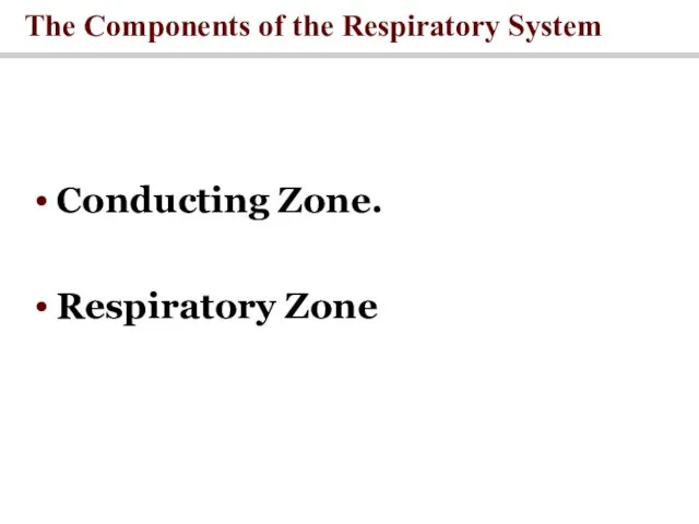
- 4. The Components of the Respiratory System Conducting Zone. Respiratory Zone
- 5. All the structures air passes through before reaching the respiratory zone. Function: Warms and humidifies inspired
- 6. Region of gas exchange between air and blood. Includes respiratory bronchioles and alveolar sacs. Respiratory Zone
- 7. The Bronchi and Lobules of the Lung
- 8. Respiratory Membrane
- 9. Respiratory Membrane
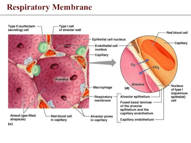
- 10. Respiratory Membrane This air-blood barrier is composed of: Alveolar and capillary walls Their fused basal laminas
- 11. Respiratory Volumes Tidal volume (TV) – air that moves into and out of the lungs with
- 12. Respiratory Capacities Inspiratory capacity (IC) – total amount of air that can be inspired after a
- 13. Respiratory Volumes and Capacities
- 14. Dead Space The volume of the airways that does not participate in gas exchange Anatomical dead
- 15. Mechanics of Breathing
- 16. The physical movement of air into and out of the lungs Pulmonary Ventilation
- 17. Movement of air depends upon Boyle’s Law Pressure and volume inverse relationship Volume depends on movement
- 18. Inspiration Inspiration Diaphragm contracts -> increased thoracic volume vertically. Intercostals contract, expanding rib cage -> increased
- 19. Expiration Expiration Due to recoil of elastic lungs. Passive. Less volume -> pressure within alveoli is
- 20. Mechanisms of Pulmonary Ventilation
- 21. Gas Exchange
- 22. Daltons Law and partial pressure Individual gases in a mixture exert pressure proportional to their abundance
- 23. Henry’s Law and the Relationship between Solubility and Pressure
- 24. Gas exchange across respiratory membrane is efficient due to: Differences in partial pressure Small diffusion distance
- 25. Gas Pickup and Delivery
- 26. An Overview of Respiratory Processes and Partial Pressures in Respiration
- 27. Gas Exchange in the Lungs and Tissues: Oxygen
- 28. Gas Transport in the Blood: Oxygen 2% in plasma 98% in hemoglobin (Hb) Blood holds O2
- 29. Carried mainly by RBCs, bound to hemoglobin The amount of oxygen hemoglobin can carried is dependent
- 30. Hemoglobin Transport of Oxygen 4 binding sites per Hb molecule 98% saturated in alveolar arteries Resting
- 31. Hemoglobin Saturation Curve
- 32. Temperature, pH, PCO2, and DPG Increase of temperature, PCO2, and DPG and decrease of pH :
- 33. The Effect of pH and Temperature on Hemoglobin Saturation
- 34. A Functional Comparison of Fetal and Adult Hemoglobin
- 35. 7% dissolved in plasma 70% carried as carbonic acid buffer system 23% bound to hemoglobin carbaminohemoglobin
- 36. Carbon Dioxide Transport in Blood
- 37. Driven by differences in partial pressure Oxygen enters blood at lungs and leaves at tissues Carbon
- 38. A Summary of the Primary Gas Transport Mechanisms
- 39. Control of Respiration
- 40. Medullary centers Respiratory rhythmicity centers set pace Dorsal respiratory group (DRG)– inspiration Ventral respiratory group (VRG)–
- 41. Pons Apneustic and pneumotaxic centers: ● regulate the respiratory rate and the depth of respiration in
- 42. Respiratory Centers and Reflex Controls
- 43. Chemoreceptors Chemoreceptors are located throughout the body (in brain and arteries). chemoreceptors are more sensitive to
- 45. Скачать презентацию
Moving air to the exchange surface of the lungs
Gas exchange
Moving air to the exchange surface of the lungs
Gas exchange
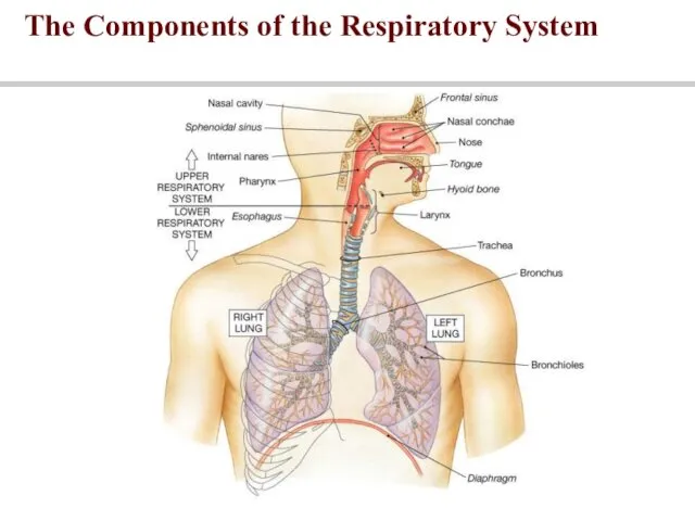
The Components of the Respiratory System
The Components of the Respiratory System
The Components of the Respiratory System
Conducting Zone.
Respiratory Zone
The Components of the Respiratory System
Conducting Zone.
Respiratory Zone
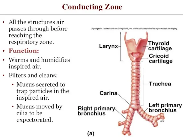
All the structures air passes through before reaching the respiratory zone.
Function:
Warms
All the structures air passes through before reaching the respiratory zone.
Function:
Warms
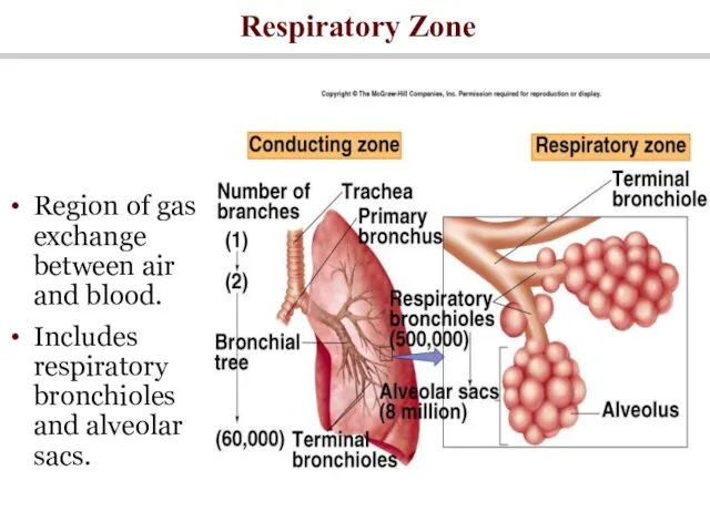
Region of gas exchange between air and blood.
Includes respiratory bronchioles and
Region of gas exchange between air and blood.
Includes respiratory bronchioles and
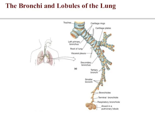
The Bronchi and Lobules of the Lung
The Bronchi and Lobules of the Lung
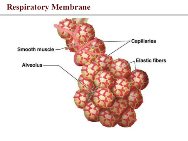
Respiratory Membrane
Respiratory Membrane
Respiratory Membrane
Respiratory Membrane
Respiratory Membrane
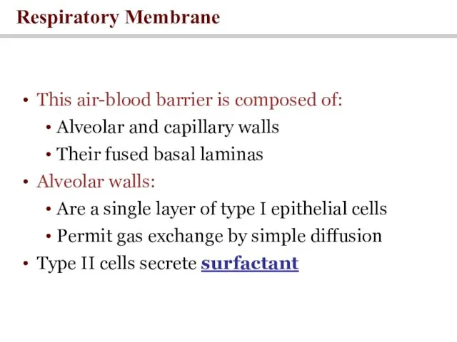
This air-blood barrier is composed of:
Alveolar and capillary walls
Their
Respiratory Membrane
This air-blood barrier is composed of:
Alveolar and capillary walls
Their
Respiratory Volumes
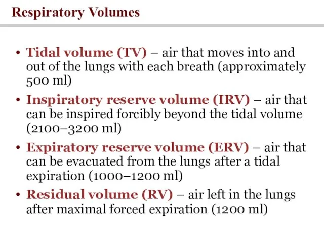
Tidal volume (TV) – air that moves into and out
Respiratory Volumes
Tidal volume (TV) – air that moves into and out
Respiratory Capacities
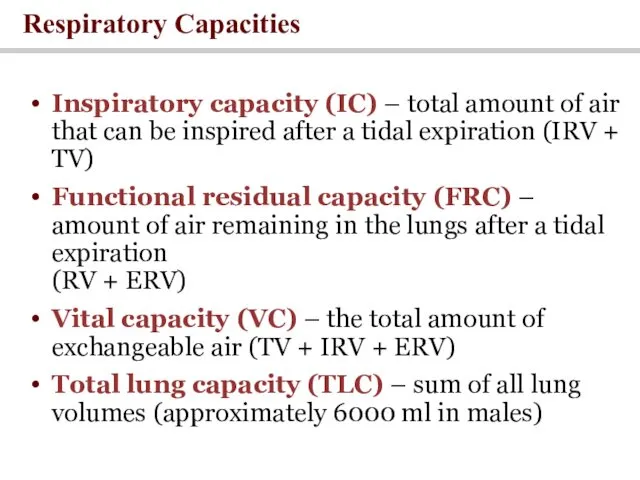
Inspiratory capacity (IC) – total amount of air that can
Respiratory Capacities
Inspiratory capacity (IC) – total amount of air that can
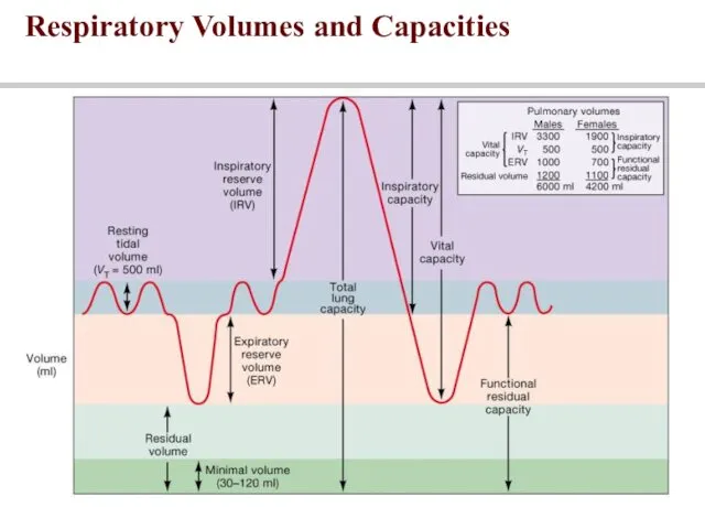
Respiratory Volumes and Capacities
Respiratory Volumes and Capacities
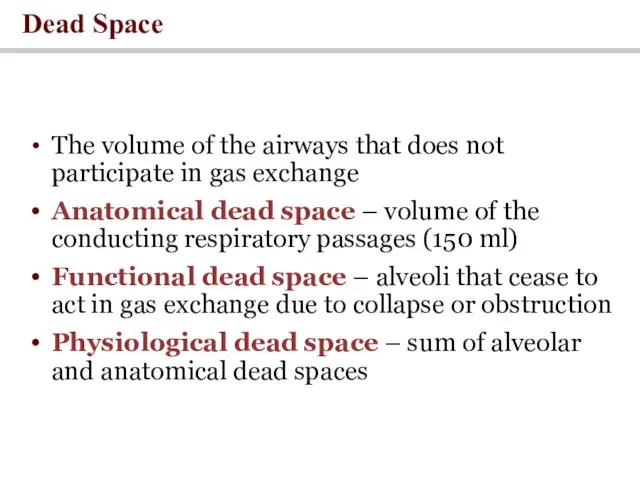
Dead Space
The volume of the airways that does not participate in
Dead Space
The volume of the airways that does not participate in
Mechanics of Breathing
Mechanics of Breathing

The physical movement of air into and out of the lungs
Pulmonary
The physical movement of air into and out of the lungs
Pulmonary
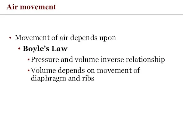
Movement of air depends upon
Boyle’s Law
Pressure and volume inverse relationship
Volume depends
Movement of air depends upon
Boyle’s Law
Pressure and volume inverse relationship
Volume depends
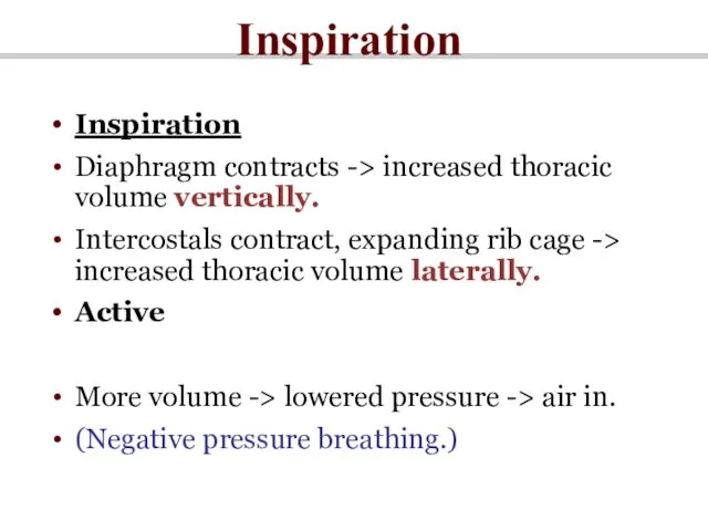
Inspiration
Inspiration
Diaphragm contracts -> increased thoracic volume vertically.
Intercostals contract, expanding rib cage
Inspiration
Inspiration
Diaphragm contracts -> increased thoracic volume vertically.
Intercostals contract, expanding rib cage
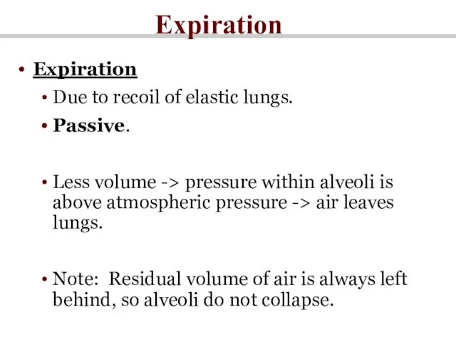
Expiration
Expiration
Due to recoil of elastic lungs.
Passive.
Less volume -> pressure within alveoli
Expiration
Expiration
Due to recoil of elastic lungs.
Passive.
Less volume -> pressure within alveoli
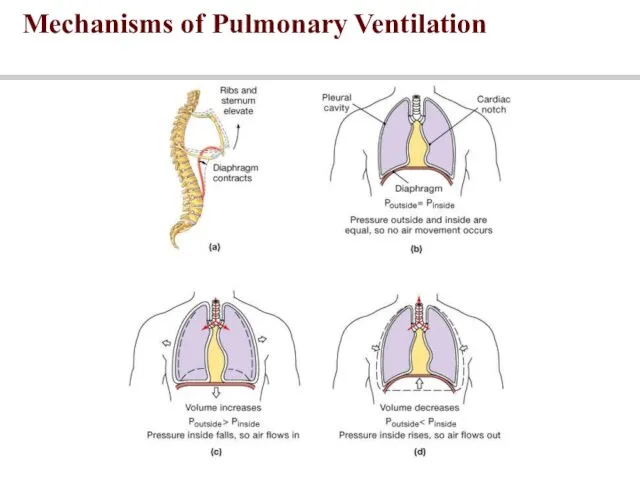
Mechanisms of Pulmonary Ventilation
Mechanisms of Pulmonary Ventilation
Gas Exchange
Gas Exchange
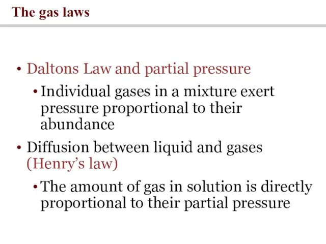
Daltons Law and partial pressure
Individual gases in a mixture exert pressure
Daltons Law and partial pressure
Individual gases in a mixture exert pressure
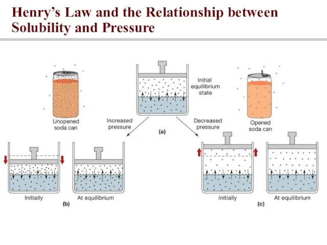
Henry’s Law and the Relationship between Solubility and Pressure
Henry’s Law and the Relationship between Solubility and Pressure
Gas exchange across respiratory membrane is efficient due to:
Differences in partial
Gas exchange across respiratory membrane is efficient due to:
Differences in partial
Gas Pickup and Delivery
Gas Pickup and Delivery
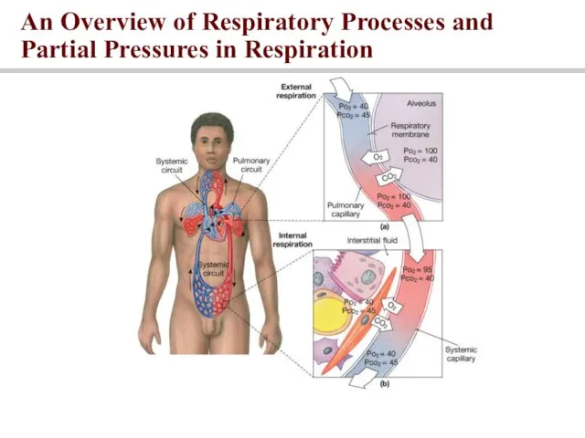
An Overview of Respiratory Processes and Partial Pressures in Respiration
An Overview of Respiratory Processes and Partial Pressures in Respiration
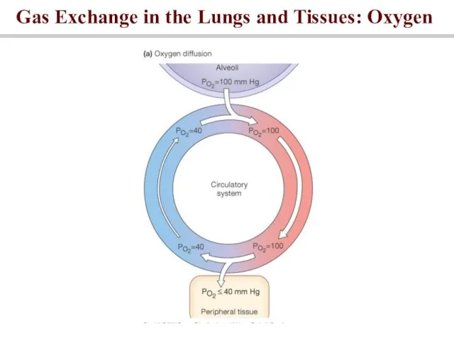
Gas Exchange in the Lungs and Tissues: Oxygen
Gas Exchange in the Lungs and Tissues: Oxygen
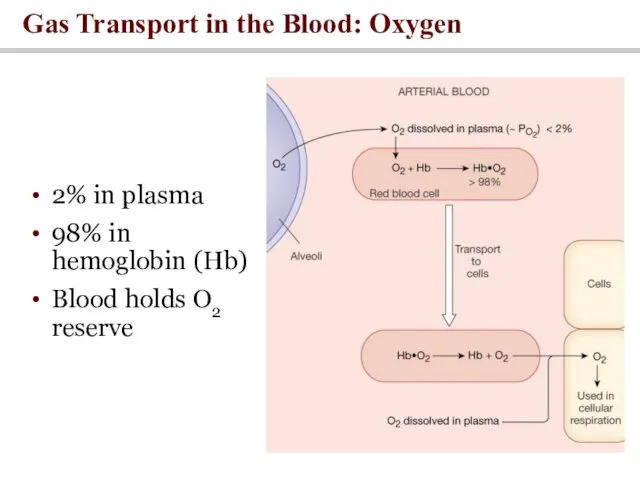
Gas Transport in the Blood: Oxygen
2% in plasma
98% in hemoglobin (Hb)
Blood
Gas Transport in the Blood: Oxygen
2% in plasma
98% in hemoglobin (Hb)
Blood
Carried mainly by RBCs, bound to hemoglobin
The amount of oxygen hemoglobin
Carried mainly by RBCs, bound to hemoglobin
The amount of oxygen hemoglobin
Hemoglobin Transport of Oxygen
4 binding sites per Hb molecule
98% saturated in
Hemoglobin Transport of Oxygen
4 binding sites per Hb molecule
98% saturated in
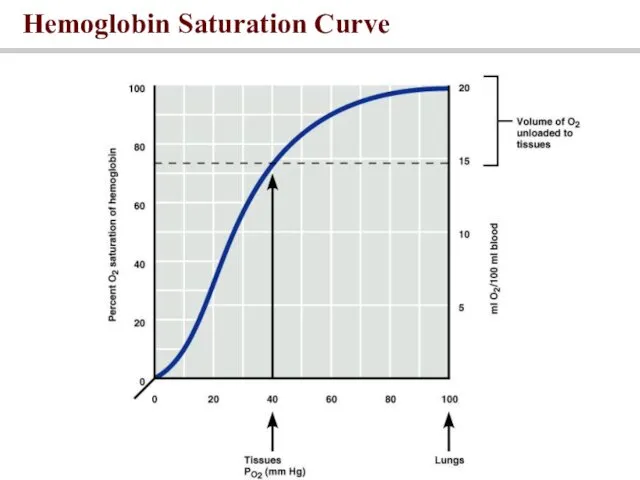
Hemoglobin Saturation Curve
Hemoglobin Saturation Curve
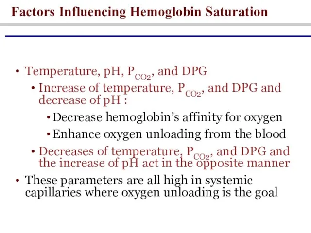
Temperature, pH, PCO2, and DPG
Increase of temperature, PCO2, and DPG and
Temperature, pH, PCO2, and DPG
Increase of temperature, PCO2, and DPG and
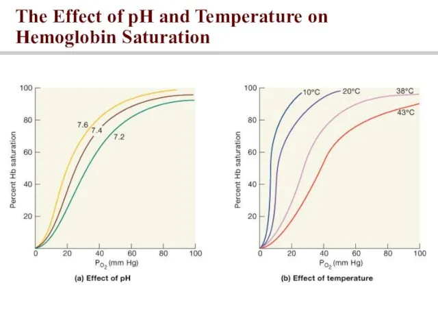
The Effect of pH and Temperature on Hemoglobin Saturation
The Effect of pH and Temperature on Hemoglobin Saturation
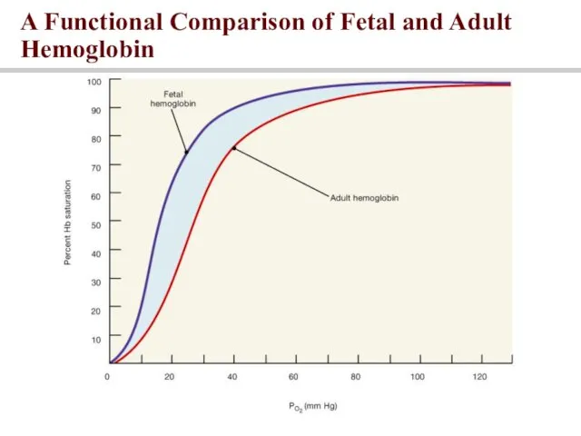
A Functional Comparison of Fetal and Adult Hemoglobin
A Functional Comparison of Fetal and Adult Hemoglobin
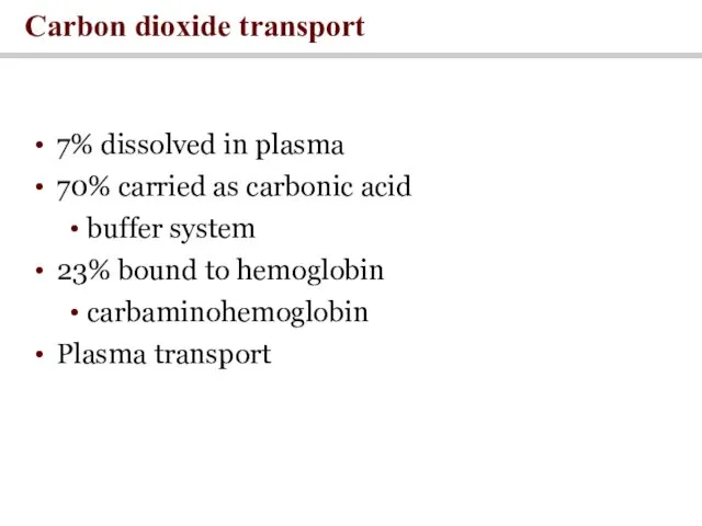
7% dissolved in plasma
70% carried as carbonic acid
buffer system
23% bound
7% dissolved in plasma
70% carried as carbonic acid
buffer system
23% bound
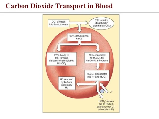
Carbon Dioxide Transport in Blood
Carbon Dioxide Transport in Blood
Driven by differences in partial pressure
Oxygen enters blood at lungs and
Driven by differences in partial pressure
Oxygen enters blood at lungs and
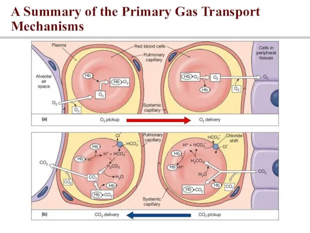
A Summary of the Primary Gas Transport Mechanisms
A Summary of the Primary Gas Transport Mechanisms
Control of Respiration
Control of Respiration
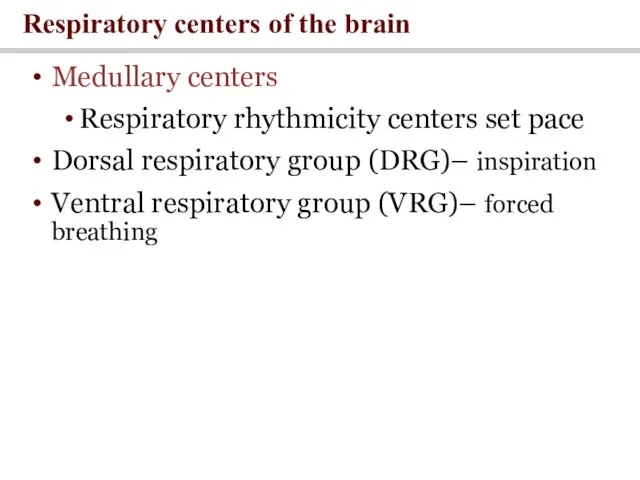
Medullary centers
Respiratory rhythmicity centers set pace
Dorsal respiratory group (DRG)– inspiration
Ventral respiratory
Medullary centers
Respiratory rhythmicity centers set pace
Dorsal respiratory group (DRG)– inspiration
Ventral respiratory
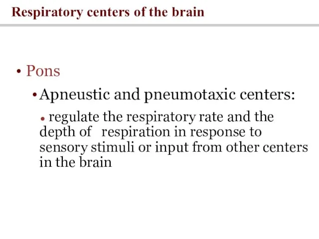
Pons
Apneustic and pneumotaxic centers:
● regulate the respiratory rate and the
Pons
Apneustic and pneumotaxic centers:
● regulate the respiratory rate and the
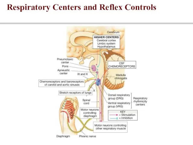
Respiratory Centers and Reflex Controls
Respiratory Centers and Reflex Controls
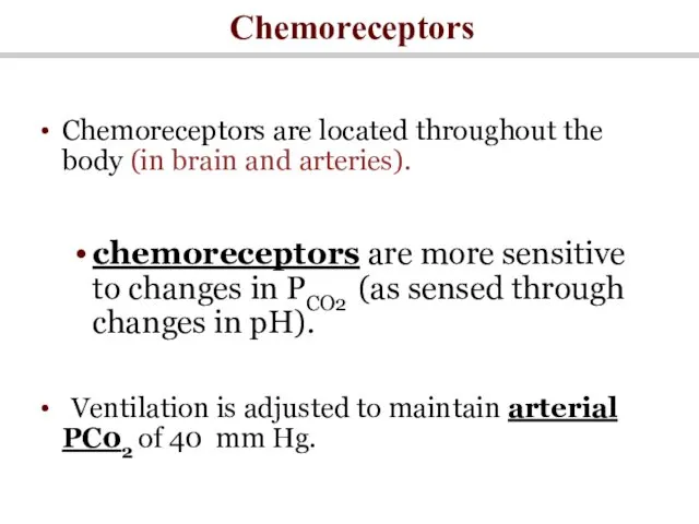
Chemoreceptors
Chemoreceptors are located throughout the body (in brain and arteries).
chemoreceptors are
Chemoreceptors
Chemoreceptors are located throughout the body (in brain and arteries).
chemoreceptors are
 Неотложная медицинская помощь и лечение при астматическом статусе
 Сифилис белая чума
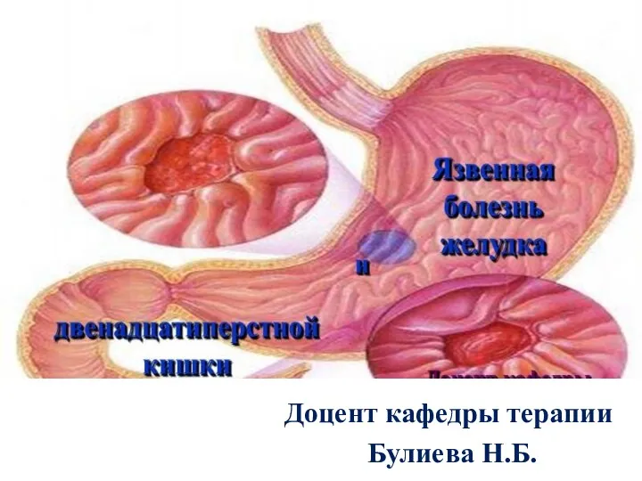 Язвенная болезнь
 Конусно-лучевая компьютерная томография в терапевтической стоматологии
 Көру мүшесінің анатомиясы, физиологиясы, патологиясы. Көз аурулары
 Травматические повреждения мягких тканей челюстно-лицевой области у детей
 Лечение животными
 Заболевание корь
 Факторы развития иммунитета у детей грудного возраста
 Починка, коррекция и реставрация полных съемных протезов
 Перелом шейки бедра
 Ревматическая лихорадка
 Імунні розлади та їх корекція при гострому гнійному перитоніті
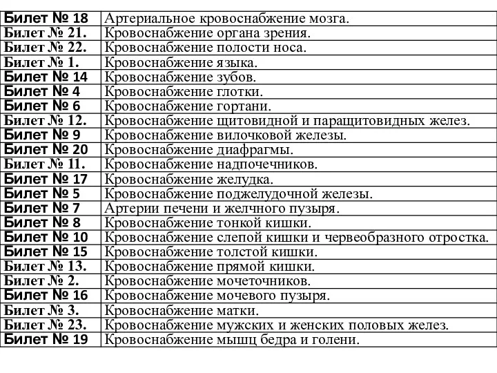 Кровоснабжение органов
 Нарушения памяти
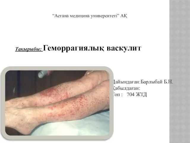 Геморрагиялық васкулит
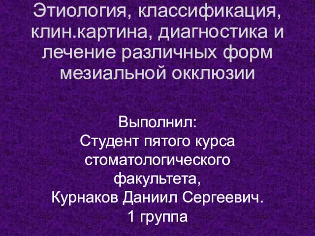 Этиология, классификация, клиническая картина, диагностика и лечение различных форм мезиальной окклюзии
 Нарушение кровообращения. Отеки
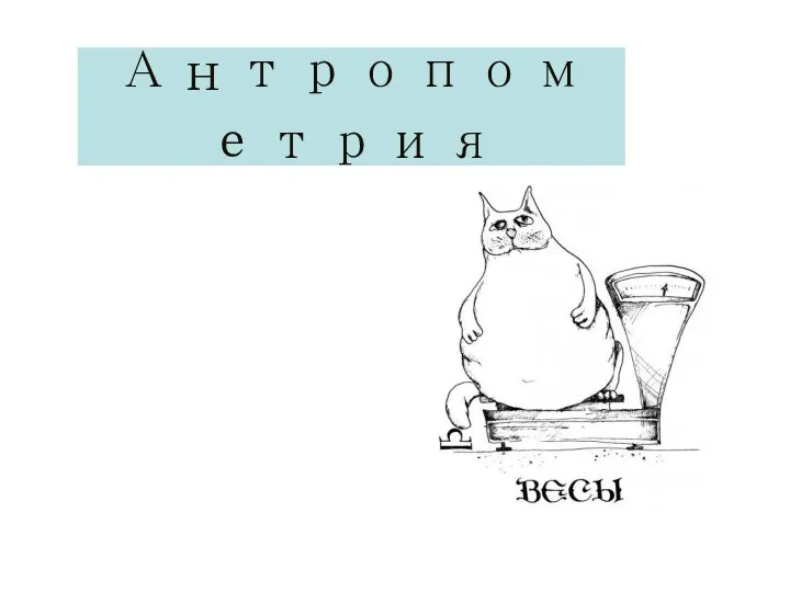 Антропометрия (Соматометрия)
 Проведение дезинфекции в ООМД (организация, осуществляющая медицинскую деятельность)
 Принципы препарирования 1,2 класса по Блэку
 Обезболивание в паллиативной медицине
 Оценка эффективности (качества) системы эпидемиологического надзора за ОВП/ полиомиелитом в Украине
 Почесуха
 Панкреонекроз. Классификация острого панкреатита
 Острая артериальная непроходимость
 Переломы. Классификация переломов
 Өзектілігі Жүрек қантамыр аурулары, атап айтсақ миокард инфаркты, стенокардия, инсульт, әлем бойынша өлім көрсеткіші жағынан