- Urolithiasis

Содержание
- 2. Urinary calculi are the third most common affliction of the urinary tract, exceeded only by urinary
- 3. The nomenclature associated with urinary stone disease arises from a variety of disciplines. .
- 4. Before the time of von Struve, the stones were referred to as guanite, because magnesium ammonium
- 5. The history of the nomenclature associated with urinary stone disease is as intriguing as that of
- 6. Urinary stones have plagued humans since the earliest records of civilization. The etiology of stones remains
- 7. Advances in the surgical treatment of urinary stones have outpaced our understanding of their etiology.
- 8. Without such follow-up and medical intervention, stone recurrence rates can be as high as 50% within
- 9. Renal & Ureteral Stones Etiology Theories to explain urinary stone disease are incomplete.
- 10. Renal & Ureteral Stones Etiology Stone formation requires supersaturated urine. Supersaturation depends on urinary pH, ionic
- 11. Renal & Ureteral Stones Etiology The activity coefficient reflects the availability of a particular ion.
- 12. Renal & Ureteral Stones Etiology Concentrations above this point are metastable and are capable of initiating
- 13. Renal & Ureteral Stones Etiology Multiplying 2 ion concentrations reveals the concentration product. The concentration products
- 14. Renal & Ureteral Stones Etiology Crystal formation is modified by a variety of other substances found
- 15. Renal & Ureteral Stones Etiology The nucleation theory suggests that urinary stones originate from crystals or
- 16. Renal & Ureteral Stones Etiology Additionally, many stone formers' 24-h urine collections are completely normal with
- 17. Renal & Ureteral Stones Etiology This theory does not have absolute validity since many people lacking
- 18. Crystal Component Stones are composed primarily of a crystalline component. Crystals of adequate size and transparency
- 19. Crystal Component Multiple steps are involved in crystal formation, including nucleation, growth, and aggregation.
- 20. Crystal Component A crystal of one type thereby serves as a nidus for the nucleation of
- 21. Crystal Component How these early crystalline structures are retained in the upper urinary tract without uneventful
- 22. Crystal Component This explanation is unsatisfactory; tubules are conical in shape and enlarge as they enter
- 23. Crystal Component The fixed particle theory postulates that formed crystals are somehow retained within cells or
- 24. Crystal Component These can be appreciated during endoscopy of the upper urinary tract.
- 25. Matrix Component The amount of the noncrystalline, matrix component of urinary stones varies with stone type,
- 26. Matrix Component Histologic inspection reveals laminations with scant calcifications.
- 27. Matrix Component The role of matrix in the initiation of ordinary urinary stones as well as
- 28. Urinary Ions Calcium Calcium is a major ion present in urinary crystals.
- 29. Diuretic medications may exert a hypocalciuric effect by further decreasing calcium excretion.
- 30. Oxalate Oxalate is a normal waste product of metabolism and is relatively insoluble.
- 31. Oxalate Once absorbed from the small bowel, oxalate is not metabolized and is excreted almost exclusively
- 32. Oxalate Normal excretion ranges from 20 to 45 mg/d and does not change significantly with age.
- 33. Oxalate Hyperoxaluria may develop in patients with bowel disorders, particularly inflammatory bowel disease, small-bowel resection, and
- 34. Oxalate The unbound oxalate is readily absorbed.
- 35. Phosphate Phosphate is an important buffer and complexes with calcium in urine.
- 36. Phosphate The small amount of phosphate filtered by the glomerulus is predominantly reabsorbed in the proximal
- 37. Uric Acid Uric acid is the by-product of purine metabolism. The pH of uric acid is
- 38. Uric Acid Rarely, a defect in xanthine oxidase results in increased levels of xanthine; the xanthine
- 39. Uric Acid This results from a deficiency of adenine phosphoribosyltransferase (APRT).
- 40. Sodium Although not identified as one of the major constituents of most urinary calculi, sodium plays
- 41. Sodium This reduces the ability of urine to inhibit calcium oxalate crystal agglomeration.
- 42. Citrate Citrate is a key factor affecting the development of calcium urinary stones.
- 43. Citrate Metabolic stimuli that consume this product (as with intracellular metabolic acidosis due to fasting, hypokalemia,
- 44. Magnesium Dietary magnesium deficiency is associated with an increased incidence of urinary stone disease.
- 45. Magnesium The exact mechanism whereby magnesium exerts its effect is undefined.
- 46. Sulfate Urinary sulfates may help prevent urinary calculi. They can complex with calcium.
- 47. Stone Varieties
- 48. Calcium Calculi Calcifications can occur and accumulate in the collecting system, resulting in nephrolithiasis. Eighty to
- 49. Calcium Calculi Hyperuricosuria is identified as a solitary defect in 8% of patients and associated with
- 50. Calcium Calculi Finally, decreased urinary citrate is found as an isolated defect in 17% of patients
- 51. Calcium Calculi Symptoms are secondary to obstruction, with resultant pain, infection, nausea, and vomiting, and rarely
- 52. Calcium Calculi Most patients with nephrolithiasis, however, do not have obvious nephrocalcinosis.
- 53. Calcium Calculi Nephrocalcinosis may result from a variety of pathologic states.
- 54. Calcium Calculi Disease processes resulting in bony destruction, including hyperparathyroidism, osteolytic lesions, and multiple myeloma, are
- 55. Absorptive Hypercalciuric Nephrolithiasis Normal calcium intake averages approximately 900-1000 mg/d.
- 56. Absorptive Hypercalciuric Nephrolithiasis This results in an increased load of calcium filtered from the glomerulus.
- 57. Absorptive Hypercalciuric Nephrolithiasis Absorptive hypercalciuria can be subdivided into 3 types.
- 58. Absorptive Hypercalciuric Nephrolithiasis Urinary calcium excretion returns to normal values with therapy.
- 59. Symptoms & Signs at Presentation
- 60. Symptomatology Pain Hematuria Pyuria
- 61. 12% of men and 5% of women will suffer from renal stones by the age of
- 62. The majority of patients with nephrolithiasis are those from 25 up to 55 years.
- 63. By localization there can be stones of the: -Calices -
- 64. Upper-tract urinary stones usually eventually cause pain. The character of the pain depends on the location.
- 65. Radiation of pain with various types of ureteral stone.
- 66. Upper right: Midureteral stone. Same as above but with more pain in the lower abdominal quadrant.
- 67. Pain Renal colic and noncolicky renal pain are the 2 types of pain originating from the
- 68. Pain This pain is due to a direct increase in intraluminal pressure, stretching nerve endings.
- 69. Pain Renal colic does not always wax and wane or come in waves like intestinal or
- 70. Pain In the ureter, however, local pain is referred to the distribution of the ilioinguinal nerve
- 71. Pain The vast majority of urinary stones present with the acute onset of pain due to
- 72. Pain The stone burden does not correlate with the severity of the symptoms. Small ureteral stones
- 73. Pain The pain frequently is abrupt in onset and severe and may awaken a patient from
- 74. Pain This movement is in contrast to the lack of movement of someone with peritoneal signs;
- 75. Renal Calyx Stones or other objects in calyces or caliceal diverticula may cause obstruction and renal
- 76. Renal Calyx Radiographic imaging may not reveal evidence of obstruction despite the patient's complaints of intermittent
- 77. Renal Calyx Caliceal calculi occasionally result in spontaneous perforation with urinoma, fistula, or abscess formation.
- 78. Renal Calyx Effective long-term treatment requires stone extraction and elimination of the obstructive component.
- 79. Renal Pelvis Stones in the renal pelvis > 1 cm in diameter commonly obstruct the ureteropelvic
- 80. Renal Pelvis Symptoms frequently occur on an intermittent basis following a drinking binge or consumption of
- 81. Renal Pelvis Partial or complete staghorn calculi that are present in the renal pelvis are not
- 82. Upper and Mid Ureter Pain associated with ureteral calculi often projects to corresponding dermatomal and spinal
- 83. Upper and Mid Ureter The pain of upper ureteral stones thus radiates to the lumbar region
- 84. Upper and Mid Ureter Stones or other objects in the upper or mid ureter often cause
- 85. Distal Ureter Calculi in the lower ureter often cause pain that radiates to the groin or
- 86. Distal Ureter Stones in the intramural ureter may mimic cystitis, urethritis, or prostatitis by causing suprapubic
- 87. Distal Ureter Strictures of the distal ureter from radiation, operative injury, or previous endoscopic procedures can
- 88. Hematuria A complete urinalysis helps to confirm the diagnosis of a urinary stone by assessing for
- 89. Infection Magnesium ammonium phosphate (struvite) stones are synonymous with infection stones.
- 90. Infection All stones, however, may be associated with infections secondary to obstruction and stasis proximal to
- 91. Infection Uropathogenic bacteria may alter ureteral peristalsis by the production of exotoxins and endotoxins.
- 92. Infection Local inflammation from infection can lead to chemoreceptor activation and perception of local pain with
- 93. Pyonephrosis Presentation is variable and may range from asymptomatic bacteriuria to florid urosepsis. Bladder urine cultures
- 94. Pyonephrosis Radiographic investigations are frequently nondiagnostic.
- 95. Pyonephrosis If unrecognized and untreated, pyonephrosis may develop into a renocutaneous fistula.
- 96. Xanthogranulomatous Pyelonephritis Xanthogranulomatous pyelonephritis is associated with upper-tract obstruction and infection.
- 97. Xanthogranulomatous Pyelonephritis Open surgical procedures, such as a simple nephrectomy for minimal or nonrenal function, can
- 98. Associated Fever Costovertebral angle tenderness may be marked with acute upper-tract obstruction; however, it cannot be
- 99. Associated Fever If retrograde manipulations are unsuccessful, insertion of a percutaneous nephrostomy tube is required.
- 100. Nausea and Vomiting Effective ureteral peristalsis requires coaptation of the ureteral walls and is most effective
- 101. Special Situations Pregnancy Renal colic is the most common nonobstetric cause of acute abdominal pain during
- 102. Special Situations Pregnancy The increased filtered load of calcium, uric acid, and sodium from the 25-50%
- 103. Special Situations Pregnancy Initial investigations can be undertaken with renal ultrasonography and limited abdominal x-rays with
- 104. Special Situations Pregnancy Treatment requires balancing the safety of the fetus with the health of the
- 105. Obesity Ultrasound examination is hindered by the attenuation of ultrasound beams.
- 106. Obesity Standard lithotripters have focal lengths less than 15 cm between the energy source and the
- 107. Obesity A preplaced heavy suture eases removal of such sheaths.
- 108. Obesity Postoperative prophylaxis for thromboembolic complications should be considered.
- 109. There are numerous theories of origination and development of urolithiasis, however, any of them does not
- 110. The known role in the etiology of urolithiasis is played by the disturbance of urate, phosphate,
- 111. It is possible to divide the numerous factors contributing to the formation of stones, into exogenous
- 112. The formation of phosphate stones is promoted also by fractures of tubular bones.
- 113. The uric acid is the end product of purine exchange.
- 114. To the internal causes, contributing to originating urolithiasis, we also attribute disturbance of a normal function
- 115. The local factors of lithogenesis
- 116. 70-80% of all stones are Ca containing. The major factor in urolithiasis in children and adults
- 117. Three conditions which contribute to the formation of struvite stones are the following: Congenital anomalies
- 118. There are four types of urate urolithiasis: Idiopathic urate urolithiasis
- 119. Formation of stones of uric acid depends on: pH of urine
- 120. Anatomical Pathology Degree of obstruction of the urinary paths Expressiveness of inflammatory process, which, as a
- 121. Complications of urolithiasis The most often complication of nephrolithiasis is the inflammatory process in the kidney,
- 122. Both chronic pyelonephrosis and pyonephrosis, as well as hydronephrosis owing to urolithiasis can entail a nephrogenic
- 123. The most severe complication of urolithiasis is prerenal anuria with the development of acute renal failure.
- 124. Diagnostics The diagnosis of urolithiasis is established, first of all, on the basis of the patient’s
- 125. Laboratory research It is necessary to remember, that the absence of pathological changes of urine does
- 126. Ultrasound investigation
- 127. X-ray examination
- 128. Retrograde ureteropyelography
- 129. Computed tomography
- 130. Differential diagnosis
- 131. Treatment
- 132. Conservative treatment
- 133. Indications for surgical intervention: Urinary obstructions with progressing damage of the kidney Persistent infection despite antibiotics
- 134. Instrumental methods of treatment
- 135. Percutaneous nephrolithotomy
- 136. Extracorporeal shock wave lithotripsy (ESWL)
- 138. Скачать презентацию
Urinary calculi are the third most common affliction of the urinary
Urinary calculi are the third most common affliction of the urinary
The nomenclature associated with urinary stone disease arises from a variety
The nomenclature associated with urinary stone disease arises from a variety
Before the time of von Struve, the stones were referred to
Before the time of von Struve, the stones were referred to
The history of the nomenclature associated with urinary stone disease is
The history of the nomenclature associated with urinary stone disease is
Urinary stones have plagued humans since the earliest records of civilization.
Urinary stones have plagued humans since the earliest records of civilization.

Advances in the surgical treatment of urinary stones have outpaced our
Advances in the surgical treatment of urinary stones have outpaced our
Without such follow-up and medical intervention, stone recurrence rates can be
Without such follow-up and medical intervention, stone recurrence rates can be
Renal & Ureteral Stones
Etiology
Theories to explain urinary stone disease are incomplete.
Renal & Ureteral Stones
Etiology
Theories to explain urinary stone disease are incomplete.
Renal & Ureteral Stones
Etiology
Stone formation requires supersaturated urine. Supersaturation depends on
Renal & Ureteral Stones
Etiology
Stone formation requires supersaturated urine. Supersaturation depends on
Renal & Ureteral Stones
Etiology
The activity coefficient reflects the availability of a
Renal & Ureteral Stones
Etiology
The activity coefficient reflects the availability of a
Renal & Ureteral Stones
Etiology
Concentrations above this point are metastable and are
Renal & Ureteral Stones
Etiology
Concentrations above this point are metastable and are
Renal & Ureteral Stones
Etiology
Multiplying 2 ion concentrations reveals the concentration product.
Renal & Ureteral Stones
Etiology
Multiplying 2 ion concentrations reveals the concentration product.
Renal & Ureteral Stones
Etiology
Crystal formation is modified by a variety of
Renal & Ureteral Stones
Etiology
Crystal formation is modified by a variety of
Renal & Ureteral Stones
Etiology
The nucleation theory suggests that urinary stones originate
Renal & Ureteral Stones
Etiology
The nucleation theory suggests that urinary stones originate
Renal & Ureteral Stones
Etiology
Additionally, many stone formers' 24-h urine collections are
Renal & Ureteral Stones
Etiology
Additionally, many stone formers' 24-h urine collections are
Renal & Ureteral Stones
Etiology
This theory does not have absolute validity since
Renal & Ureteral Stones
Etiology
This theory does not have absolute validity since
Crystal Component
Stones are composed primarily of a crystalline component.
Crystals of
Crystal Component
Stones are composed primarily of a crystalline component.
Crystals of
Crystal Component
Multiple steps are involved in crystal formation, including nucleation, growth,
Crystal Component
Multiple steps are involved in crystal formation, including nucleation, growth,
Crystal Component
A crystal of one type thereby serves as a nidus
Crystal Component
A crystal of one type thereby serves as a nidus
Crystal Component
How these early crystalline structures are retained in the upper
Crystal Component
How these early crystalline structures are retained in the upper

Crystal Component
This explanation is unsatisfactory; tubules are conical in shape and
Crystal Component
This explanation is unsatisfactory; tubules are conical in shape and
Crystal Component
The fixed particle theory postulates that formed crystals are somehow
Crystal Component
The fixed particle theory postulates that formed crystals are somehow
Crystal Component
These can be appreciated during endoscopy of the upper urinary
Crystal Component
These can be appreciated during endoscopy of the upper urinary
Matrix Component
The amount of the noncrystalline, matrix component of urinary stones
Matrix Component
The amount of the noncrystalline, matrix component of urinary stones
Matrix Component
Histologic inspection reveals laminations with scant calcifications.
Matrix Component
Histologic inspection reveals laminations with scant calcifications.
Matrix Component
The role of matrix in the initiation of ordinary urinary
Matrix Component
The role of matrix in the initiation of ordinary urinary
Urinary Ions
Calcium
Calcium is a major ion present in urinary crystals.
Urinary Ions
Calcium
Calcium is a major ion present in urinary crystals.
Diuretic medications may exert a hypocalciuric effect by further decreasing calcium
Diuretic medications may exert a hypocalciuric effect by further decreasing calcium
Oxalate
Oxalate is a normal waste product of metabolism and is relatively
Oxalate
Oxalate is a normal waste product of metabolism and is relatively

Oxalate
Once absorbed from the small bowel, oxalate is not metabolized and
Oxalate
Once absorbed from the small bowel, oxalate is not metabolized and
Oxalate
Normal excretion ranges from 20 to 45 mg/d and does not
Oxalate
Normal excretion ranges from 20 to 45 mg/d and does not
Oxalate
Hyperoxaluria may develop in patients with bowel disorders, particularly inflammatory bowel
Oxalate
Hyperoxaluria may develop in patients with bowel disorders, particularly inflammatory bowel
Oxalate
The unbound oxalate is readily absorbed.
Oxalate
The unbound oxalate is readily absorbed.
Phosphate
Phosphate is an important buffer and complexes with calcium in urine.
Phosphate
Phosphate is an important buffer and complexes with calcium in urine.
Phosphate
The small amount of phosphate filtered by the glomerulus is predominantly
Phosphate
The small amount of phosphate filtered by the glomerulus is predominantly
Uric Acid
Uric acid is the by-product of purine metabolism. The pH
Uric Acid
Uric acid is the by-product of purine metabolism. The pH
Uric Acid
Rarely, a defect in xanthine oxidase results in increased levels
Uric Acid
Rarely, a defect in xanthine oxidase results in increased levels
Uric Acid
This results from a deficiency of adenine phosphoribosyltransferase (APRT).
Uric Acid
This results from a deficiency of adenine phosphoribosyltransferase (APRT).
Sodium
Although not identified as one of the major constituents of most
Sodium
Although not identified as one of the major constituents of most

Sodium
This reduces the ability of urine to inhibit calcium oxalate crystal
Sodium
This reduces the ability of urine to inhibit calcium oxalate crystal
Citrate
Citrate is a key factor affecting the development of calcium urinary
Citrate
Citrate is a key factor affecting the development of calcium urinary
Citrate
Metabolic stimuli that consume this product (as with intracellular metabolic acidosis
Citrate
Metabolic stimuli that consume this product (as with intracellular metabolic acidosis

Magnesium
Dietary magnesium deficiency is associated with an increased incidence of urinary
Magnesium
Dietary magnesium deficiency is associated with an increased incidence of urinary
Magnesium
The exact mechanism whereby magnesium exerts its effect is undefined.
Magnesium
The exact mechanism whereby magnesium exerts its effect is undefined.
Sulfate
Urinary sulfates may help prevent urinary calculi. They can complex with
Sulfate
Urinary sulfates may help prevent urinary calculi. They can complex with

Stone Varieties
Stone Varieties
Calcium Calculi
Calcifications can occur and accumulate in the collecting system, resulting
Calcium Calculi
Calcifications can occur and accumulate in the collecting system, resulting

Calcium Calculi
Hyperuricosuria is identified as a solitary defect in 8% of
Calcium Calculi
Hyperuricosuria is identified as a solitary defect in 8% of
Calcium Calculi
Finally, decreased urinary citrate is found as an isolated defect
Calcium Calculi
Finally, decreased urinary citrate is found as an isolated defect
Calcium Calculi
Symptoms are secondary to obstruction, with resultant pain, infection, nausea,
Calcium Calculi
Symptoms are secondary to obstruction, with resultant pain, infection, nausea,

Calcium Calculi
Most patients with nephrolithiasis, however, do not have obvious nephrocalcinosis.
Calcium Calculi
Most patients with nephrolithiasis, however, do not have obvious nephrocalcinosis.
Calcium Calculi
Nephrocalcinosis may result from a variety of pathologic states.
Calcium Calculi
Nephrocalcinosis may result from a variety of pathologic states.
Calcium Calculi
Disease processes resulting in bony destruction, including hyperparathyroidism, osteolytic lesions,
Calcium Calculi
Disease processes resulting in bony destruction, including hyperparathyroidism, osteolytic lesions,
Absorptive Hypercalciuric Nephrolithiasis
Normal calcium intake averages approximately 900-1000 mg/d.
Absorptive Hypercalciuric Nephrolithiasis
Normal calcium intake averages approximately 900-1000 mg/d.

Absorptive Hypercalciuric Nephrolithiasis
This results in an increased load of calcium filtered
Absorptive Hypercalciuric Nephrolithiasis
This results in an increased load of calcium filtered
Absorptive Hypercalciuric Nephrolithiasis
Absorptive hypercalciuria can be subdivided into 3 types.
Absorptive Hypercalciuric Nephrolithiasis
Absorptive hypercalciuria can be subdivided into 3 types.
Absorptive Hypercalciuric Nephrolithiasis
Urinary calcium excretion returns to normal values with therapy.
Absorptive Hypercalciuric Nephrolithiasis
Urinary calcium excretion returns to normal values with therapy.

Symptoms & Signs at Presentation
Symptoms & Signs at Presentation
Symptomatology
Pain
Hematuria
Pyuria
Symptomatology
Pain
Hematuria
Pyuria
12% of men and 5% of women will suffer from renal
12% of men and 5% of women will suffer from renal

The majority of patients with nephrolithiasis are those from 25 up
The majority of patients with nephrolithiasis are those from 25 up

By localization there can be stones of the:
-Calices
-
By localization there can be stones of the:
-Calices
-
Upper-tract urinary stones usually eventually cause pain.
The character of the
Upper-tract urinary stones usually eventually cause pain.
The character of the
Radiation of pain with various types of ureteral stone.
Radiation of pain with various types of ureteral stone.
Upper right: Midureteral stone. Same as above but with more pain
Upper right: Midureteral stone. Same as above but with more pain
Pain
Renal colic and noncolicky renal pain are the 2 types of
Pain
Renal colic and noncolicky renal pain are the 2 types of
Pain
This pain is due to a direct increase in intraluminal pressure,
Pain
This pain is due to a direct increase in intraluminal pressure,

Pain
Renal colic does not always wax and wane or come in
Pain
Renal colic does not always wax and wane or come in
Pain
In the ureter, however, local pain is referred to the distribution
Pain
In the ureter, however, local pain is referred to the distribution
Pain
The vast majority of urinary stones present with the acute onset
Pain
The vast majority of urinary stones present with the acute onset

Pain
The stone burden does not correlate with the severity of the
Pain
The stone burden does not correlate with the severity of the
Pain
The pain frequently is abrupt in onset and severe and may
Pain
The pain frequently is abrupt in onset and severe and may
Pain
This movement is in contrast to the lack of movement of
Pain
This movement is in contrast to the lack of movement of

Renal Calyx
Stones or other objects in calyces or caliceal diverticula may
Renal Calyx
Stones or other objects in calyces or caliceal diverticula may
Renal Calyx
Radiographic imaging may not reveal evidence of obstruction despite the
Renal Calyx
Radiographic imaging may not reveal evidence of obstruction despite the

Renal Calyx
Caliceal calculi occasionally result in spontaneous perforation with urinoma, fistula,
Renal Calyx
Caliceal calculi occasionally result in spontaneous perforation with urinoma, fistula,
Renal Calyx
Effective long-term treatment requires stone extraction and elimination of the
Renal Calyx
Effective long-term treatment requires stone extraction and elimination of the
Renal Pelvis
Stones in the renal pelvis > 1 cm in diameter
Renal Pelvis
Stones in the renal pelvis > 1 cm in diameter
Renal Pelvis
Symptoms frequently occur on an intermittent basis following a drinking
Renal Pelvis
Symptoms frequently occur on an intermittent basis following a drinking

Renal Pelvis
Partial or complete staghorn calculi that are present in the
Renal Pelvis
Partial or complete staghorn calculi that are present in the
Upper and Mid Ureter
Pain associated with ureteral calculi often projects to
Upper and Mid Ureter
Pain associated with ureteral calculi often projects to

Upper and Mid Ureter
The pain of upper ureteral stones thus radiates
Upper and Mid Ureter
The pain of upper ureteral stones thus radiates
Upper and Mid Ureter
Stones or other objects in the upper or
Upper and Mid Ureter
Stones or other objects in the upper or

Distal Ureter
Calculi in the lower ureter often cause pain that radiates
Distal Ureter
Calculi in the lower ureter often cause pain that radiates
Distal Ureter
Stones in the intramural ureter may mimic cystitis, urethritis, or
Distal Ureter
Stones in the intramural ureter may mimic cystitis, urethritis, or

Distal Ureter
Strictures of the distal ureter from radiation, operative injury, or
Distal Ureter
Strictures of the distal ureter from radiation, operative injury, or
Hematuria
A complete urinalysis helps to confirm the diagnosis of a urinary
Hematuria
A complete urinalysis helps to confirm the diagnosis of a urinary
Infection
Magnesium ammonium phosphate (struvite) stones are synonymous with infection stones.
Infection
Magnesium ammonium phosphate (struvite) stones are synonymous with infection stones.
Infection
All stones, however, may be associated with infections secondary to obstruction
Infection
All stones, however, may be associated with infections secondary to obstruction
Infection
Uropathogenic bacteria may alter ureteral peristalsis by the production of exotoxins
Infection
Uropathogenic bacteria may alter ureteral peristalsis by the production of exotoxins
Infection
Local inflammation from infection can lead to chemoreceptor activation and perception
Infection
Local inflammation from infection can lead to chemoreceptor activation and perception
Pyonephrosis
Presentation is variable and may range from asymptomatic bacteriuria to florid
Pyonephrosis
Presentation is variable and may range from asymptomatic bacteriuria to florid

Pyonephrosis
Radiographic investigations are frequently nondiagnostic.
Pyonephrosis
Radiographic investigations are frequently nondiagnostic.
Pyonephrosis
If unrecognized and untreated, pyonephrosis may develop into a renocutaneous fistula.
Pyonephrosis
If unrecognized and untreated, pyonephrosis may develop into a renocutaneous fistula.
Xanthogranulomatous Pyelonephritis
Xanthogranulomatous pyelonephritis is associated with upper-tract obstruction and infection.
Xanthogranulomatous Pyelonephritis
Xanthogranulomatous pyelonephritis is associated with upper-tract obstruction and infection.
Xanthogranulomatous Pyelonephritis
Open surgical procedures, such as a simple nephrectomy for minimal
Xanthogranulomatous Pyelonephritis
Open surgical procedures, such as a simple nephrectomy for minimal
Associated Fever
Costovertebral angle tenderness may be marked with acute upper-tract obstruction;
Associated Fever
Costovertebral angle tenderness may be marked with acute upper-tract obstruction;
Associated Fever
If retrograde manipulations are unsuccessful, insertion of a percutaneous nephrostomy
Associated Fever
If retrograde manipulations are unsuccessful, insertion of a percutaneous nephrostomy
Nausea and Vomiting
Effective ureteral peristalsis requires coaptation of the ureteral walls
Nausea and Vomiting
Effective ureteral peristalsis requires coaptation of the ureteral walls
Special Situations
Pregnancy
Renal colic is the most common nonobstetric cause of acute
Special Situations
Pregnancy
Renal colic is the most common nonobstetric cause of acute
Special Situations
Pregnancy
The increased filtered load of calcium, uric acid, and sodium
Special Situations
Pregnancy
The increased filtered load of calcium, uric acid, and sodium
Special Situations
Pregnancy
Initial investigations can be undertaken with renal ultrasonography and limited
Special Situations
Pregnancy
Initial investigations can be undertaken with renal ultrasonography and limited

Special Situations
Pregnancy
Treatment requires balancing the safety of the fetus with the
Special Situations
Pregnancy
Treatment requires balancing the safety of the fetus with the
Obesity
Ultrasound examination is hindered by the attenuation of ultrasound beams.
Obesity
Ultrasound examination is hindered by the attenuation of ultrasound beams.

Obesity
Standard lithotripters have focal lengths less than 15 cm between the
Obesity
Standard lithotripters have focal lengths less than 15 cm between the
Obesity
A preplaced heavy suture eases removal of such sheaths.
Obesity
A preplaced heavy suture eases removal of such sheaths.
Obesity
Postoperative prophylaxis for thromboembolic complications should be considered.
Obesity
Postoperative prophylaxis for thromboembolic complications should be considered.
There are numerous theories of origination and development of urolithiasis, however,
There are numerous theories of origination and development of urolithiasis, however,
The known role in the etiology of urolithiasis is played by
The known role in the etiology of urolithiasis is played by
It is possible to divide the numerous factors contributing to the
It is possible to divide the numerous factors contributing to the
The formation of phosphate stones is promoted also by fractures of
The formation of phosphate stones is promoted also by fractures of
The uric acid is the end product of purine exchange.
To the internal causes, contributing to originating urolithiasis, we also attribute
To the internal causes, contributing to originating urolithiasis, we also attribute
The local factors of lithogenesis
The local factors of lithogenesis
70-80% of all stones are Ca containing. The major factor in
70-80% of all stones are Ca containing. The major factor in
Three conditions which contribute to the formation of struvite stones are
Three conditions which contribute to the formation of struvite stones are
There are four types of urate urolithiasis:
Idiopathic urate urolithiasis
There are four types of urate urolithiasis:
Idiopathic urate urolithiasis
Formation of stones of uric acid depends on:
pH of urine
Formation of stones of uric acid depends on:
pH of urine
Anatomical Pathology
Degree of obstruction of the urinary paths
Expressiveness of inflammatory process,
Anatomical Pathology
Degree of obstruction of the urinary paths
Expressiveness of inflammatory process,
Complications of urolithiasis
The most often complication of nephrolithiasis is the inflammatory
Complications of urolithiasis
The most often complication of nephrolithiasis is the inflammatory
Both chronic pyelonephrosis and pyonephrosis, as well as hydronephrosis owing to
Both chronic pyelonephrosis and pyonephrosis, as well as hydronephrosis owing to
The most severe complication of urolithiasis is prerenal anuria with the
The most severe complication of urolithiasis is prerenal anuria with the
Diagnostics
The diagnosis of urolithiasis is established, first of all, on the
Diagnostics
The diagnosis of urolithiasis is established, first of all, on the
Laboratory research
It is necessary to remember, that the absence of pathological
Laboratory research
It is necessary to remember, that the absence of pathological
Ultrasound investigation
Ultrasound investigation
X-ray examination
X-ray examination
Retrograde ureteropyelography
Retrograde ureteropyelography
Computed tomography
Computed tomography

Differential diagnosis
Differential diagnosis
Treatment
Treatment
Conservative treatment
Conservative treatment

Indications for surgical intervention:
Urinary obstructions with progressing damage of the kidney
Persistent
Indications for surgical intervention:
Urinary obstructions with progressing damage of the kidney
Persistent
Instrumental methods of treatment
Instrumental methods of treatment

Percutaneous nephrolithotomy
Percutaneous nephrolithotomy
Extracorporeal shock wave lithotripsy (ESWL)
Extracorporeal shock wave lithotripsy (ESWL)
 Полиомиелит. Протекание болезни полиомиелита
 Дисфункционалдық жатырдан қан кету
 Интерпретация данных методов инструментальной диагностики ЭЭГ психических расстройств, связанных с органическим поражением ЦНС
 Временная остановка наружного кровотечения. Ошибки на догоспитальном этапе
 Акушерлік перитонит. Жайылған септикалық инфекция
 Синдром хронической болезни почек
 Балалар ауруларын біріктіріп жүргізу жоспарын құрастыру(жөтел және қиындаған тыныс)
 Иммундық процестердің бұзылуы. Аллергия, анафилаксия, СПИД
 Сестринский процесс при гломерулонефритах
 Гинекологиялық науқастардан анамнез жинау
 Роль ствола мозга в регуляции двигательных функций
 Хронический гастрит
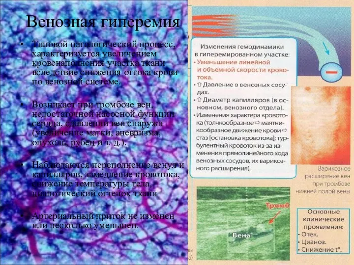 Венозная гиперемия
 Электронная медицинская аппаратура
 Лечение заболеваний и травм коленных суставов
 Диагностические и профилактические мероприятия болезней вымени
 ЛФК при заболеваниях органов пищеварения
 Первая помощь при неотложных состояниях: закон и порядок
 Нарушение кровообращения. Отеки
 Лайелл синдромы, Стивен-Джонсон синдромы
 Профессиональные нейротоксикозы
 Азық қорыту жүйесін зерттеу. Азық қабылдау және су ішудің бұзылуы
 IgA нефропатия. Клиникасы:
 Биохимия почек и мочи. (Лекция 10)
 Укусы ядовитых животных
 Көз жасы мүшесінің патологиясы. Дакриоцистит, жас нүктесінің тарылуы, жас нүктесінің сырт айналуы
 Парвавирусный энтерит собак
 Наследственные болезни обмена веществ