- Viral Hemorrhagic Fever

Содержание
- 2. Overview Organism History Epidemiology Transmission Disease in Humans Disease in Animals Prevention and Control
- 3. What is Viral Hemorrhagic Fever? Severe multisystem syndrome Damage to overall vascular system Symptoms often accompanied
- 4. The Organisms
- 5. Viral Hemorrhagic Fever Viruses of four distinct families Arenaviruses Filoviruses Bunyaviruses Flaviviruses RNA viruses Enveloped in
- 6. Classification
- 7. Arenaviridae Junin virus Machupo virus Guanarito virus Lassa virus Sabia virus
- 8. Arenaviridae History 1933: The first arenavirus was isolated 1958: Junin virus - Argentina First to cause
- 9. Arenaviridae Transmission Virus transmission and amplification occurs in rodents Shed virus through urine, feces, and other
- 10. Arenaviridae Epidemiology West Africa Lassa South America Junin, Machupo, Guanarito, and Sabia Contact with rodent excreta
- 11. Arenaviridae in Humans Incubation period: 10–14 days Prodromal period: Fever and malaise 2–4 days Hemorrhagic stage:
- 12. Bunyaviridae Rift Valley Fever virus Crimean-Congo Hemorrhagic Fever virus Hantavirus
- 13. Bunyaviridae History 1930: Rift Valley Fever – Egypt Epizootic in sheep 1940s: CCHF - Crimean peninsula
- 14. Bunyaviridae Transmission Arthropod vector Exception – Hantaviruses RVF – Aedes mosquito CCHF – Ixodid tick Hantavirus
- 15. Bunyaviridae Epidemiology RVF - sub-Saharan Africa and Saudi Arabia and Yemen 1% case fatality rate CCHF
- 16. Bunyaviridae Humans Rift Valley Fever Incubation period – 2-5 days 0.5% - Hemorrhagic Fever 0.5% -
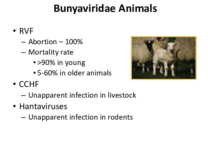
- 17. Bunyaviridae Animals RVF Abortion – 100% Mortality rate >90% in young 5-60% in older animals CCHF
- 18. Filoviridae Marburg virus Ebola virus
- 19. Filoviridae History 1967: Marburg virus European laboratory workers in Germany and former Yugoslavia 1976: Ebola virus
- 20. Filoviridae Transmission Reservoir is UNKNOWN Bats implicated with Marburg Intimate contact Nosicomial transmission Reuse of needles
- 21. Filoviridae Epidemiology Marburg – Africa Case fatality – 23-33% Ebola - Sudan, Zaire and Côte d'Ivoire
- 22. Filoviridae Humans Most severe hemorrhagic fever Incubation period: 4–10 days Abrupt onset Fever, chills, malaise, and
- 23. Filoviridae Animals Hemorrhagic fever Same clinical course as humans Ebola Reston High primate mortality - ~82%
- 24. Flaviviridae Dengue virus Yellow Fever virus Omsk Hemorrhagic Fever virus Kyassnur Forest Disease virus
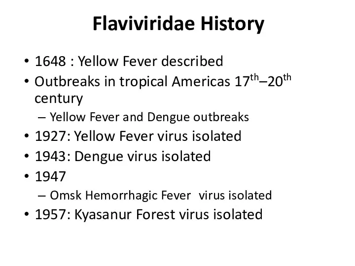
- 25. Flaviviridae History 1648 : Yellow Fever described Outbreaks in tropical Americas 17th–20th century Yellow Fever and
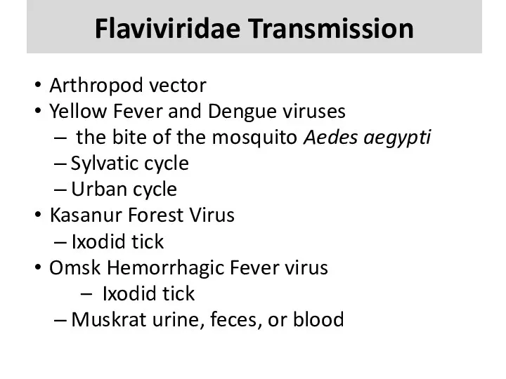
- 26. Flaviviridae Transmission Arthropod vector Yellow Fever and Dengue viruses the bite of the mosquito Aedes aegypti
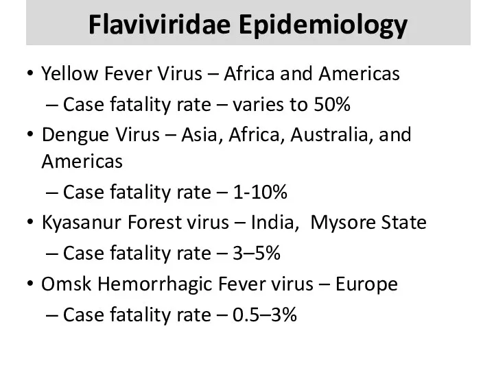
- 27. Flaviviridae Epidemiology Yellow Fever Virus – Africa and Americas Case fatality rate – varies to 50%
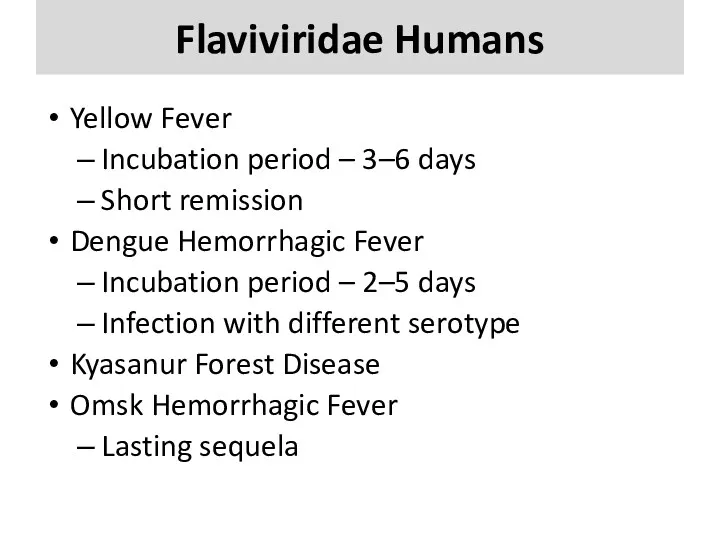
- 28. Flaviviridae Humans Yellow Fever Incubation period – 3–6 days Short remission Dengue Hemorrhagic Fever Incubation period
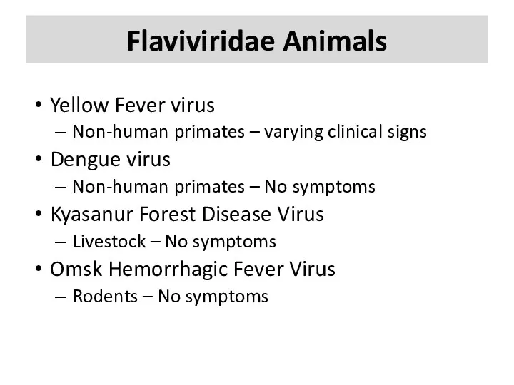
- 29. Flaviviridae Animals Yellow Fever virus Non-human primates – varying clinical signs Dengue virus Non-human primates –
- 30. Disease in Humans
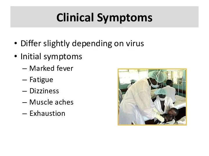
- 31. Clinical Symptoms Differ slightly depending on virus Initial symptoms Marked fever Fatigue Dizziness Muscle aches Exhaustion
- 32. Clinical Symptoms More severe Bleeding under skin Petechiae, echymoses, conjunctivitis Bleeding in internal organs Bleeding from
- 33. Diagnosis Specimens must be sent to CDC U.S. Army Medical Research Institute of Infectious Disease (USAMRIID)
- 34. Treatment Supportive treatment: maintaining fluid and electrolyte balance, circulatory volume, BP and treating for any complicating
- 35. Prevention and Control
- 36. Prevention and Control Avoid contact with host species Rodents Control rodent populations Discourage rodents from entering
- 37. Prevention and Control Vaccine available for Yellow fever Experimental vaccines under study Argentine HF, Rift Valley
- 38. Prevention and Control Protective clothing Disposable gowns, gloves, masks and shoe covers, protective eyewear when splashing
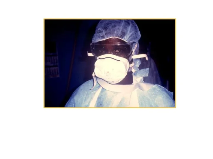
- 39. Protective equipment worn by a nurse during Ebola outbreak in Zaire, 1995
- 40. Prevention and Control Anyone suspected of having a VHF must use a chemical toilet Disinfect and
- 41. VHF Agents as Biological Weapons Outbreak of undifferentiated febrile illness 2-21 days following attack Could include
- 43. Скачать презентацию
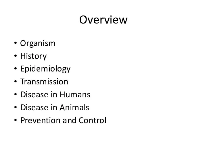
Overview
Organism
History
Epidemiology
Transmission
Disease in Humans
Disease in Animals
Prevention and Control
Overview
Organism
History
Epidemiology
Transmission
Disease in Humans
Disease in Animals
Prevention and Control
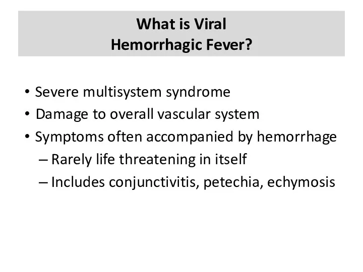
What is Viral
Hemorrhagic Fever?
Severe multisystem syndrome
Damage to overall vascular
What is Viral
Hemorrhagic Fever?
Severe multisystem syndrome
Damage to overall vascular

The Organisms
The Organisms
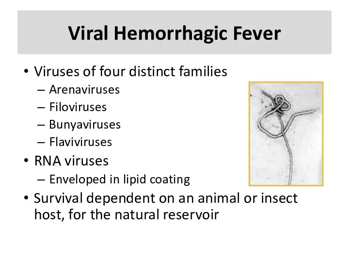
Viral Hemorrhagic Fever
Viruses of four distinct families
Arenaviruses
Filoviruses
Bunyaviruses
Flaviviruses
RNA viruses
Enveloped in
Viral Hemorrhagic Fever
Viruses of four distinct families
Arenaviruses
Filoviruses
Bunyaviruses
Flaviviruses
RNA viruses
Enveloped in
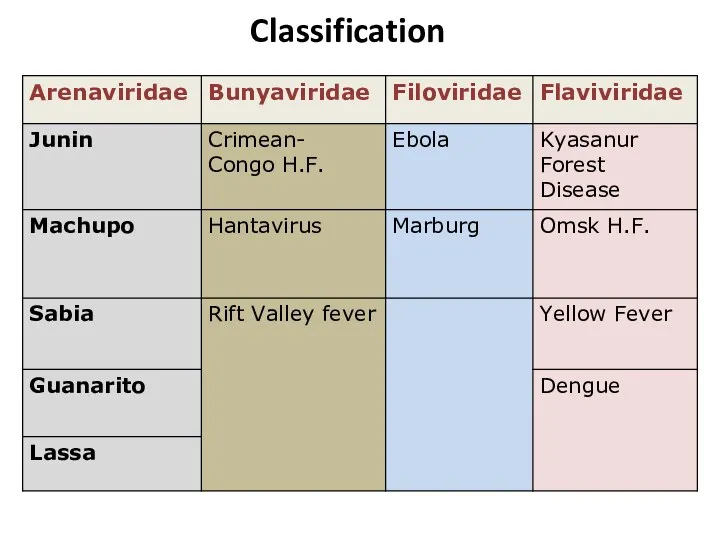
Classification
Classification
Arenaviridae
Junin virus
Machupo virus
Guanarito virus
Lassa virus
Sabia virus
Arenaviridae
Junin virus
Machupo virus
Guanarito virus
Lassa virus
Sabia virus
Arenaviridae History
1933: The first arenavirus was isolated
1958: Junin virus -
Arenaviridae History
1933: The first arenavirus was isolated
1958: Junin virus -
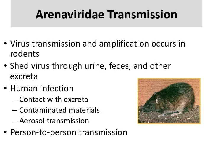
Arenaviridae Transmission
Virus transmission and amplification occurs in rodents
Shed virus through urine,
Arenaviridae Transmission
Virus transmission and amplification occurs in rodents
Shed virus through urine,
Arenaviridae Epidemiology
West Africa
Lassa
South America
Junin, Machupo, Guanarito, and Sabia
Contact with
Arenaviridae Epidemiology
West Africa
Lassa
South America
Junin, Machupo, Guanarito, and Sabia
Contact with
Arenaviridae in Humans
Incubation period: 10–14 days
Prodromal period: Fever and malaise 2–4
Arenaviridae in Humans
Incubation period: 10–14 days
Prodromal period: Fever and malaise 2–4
Bunyaviridae
Rift Valley Fever virus
Crimean-Congo Hemorrhagic Fever virus
Hantavirus
Bunyaviridae
Rift Valley Fever virus
Crimean-Congo Hemorrhagic Fever virus
Hantavirus
Bunyaviridae History
1930: Rift Valley Fever – Egypt
Epizootic in sheep
1940s: CCHF
Bunyaviridae History
1930: Rift Valley Fever – Egypt
Epizootic in sheep
1940s: CCHF
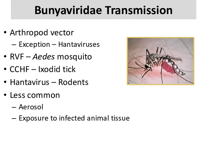
Bunyaviridae Transmission
Arthropod vector
Exception – Hantaviruses
RVF – Aedes mosquito
CCHF – Ixodid
Bunyaviridae Transmission
Arthropod vector
Exception – Hantaviruses
RVF – Aedes mosquito
CCHF – Ixodid
Bunyaviridae Epidemiology
RVF - sub-Saharan Africa and Saudi Arabia and Yemen
1%
Bunyaviridae Epidemiology
RVF - sub-Saharan Africa and Saudi Arabia and Yemen
1%
Bunyaviridae Humans
Rift Valley Fever
Incubation period – 2-5 days
0.5% -
Bunyaviridae Humans
Rift Valley Fever
Incubation period – 2-5 days
0.5% -
Bunyaviridae Animals
RVF
Abortion – 100%
Mortality rate
>90% in young
5-60% in older animals
CCHF
Unapparent
Bunyaviridae Animals
RVF
Abortion – 100%
Mortality rate
>90% in young
5-60% in older animals
CCHF
Unapparent
Filoviridae
Marburg virus
Ebola virus
Filoviridae
Marburg virus
Ebola virus
Filoviridae History
1967: Marburg virus
European laboratory workers in Germany and former Yugoslavia
Filoviridae History
1967: Marburg virus
European laboratory workers in Germany and former Yugoslavia
Filoviridae Transmission
Reservoir is UNKNOWN
Bats implicated with Marburg
Intimate contact
Nosicomial transmission
Reuse of
Filoviridae Transmission
Reservoir is UNKNOWN
Bats implicated with Marburg
Intimate contact
Nosicomial transmission
Reuse of
Filoviridae Epidemiology
Marburg – Africa
Case fatality – 23-33%
Ebola - Sudan, Zaire and
Filoviridae Epidemiology
Marburg – Africa
Case fatality – 23-33%
Ebola - Sudan, Zaire and
Filoviridae Humans
Most severe hemorrhagic fever
Incubation period: 4–10 days
Abrupt onset
Fever, chills, malaise,
Filoviridae Humans
Most severe hemorrhagic fever
Incubation period: 4–10 days
Abrupt onset
Fever, chills, malaise,
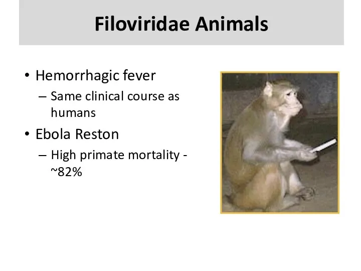
Filoviridae Animals
Hemorrhagic fever
Same clinical course as humans
Ebola Reston
High primate mortality -
Filoviridae Animals
Hemorrhagic fever
Same clinical course as humans
Ebola Reston
High primate mortality -
Flaviviridae
Dengue virus
Yellow Fever virus
Omsk Hemorrhagic Fever virus
Kyassnur Forest Disease virus
Flaviviridae
Dengue virus
Yellow Fever virus
Omsk Hemorrhagic Fever virus
Kyassnur Forest Disease virus
Flaviviridae History
1648 : Yellow Fever described
Outbreaks in tropical Americas 17th–20th century
Yellow
Flaviviridae History
1648 : Yellow Fever described
Outbreaks in tropical Americas 17th–20th century
Yellow
Flaviviridae Transmission
Arthropod vector
Yellow Fever and Dengue viruses
the bite of the
Flaviviridae Transmission
Arthropod vector
Yellow Fever and Dengue viruses
the bite of the
Flaviviridae Epidemiology
Yellow Fever Virus – Africa and Americas
Case fatality rate –
Flaviviridae Epidemiology
Yellow Fever Virus – Africa and Americas
Case fatality rate –
Flaviviridae Humans
Yellow Fever
Incubation period – 3–6 days
Short remission
Dengue Hemorrhagic Fever
Incubation
Flaviviridae Humans
Yellow Fever
Incubation period – 3–6 days
Short remission
Dengue Hemorrhagic Fever
Incubation
Flaviviridae Animals
Yellow Fever virus
Non-human primates – varying clinical signs
Dengue virus
Non-human primates
Flaviviridae Animals
Yellow Fever virus
Non-human primates – varying clinical signs
Dengue virus
Non-human primates

Disease in Humans
Disease in Humans
Clinical Symptoms
Differ slightly depending on virus
Initial symptoms
Marked fever
Fatigue
Dizziness
Muscle aches
Exhaustion
Clinical Symptoms
Differ slightly depending on virus
Initial symptoms
Marked fever
Fatigue
Dizziness
Muscle aches
Exhaustion
Clinical Symptoms
More severe
Bleeding under skin
Petechiae, echymoses, conjunctivitis
Bleeding in internal organs
Bleeding
Clinical Symptoms
More severe
Bleeding under skin
Petechiae, echymoses, conjunctivitis
Bleeding in internal organs
Bleeding
Diagnosis
Specimens must be sent to
CDC
U.S. Army Medical Research Institute of Infectious
Diagnosis
Specimens must be sent to
CDC
U.S. Army Medical Research Institute of Infectious
Treatment
Supportive treatment: maintaining fluid and electrolyte balance, circulatory volume, BP and
Treatment
Supportive treatment: maintaining fluid and electrolyte balance, circulatory volume, BP and
Prevention and Control
Prevention and Control
Prevention and Control
Avoid contact with host species
Rodents
Control rodent populations
Discourage rodents from
Prevention and Control
Avoid contact with host species
Rodents
Control rodent populations
Discourage rodents from
Prevention and Control
Vaccine available for Yellow fever
Experimental vaccines under study
Argentine
Prevention and Control
Vaccine available for Yellow fever
Experimental vaccines under study
Argentine
Prevention and Control
Protective clothing
Disposable gowns, gloves, masks and shoe covers, protective
Prevention and Control
Protective clothing
Disposable gowns, gloves, masks and shoe covers, protective
Protective equipment worn by a nurse during Ebola outbreak in Zaire,
Protective equipment worn by a nurse during Ebola outbreak in Zaire,
Prevention and Control
Anyone suspected of having a VHF must use a
Prevention and Control
Anyone suspected of having a VHF must use a
VHF Agents as
Biological Weapons
Outbreak of undifferentiated febrile illness 2-21 days
VHF Agents as
Biological Weapons
Outbreak of undifferentiated febrile illness 2-21 days
 История ФР. Лекция № 2 (картинки)
 ЦПР - Анализ 17-18 и МЕТОДЫ
 Самостоятельная работа интерна. Нарушения пищевого поведения
 Основы трансфузиологии
 Туберкулез внелегочной локализации
 Наркомания - острая проблема современности
 Сүйел. Жұқпалы моллюск. Сақтық шаралары
 Туберкулинді диагностика
 Наркомания и ее вред
 Стратегия управляемой нейромиорелаксации в клинической анестезиологии
 Guideline for the Prevention, Detection, Evaluation, and Management of High Blood Pressure in Adults
 Аддисон ауруы
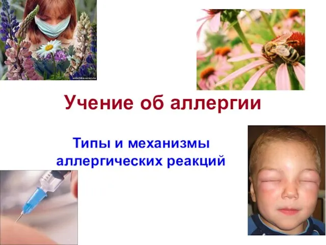 Учение об аллергии
 Болезни органов дыхательной системы
 Хирургическая инфекция
 Хирургия мягких тканей. Закрытие дефектов неправильной формы
 Психотические расстройства вызванные употреблением ПАВ
 Ауыздың ойылуы (стоматит)
 Клиническая фармакология лекарственных средств для лечения бронхообструктивного синдрома
 Подготовка больных к различным видам оперативных вмешательств
 Болезнь Виллебранда. Клинический случай
 Ќанныѕ тамыр ішілік шашыраѕќы ўю синдромы
 Чувствительность: общие понятия
 Выпот в полость перикарда
 Острый коронарный синдром. Реваскуляризация миокарда
 Заболевания артерий
 Дезинфекционно-стерилизационные мероприятия в учреждениях стоматологического профиля
 Клиническая энзимология