- Clinical anatomy of the head

Содержание
- 2. Topographic anatomy & operative surgery TOPOGRAPHIC ANATOMY is a science which studies relations between organs and
- 3. Cranial bones The cranial bones enclose and protect the brain and associated sensory organs. They consist
- 4. SKULL The human skull, consisting of 8 cranial and 14 facial bones, contains several cavities that
- 5. The eight bones of the cranium articulate firmly with one another to enclose and protect the
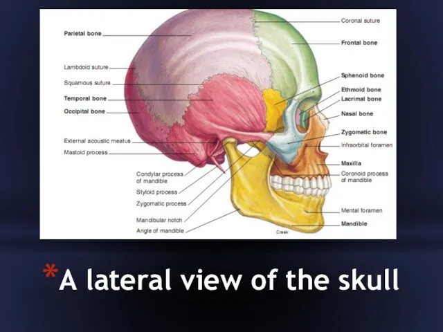
- 6. A lateral view of the skull
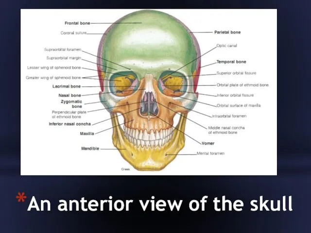
- 7. An anterior view of the skull
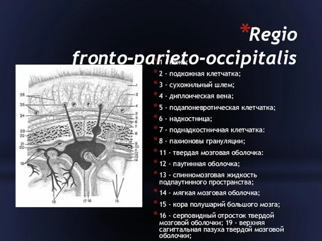
- 8. Regio fronto-parieto-occipitalis 1 - кожа; 2 - подкожная клетчатка; 3 - сухожильный шлем; 4 - диплоическая
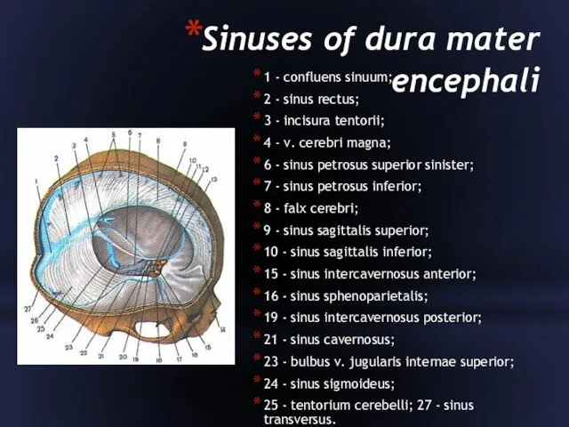
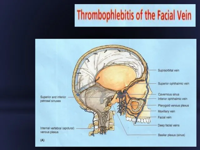
- 9. Sinuses of dura mater encephali 1 - confluens sinuum; 2 - sinus rectus; 3 - incisura
- 10. Regio temporalis
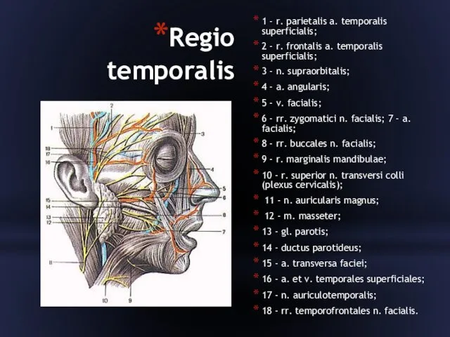
- 11. Regio temporalis 1 - r. parietalis a. temporalis superficialis; 2 - r. frontalis a. temporalis superficialis;
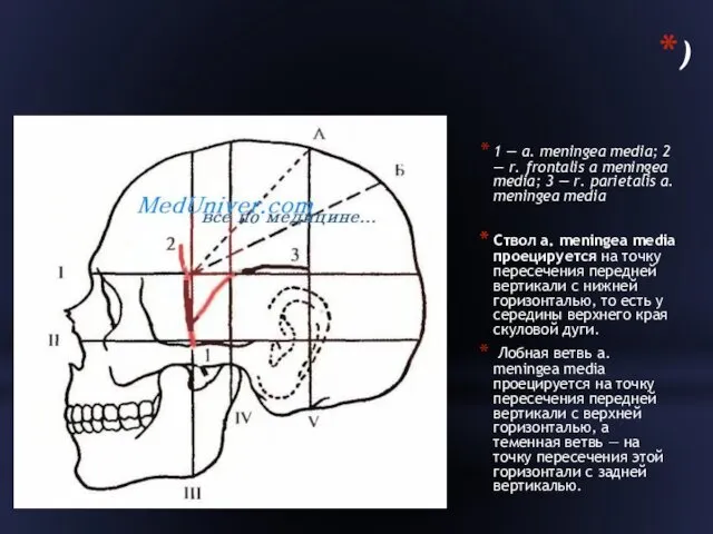
- 12. ) 1 — a. meningea media; 2 — r. frontalis a meningea media; 3 — r.
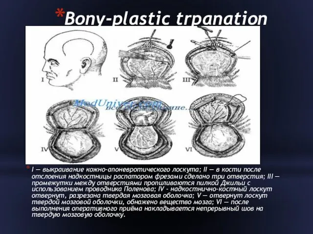
- 13. Bony-plastic trpanation I — выкраивание кожно-апоневротического лоскута; II — в кости после отслоения надкостницы распатором фрезами
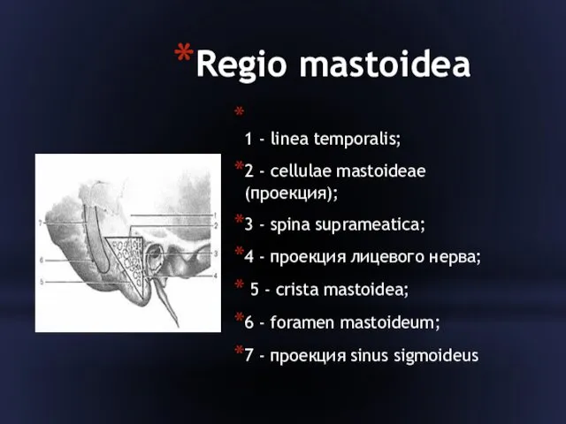
- 14. Regio mastoidea 1 - linea temporalis; 2 - cellulae mastoideae (проекция); 3 - spina suprameatica; 4
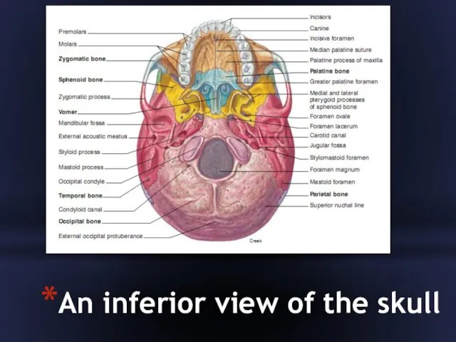
- 15. An inferior view of the skull
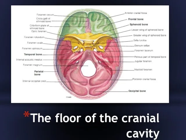
- 16. The floor of the cranial cavity
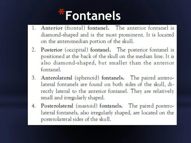
- 17. Fontanels
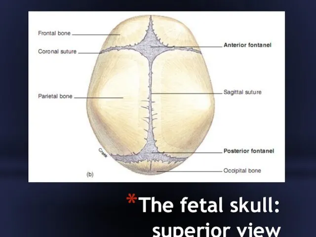
- 18. The fetal skull: superior view
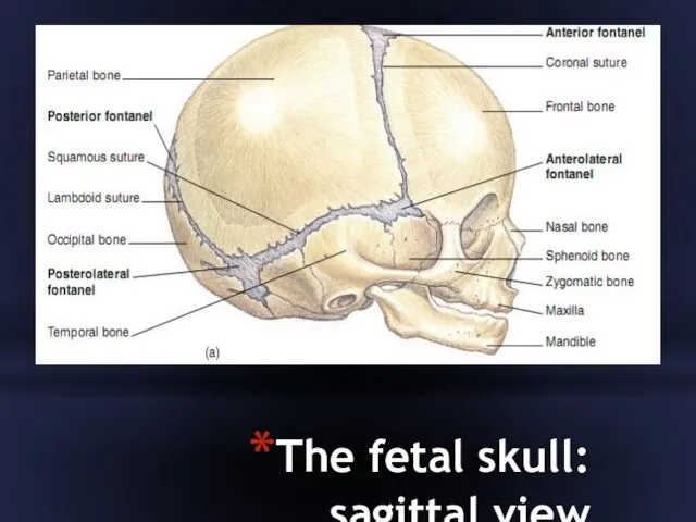
- 19. The fetal skull: sagittal view
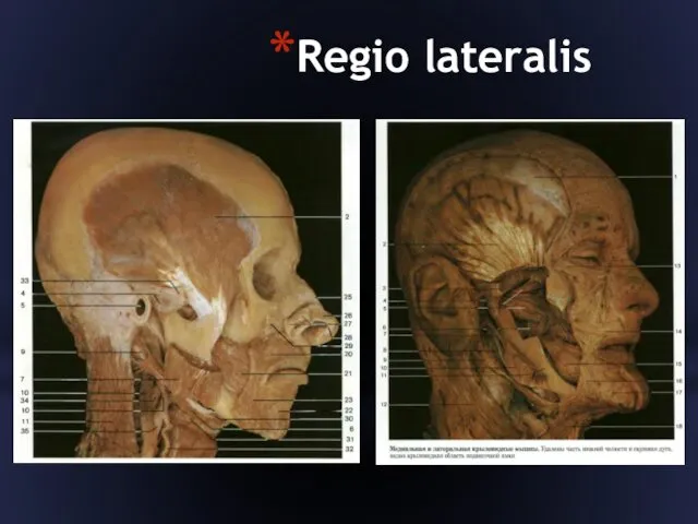
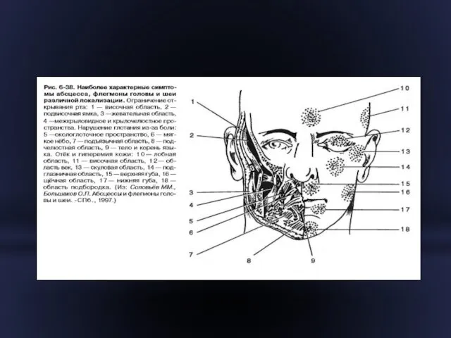
- 20. Regio lateralis
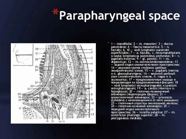
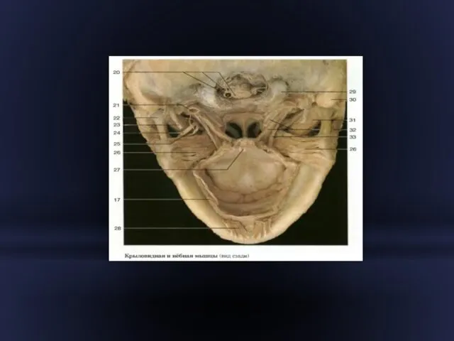
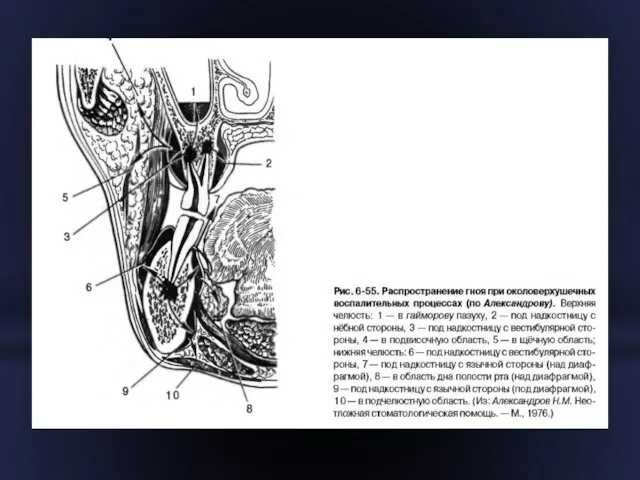
- 22. Parapharyngeal space 1 — mandibula; 2 — m. masseter; 3 — ductus parotideus; 4 — fascia
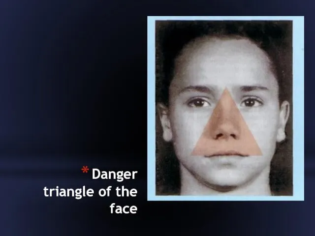
- 24. Danger triangle of the face
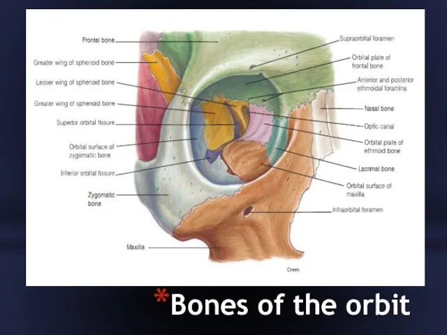
- 29. Bones of the orbit
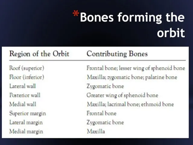
- 30. Bones forming the orbit
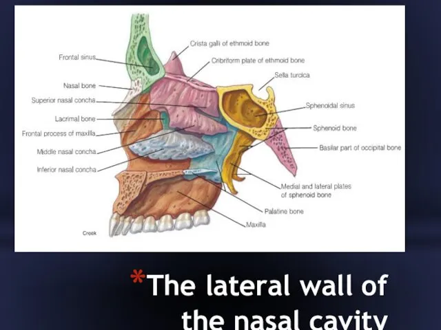
- 31. The lateral wall of the nasal cavity
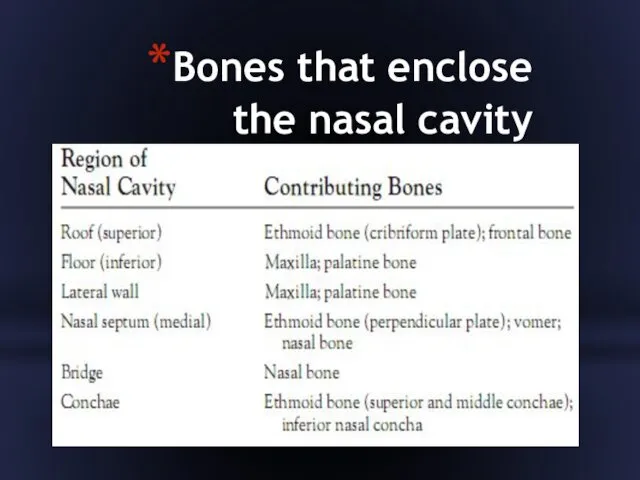
- 32. Bones that enclose the nasal cavity
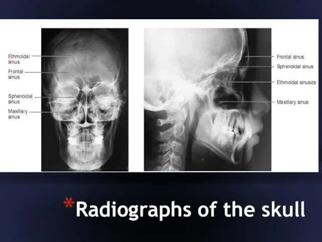
- 33. Radiographs of the skull
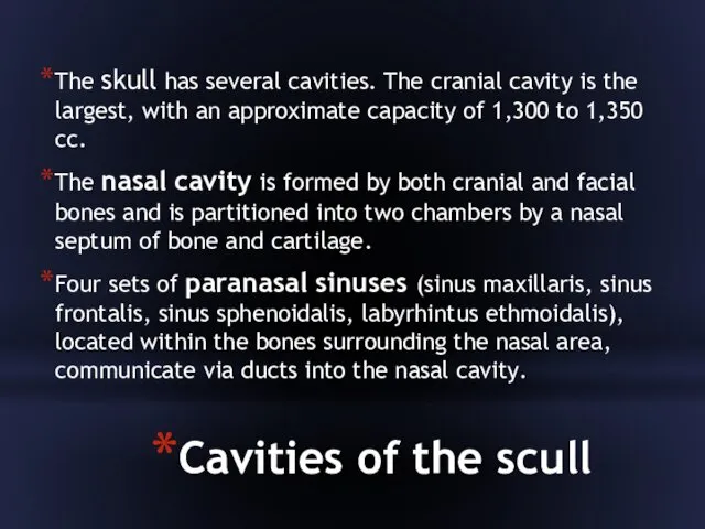
- 34. Cavities of the scull The skull has several cavities. The cranial cavity is the largest, with
- 35. Cavities of the scull Middle and inner-ear cavities are positioned inferior to the cranial cavity and
- 36. An inferolateral view of the skull
- 37. A sagittal view of the skull
- 39. Скачать презентацию
Topographic anatomy & operative surgery
TOPOGRAPHIC ANATOMY is a science which studies relations between
Topographic anatomy & operative surgery
TOPOGRAPHIC ANATOMY is a science which studies relations between
Cranial bones
The cranial bones enclose and protect the brain and associated sensory organs.
They
Cranial bones
The cranial bones enclose and protect the brain and associated sensory organs.
They
SKULL
The human skull, consisting of 8 cranial and 14 facial bones, contains several
SKULL
The human skull, consisting of 8 cranial and 14 facial bones, contains several
The eight bones of the cranium articulate firmly with one another to enclose
The eight bones of the cranium articulate firmly with one another to enclose
A lateral view of the skull
A lateral view of the skull
An anterior view of the skull
An anterior view of the skull
Regio fronto-parieto-occipitalis
1 - кожа;
2 - подкожная клетчатка;
3 - сухожильный шлем;
4
Regio fronto-parieto-occipitalis
1 - кожа;
2 - подкожная клетчатка;
3 - сухожильный шлем;
4
Sinuses of dura mater encephali
1 - confluens sinuum;
2 - sinus rectus;
3
Sinuses of dura mater encephali
1 - confluens sinuum;
2 - sinus rectus;
3
Regio temporalis
Regio temporalis
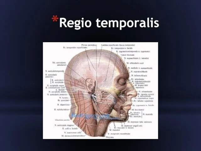
Regio temporalis
1 - r. parietalis a. temporalis superficialis;
2 - r. frontalis a.
Regio temporalis
1 - r. parietalis a. temporalis superficialis;
2 - r. frontalis a.
)
1 — a. meningea media; 2 — r. frontalis a meningea media; 3
)
1 — a. meningea media; 2 — r. frontalis a meningea media; 3
Bony-plastic trpanation
I — выкраивание кожно-апоневротического лоскута; II — в кости после отслоения
Bony-plastic trpanation
I — выкраивание кожно-апоневротического лоскута; II — в кости после отслоения
Regio mastoidea
1 - linea temporalis;
2 - cellulae mastoideae (проекция);
3 - spina
Regio mastoidea
1 - linea temporalis;
2 - cellulae mastoideae (проекция);
3 - spina
An inferior view of the skull
An inferior view of the skull
The floor of the cranial cavity
The floor of the cranial cavity
Fontanels
Fontanels
The fetal skull: superior view
The fetal skull: superior view
The fetal skull: sagittal view
The fetal skull: sagittal view
Regio lateralis
Regio lateralis

Parapharyngeal space
1 — mandibula; 2 — m. masseter; 3 — ductus parotideus; 4
Parapharyngeal space
1 — mandibula; 2 — m. masseter; 3 — ductus parotideus; 4
Danger triangle of the face
Danger triangle of the face
Bones of the orbit
Bones of the orbit
Bones forming the orbit
Bones forming the orbit
The lateral wall of the nasal cavity
The lateral wall of the nasal cavity
Bones that enclose the nasal cavity
Bones that enclose the nasal cavity
Radiographs of the skull
Radiographs of the skull
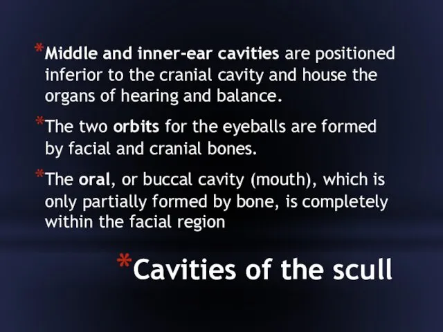
Cavities of the scull
The skull has several cavities. The cranial cavity is the
Cavities of the scull
The skull has several cavities. The cranial cavity is the
Cavities of the scull
Middle and inner-ear cavities are positioned inferior to the cranial
Cavities of the scull
Middle and inner-ear cavities are positioned inferior to the cranial
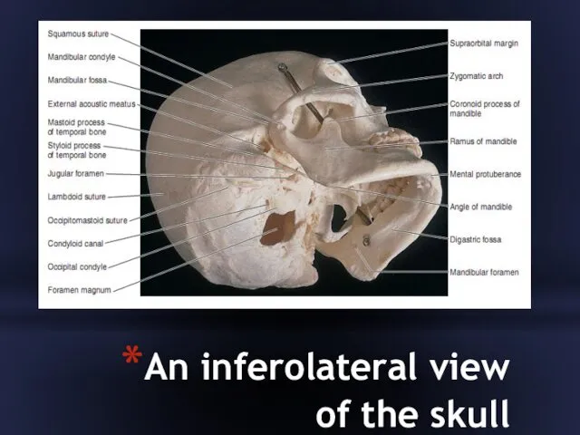
An inferolateral view of the skull
An inferolateral view of the skull
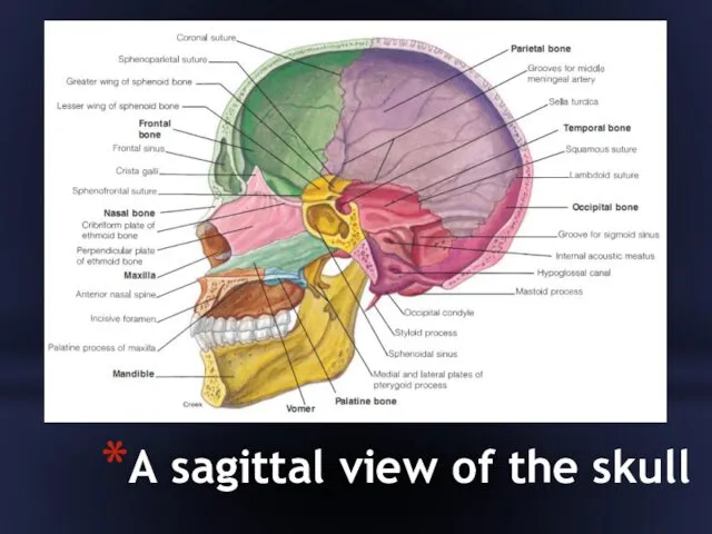
A sagittal view of the skull
A sagittal view of the skull
 Зрительные диктанты часть 3.
 Экологическая этика
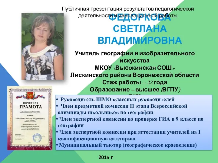 ПУБЛИЧНАЯ ПРЕЗЕНТАЦИЯ - 2015г.
 Составные задачи на нахождение суммы, в которых неизвестно одно из слагаемых
 Программа Сметное дело
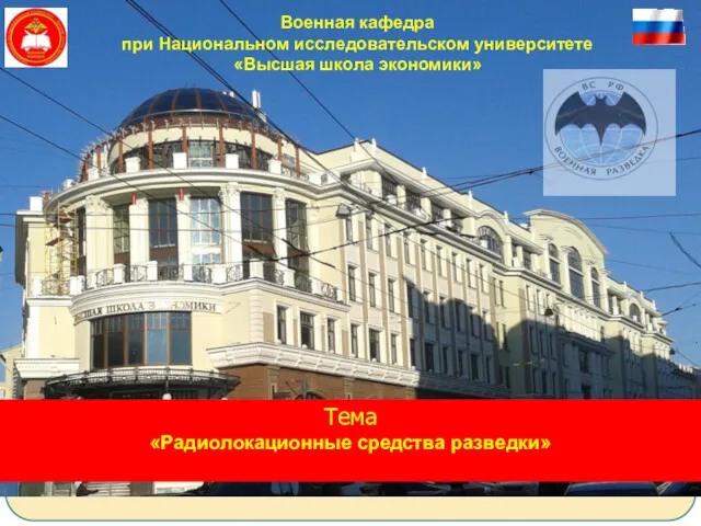 Радиолокационные средства разведки
 Денсаулық пен өмірлік дағдыларды қалыптасыру, сондай-ақ кәмелетке толмағандар арасында суицидтің алдын алу бағдарламасы
 Мой родной город - Колпино
 Буддийский храм в Петербурге - уникальное сооружение в Европе
 Архитектурные конструкции общественных зданий (часть 1 - общая типология)
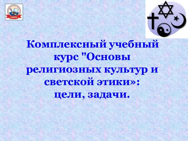 Основы религиозных культур и светской этики
 Научное исследование, учебное исследование, проектная деятельность
 Магистратура ВятГУ
 21- ғасыр Көшбасшысы
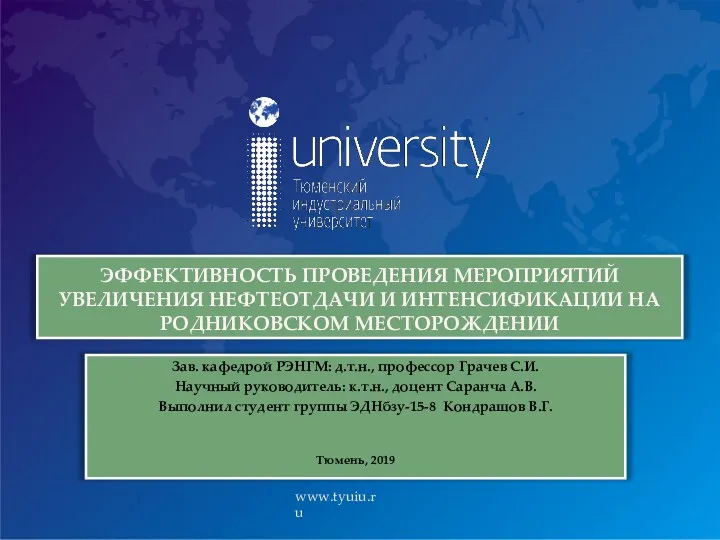 Эффективность проведения мероприятий увеличения нефтеотдачи и интенсификации на Родниковском месторождении
 Особенности организационных структур в международном маркетинге
 Инвертор сварочный IW-140/6ATL
 Проектная работа Лучшее электронное портфолио
 Растительный и животный мир России.
 Воспитательная система класса Азбука вежливости.
 презентация по кружку РАДУГА
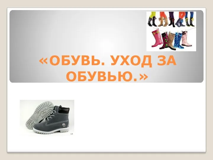 Конспект занятия к образовательной программе по трудовому воспитанию и профориентации Обувь. Уход за обувью
 Принципы имитационного моделирования. Математические методы моделирования
 Изменения в сердечно-сосудистой системе женщины во время беременности:
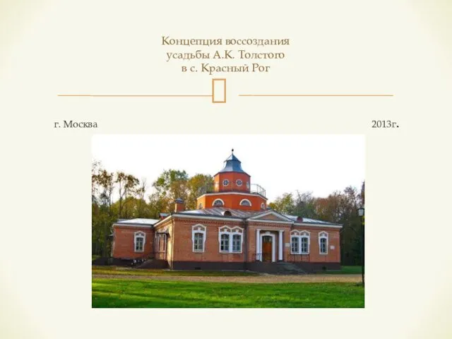 Концепция воссоздания усадьбы А.К. Толстого в селе Красный Рог
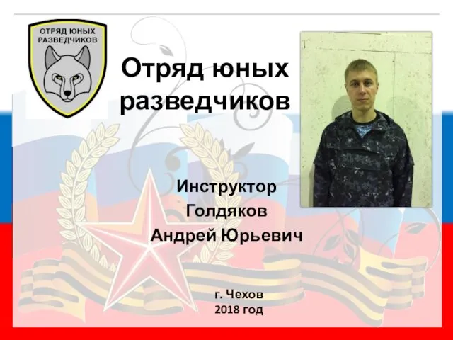 Отряд юных разведчиков
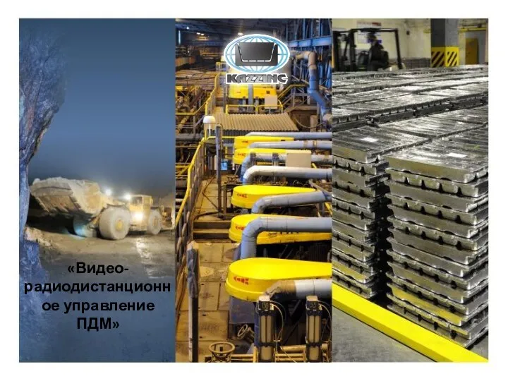 Видеорадиодистанционное управление ПДМ
 Развитие интереса к элементарной познавательно–исследовательской деятельности у детей раннего дошкольного возраста