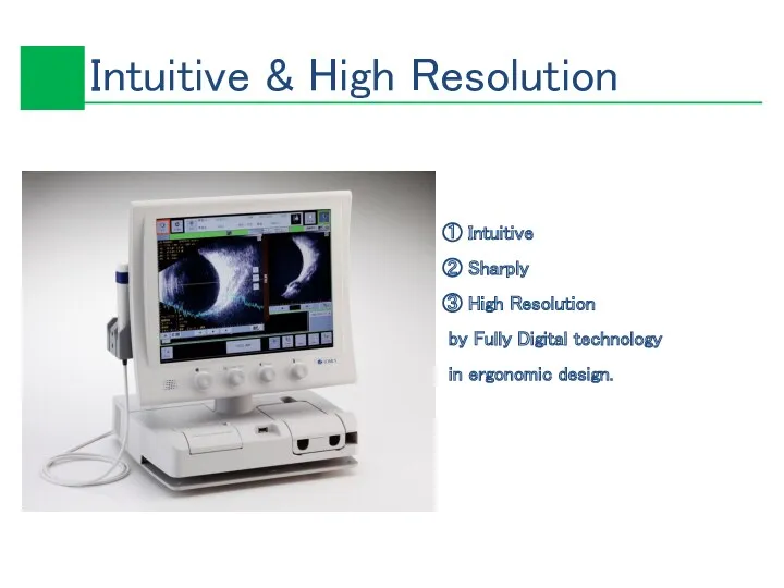
- Ultrasonic B-Scanner UD-8000. Inspire by Digital Tomey Corporation

Содержание
- 2. ① Intuitive ② Sharply ③ High Resolution by Fully Digital technology in ergonomic design.
- 3. Adjustable focusing by long ultrasound beam Extra High-resolution 6 Gradual Reception Dynamic Focus ① Anullar Array
- 4. ② 15MHz 20MHz by one probe
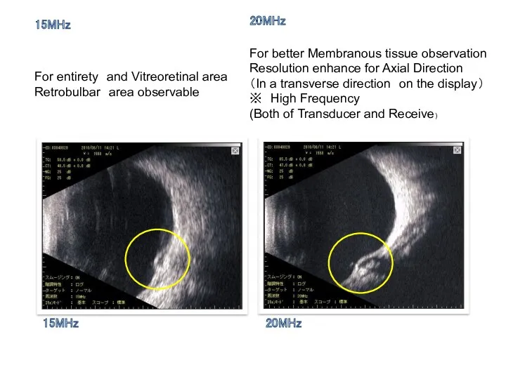
- 5. 15MHz 20MHz For entirety and Vitreoretinal area Retrobulbar area observable For better Membranous tissue observation Resolution
- 6. ③ Harmonic For Optic disc, Macular area disease and ailments observable resolution enhance for Lateral Direction
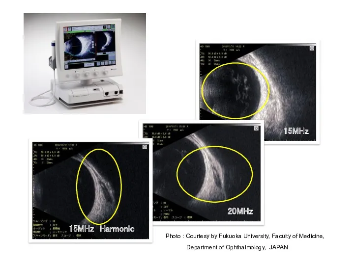
- 7. 15MHz 20MHz 15MHz Harmonic Photo : Courtesy by Fukuoka University, Faculty of Medicine, Department of Ophthalmology,
- 8. ④ Indicate of probe direction
- 9. ⑤ New designed probe
- 10. ① Standard mode ② High Sensitive Mode ※ Deduct noise, improve SNR Frame rate 】 ①
- 11. Standard Eye ball or Wide Orbit Display Range】 Standard : 42 mm Wide : 54 mm
- 12. You can select 4 eye types Micro ( Child ) Normal Long Retoro( Normal ) ③
- 13. Select eye type On screen annotation
- 14. ① Play back movie ⑦ Vector A-Mode ② Zoom ⑧ Length measurement ③ Area calculation ⑨
- 15. ① Movie ( Play Back) Play back movie by 20 seconds or 400 frames still images.
- 16. ② Vector A-Mode Temporary A-Biometry by Compare with pathology wave form and retina wave form.
- 17. 5 steps Zoom 100%, 125%, 150%, 200%, 300% Intuitive navigation monitor ③ Zoom
- 18. ④ Length calculation Up to 3 spots on same time calculation
- 19. ⑤ Area calculation Up to 2 spots on same time calculation
- 20. ⑥ Angle calculation Up to 2 spots on same time calculation
- 21. ⑦ Dual/Quad Display
- 22. ⑧ R(OD)/L(OS) Display
- 23. ⑨ In put Comment In put comment on still picture or movie
- 24. ⑩ Print out by still image part only
- 25. ⑪ Thumbnail images
- 26. ⑫ Group Export / Group Save
- 27. ①Still image Save Echo or JPEG format ( Re-Display, Edit possible ) ② Movie Save in
- 28. USB memory save capacity ID : 32,750 Patients (Max) Group Save : 64 Groups/Patient 20 still
- 29. UBM 60Mhz B-Scan (Estimate) Still picture 32GB Approximately 56,700 Pictures 16GB Approximately 28,300 Pictures 8GB Approximately
- 30. ① Disposable ② No eye cup ③ Sitting position
- 34. Angle Analysis Parameter 】
- 35. Iris Analysis
- 36. Iris Analysis Parameter 】
- 37. AL-4000 ( Biometry/Pachymetry) ① A-Biometry ② IOL Power calculation ③ Pachymetry USB or Bluetooth communication
- 38. ① A-Biometery One eye Both eye(R/L)
- 39. ② IOL Power calculation One eye Both eye(R/L)
- 40. ③ Pachymetry One eye Both eye(R/L)
- 41. ? Focus : Dynamic Focus ? Frame rate Basic mode : 22 frame / sec High
- 44. Скачать презентацию
① Intuitive
② Sharply
③ High Resolution
by Fully Digital technology
in ergonomic
① Intuitive
② Sharply
③ High Resolution
by Fully Digital technology
in ergonomic
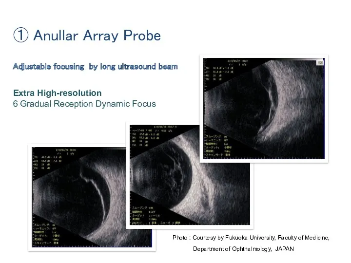
Adjustable focusing by long ultrasound beam
Extra High-resolution
6 Gradual Reception Dynamic
Adjustable focusing by long ultrasound beam
Extra High-resolution
6 Gradual Reception Dynamic
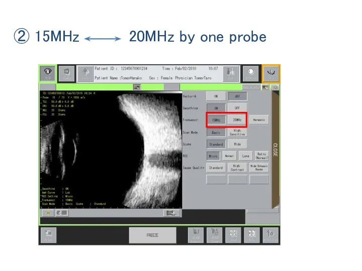
② 15MHz 20MHz by one probe
② 15MHz 20MHz by one probe
15MHz
20MHz
For entirety and Vitreoretinal area
Retrobulbar area observable
For better Membranous tissue observation
Resolution enhance
15MHz
20MHz
For entirety and Vitreoretinal area
Retrobulbar area observable
For better Membranous tissue observation
Resolution enhance
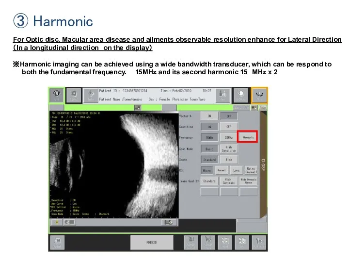
③ Harmonic
For Optic disc, Macular area disease and ailments observable resolution
③ Harmonic
For Optic disc, Macular area disease and ailments observable resolution
15MHz
20MHz
15MHz Harmonic
Photo : Courtesy by Fukuoka University, Faculty of Medicine,
15MHz
20MHz
15MHz Harmonic
Photo : Courtesy by Fukuoka University, Faculty of Medicine,
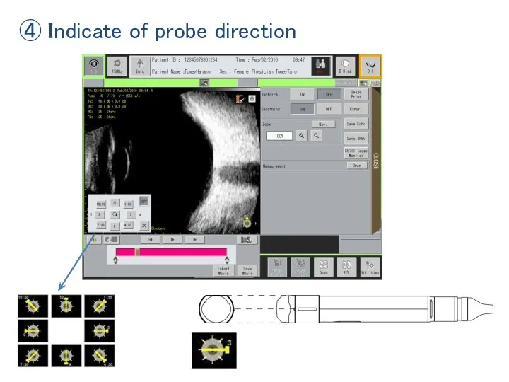
④ Indicate of probe direction
④ Indicate of probe direction
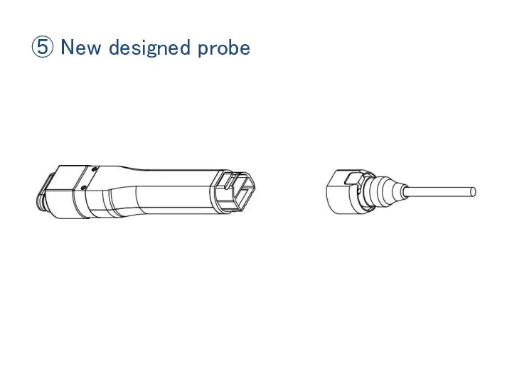
⑤ New designed probe
⑤ New designed probe
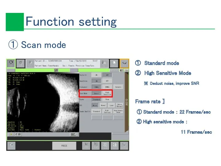
① Standard mode
② High Sensitive Mode
※ Deduct noise, improve SNR
Frame rate
① Standard mode
② High Sensitive Mode
※ Deduct noise, improve SNR
Frame rate
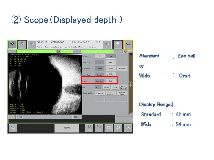
Standard Eye ball
or
Wide Orbit
Display Range】
Standard : 42 mm
Wide :
Standard Eye ball
or
Wide Orbit
Display Range】
Standard : 42 mm
Wide :
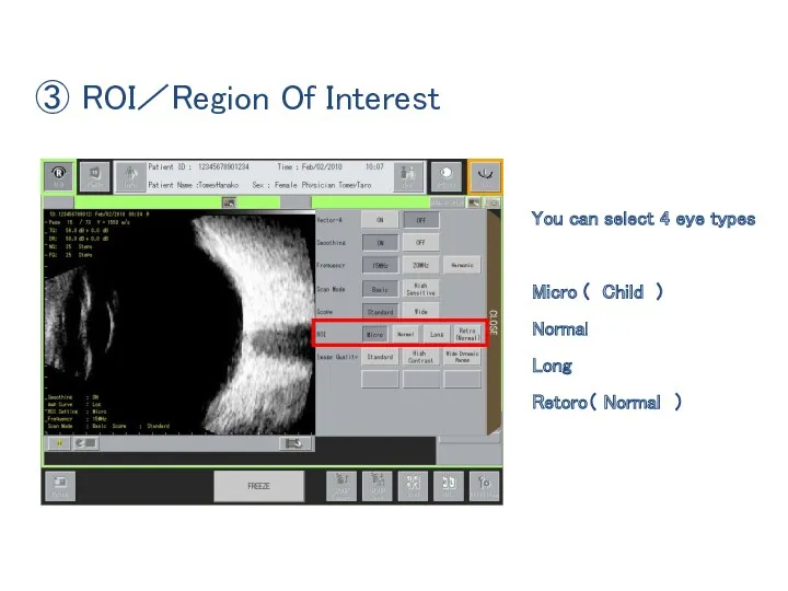
You can select 4 eye types
Micro ( Child )
Normal
Long
Retoro( Normal )
③ ROI/Region Of Interest
You can select 4 eye types
Micro ( Child )
Normal
Long
Retoro( Normal )
③ ROI/Region Of Interest
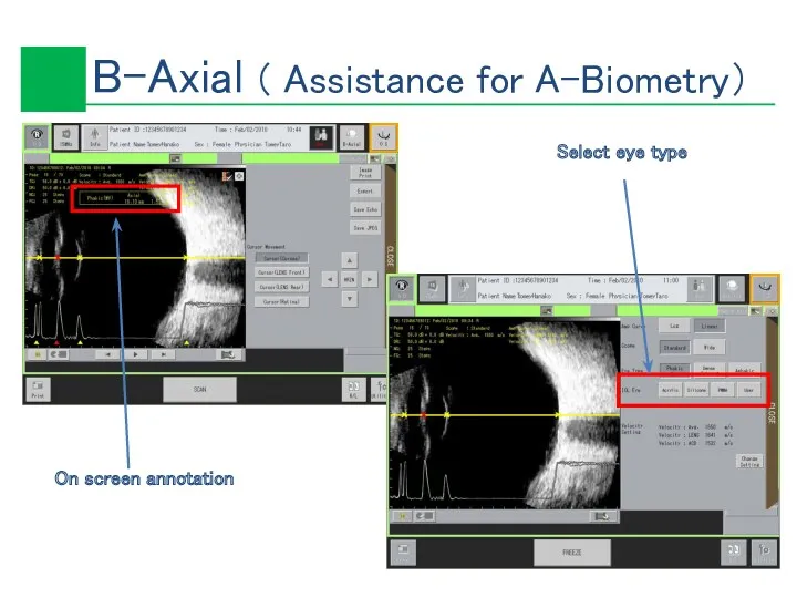
Select eye type
On screen annotation
Select eye type
On screen annotation
① Play back movie ⑦ Vector A-Mode
② Zoom ⑧ Length measurement
③
① Play back movie ⑦ Vector A-Mode
② Zoom ⑧ Length measurement
③
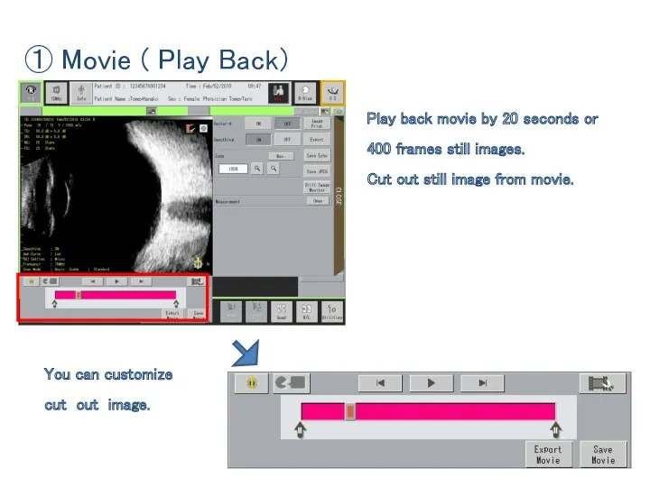
① Movie ( Play Back)
Play back movie by 20 seconds or
① Movie ( Play Back)
Play back movie by 20 seconds or
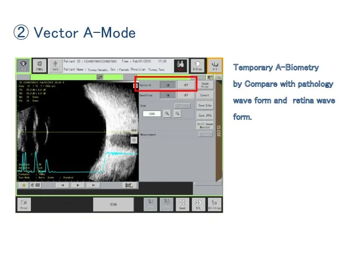
② Vector A-Mode
Temporary A-Biometry
by Compare with pathology
wave form and retina wave form.
② Vector A-Mode
Temporary A-Biometry
by Compare with pathology
wave form and retina wave form.
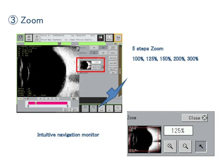
5 steps Zoom
100%, 125%, 150%, 200%, 300%
Intuitive navigation monitor
③
5 steps Zoom
100%, 125%, 150%, 200%, 300%
Intuitive navigation monitor
③
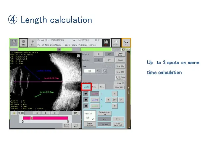
④ Length calculation
Up to 3 spots on same time calculation
④ Length calculation
Up to 3 spots on same time calculation
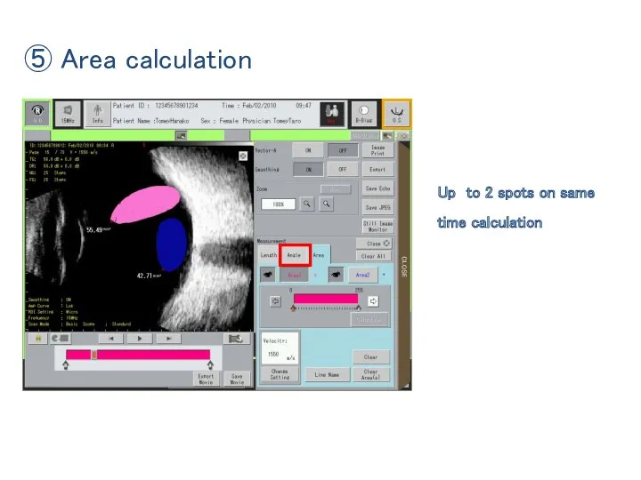
⑤ Area calculation
Up to 2 spots on same time calculation
⑤ Area calculation
Up to 2 spots on same time calculation
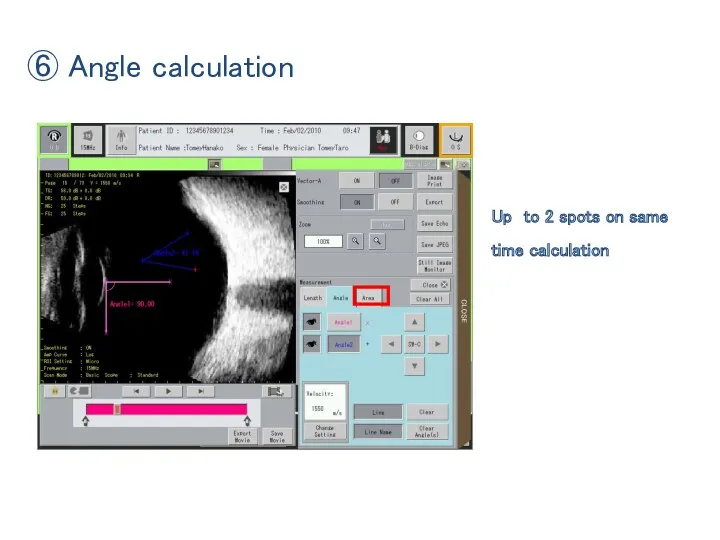
⑥ Angle calculation
Up to 2 spots on same time calculation
⑥ Angle calculation
Up to 2 spots on same time calculation
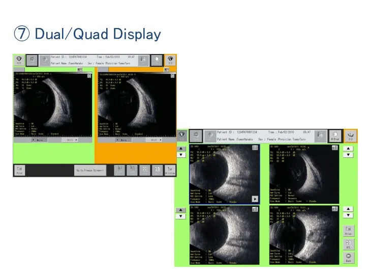
⑦ Dual/Quad Display
⑦ Dual/Quad Display
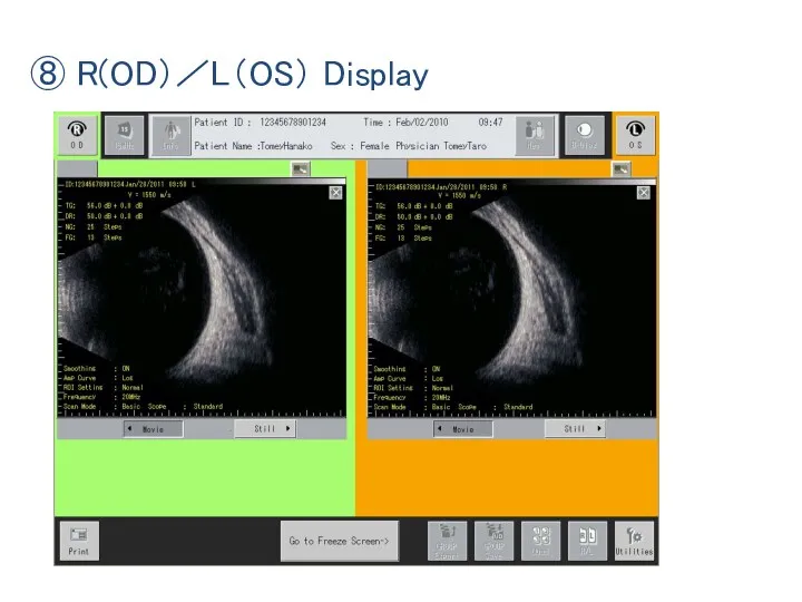
⑧ R(OD)/L(OS) Display
⑧ R(OD)/L(OS) Display
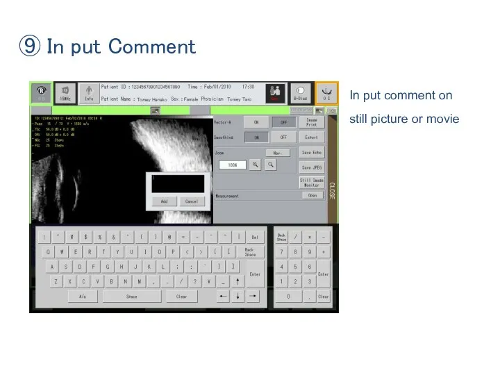
⑨ In put Comment
In put comment on
still picture or movie
⑨ In put Comment
In put comment on
still picture or movie
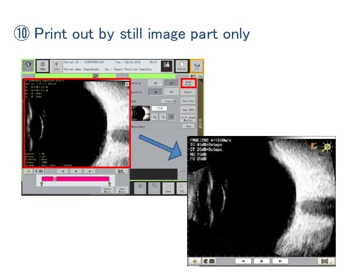
⑩ Print out by still image part only
⑩ Print out by still image part only
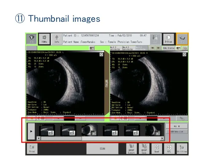
⑪ Thumbnail images
⑪ Thumbnail images
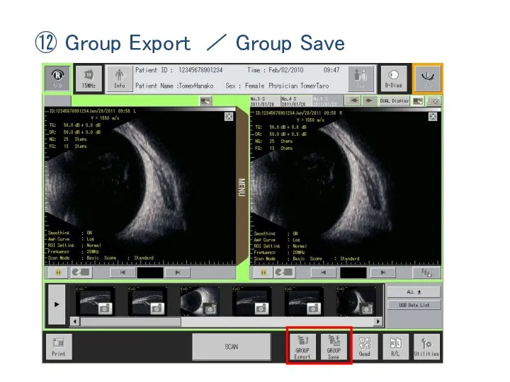
⑫ Group Export / Group Save
⑫ Group Export / Group Save
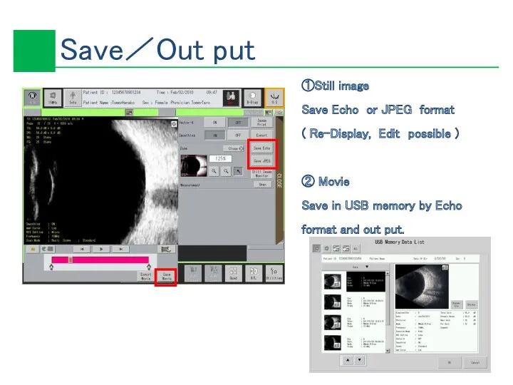
①Still image
Save Echo or JPEG format
( Re-Display, Edit possible )
①Still image
Save Echo or JPEG format
( Re-Display, Edit possible )

USB memory save capacity
ID : 32,750 Patients (Max)
Group Save : 64
USB memory save capacity
ID : 32,750 Patients (Max)
Group Save : 64
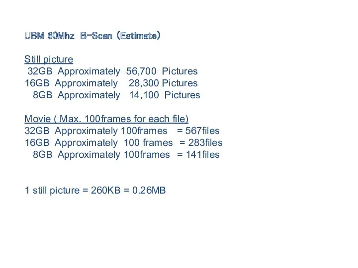
UBM 60Mhz B-Scan (Estimate)
Still picture
32GB Approximately 56,700 Pictures
16GB Approximately 28,300
Still picture
32GB Approximately 56,700 Pictures
16GB Approximately 28,300
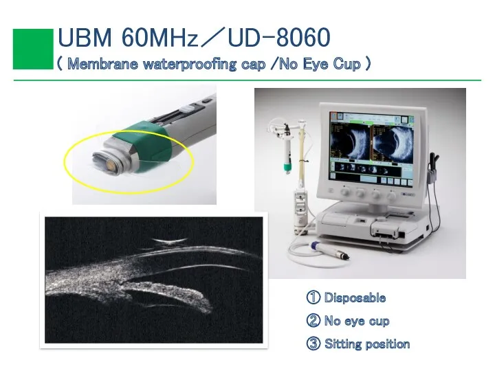
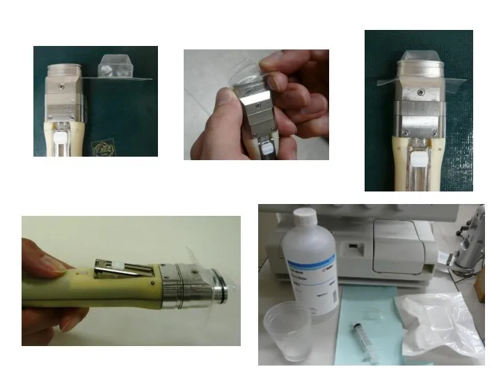
① Disposable
② No eye cup
③ Sitting position
① Disposable
② No eye cup
③ Sitting position

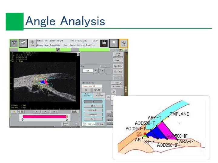
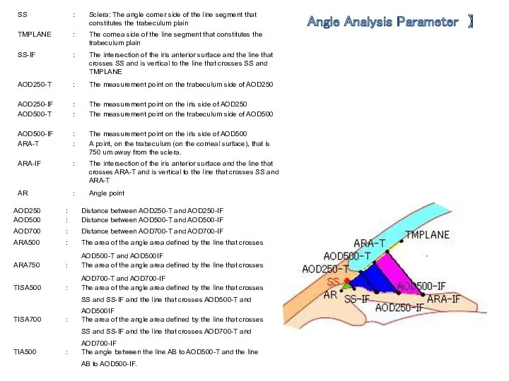
Angle Analysis Parameter 】
Angle Analysis Parameter 】
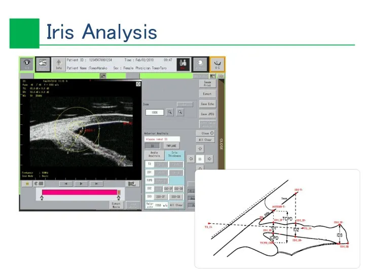
Iris Analysis
Iris Analysis
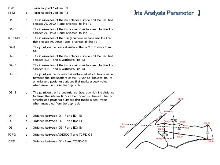
Iris Analysis Parameter 】
Iris Analysis Parameter 】
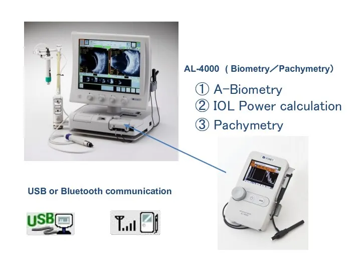
AL-4000 ( Biometry/Pachymetry)
① A-Biometry
② IOL Power calculation
③ Pachymetry
USB or Bluetooth communication
AL-4000 ( Biometry/Pachymetry)
① A-Biometry
② IOL Power calculation
③ Pachymetry
USB or Bluetooth communication
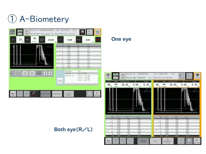
① A-Biometery
One eye
Both eye(R/L)
① A-Biometery
One eye
Both eye(R/L)
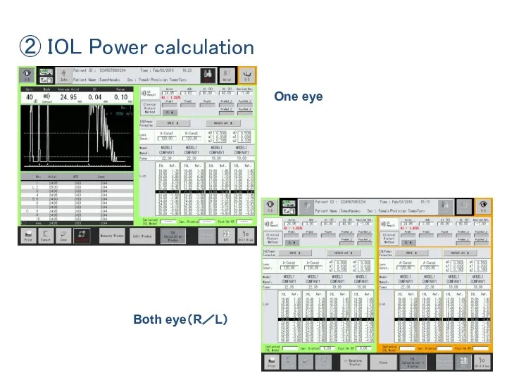
② IOL Power calculation
One eye
Both eye(R/L)
② IOL Power calculation
One eye
Both eye(R/L)
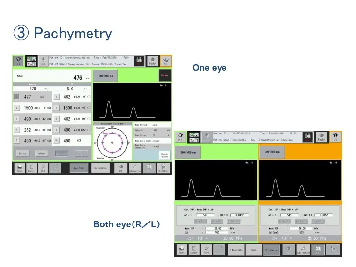
③ Pachymetry
One eye
Both eye(R/L)
③ Pachymetry
One eye
Both eye(R/L)
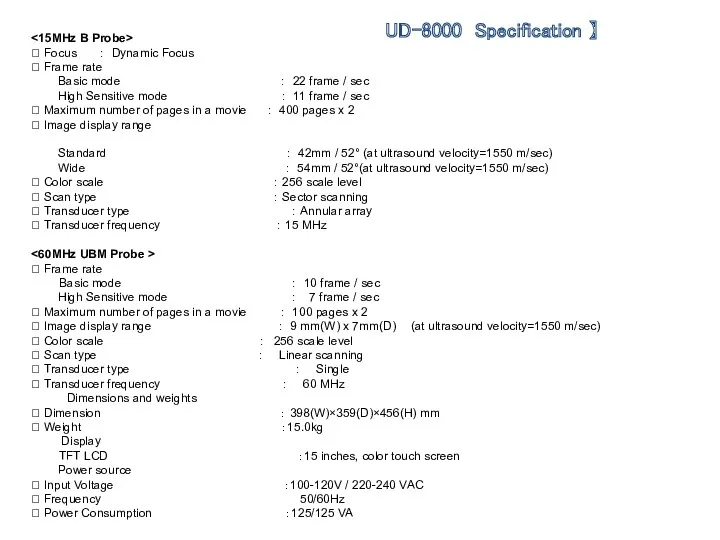
<15MHz B Probe>
? Focus : Dynamic Focus
? Frame rate
Basic mode
<15MHz B Probe>
? Focus : Dynamic Focus
? Frame rate
Basic mode
 Конкурс знатоков химии и биологии
 Основы производства комбикормов
 Логические схемы
 Приоритетный региональный проект Муниципальные дороги
 Ох уж эта математика
 Совершенствование системы адаптации сотрудников и её влияние на устойчивость персонала ПАО МинБанк
 Основы конституционного права Республики Индия
 Моя семья
 Создание однотабличной базы данных
 Мұнай ұңғымаларында құм түзілуін болдырмау әдістері
 Разработка нефтяных месторождений. Часть 2
 Материальная часть и правила стрельбы из АК-74
 Полные и краткие формы качественных прилагательных, их роль в предложении
 Разработка алгоритма ориентации роботизированных платформ с учетом датчиков
 Светодиодные лампы
 The Personal Computer and its devices
 Светлейший князь Григо́рий Алекса́ндрович Потёмкин-Таври́ческий
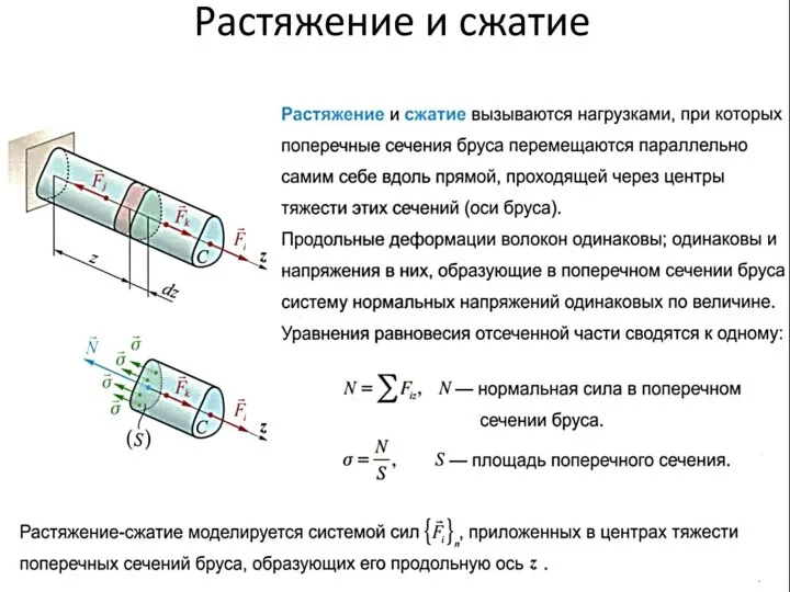 Растяжение и сжатие
 Тульский Кремль
 Сказочная страна. Игра
 Порядок составления, уточнения и использования списков избирателей на выборах губернатора Свердловской обл. 10 сентября 2017 г
 Проект 1 Б класса Исследование: Моё родословие
 Социальные статусы и роли
 General Overview 04-23-2018
 В гостях у сказки
 Курманжан Датка
 Чона металургія України
 Горячий цех на предприятиях общественного питания