- Acute renal failure (ARF)

Содержание
- 2. DEFINITION Acute renal failure (ARF) is an abrupt and sudden reduction in renal function resulting in
- 3. DEFINITION It is usually associated with oliguria (urine output BUN & creatinine values are elevated
- 4. FUNCTIONS OF THE KIDNEY’S Urine Formation: Formed in the nephrons through a complex three-step process: Glomerular
- 5. KIDNEY FUNCTIONS Control of water balance: Normal ingestion of water daily is 1-2L and normally all
- 6. FUNCTIONS OF THE KIDNEY’S Regulation of electrolytes: volume of electrolytes excreted per day is exactly equal
- 7. FUNCTIONS OF THE KIDNEY’S Control of blood pressure: BP monitored by the vasa recta. Juxtaglomerular cells,
- 8. KIDNEY FUNCTIONS Regulation of acid-base balance: elimination of sulphuric and phosphoric acid
- 9. ANATOMY OF THE KIDNEY
- 10. NEPHRON
- 11. RENIN-ANGIOTENSIN SYSTEM
- 12. PATHOPHYSIOLOGY Glomerular filtration is caused by difference between glomerular pressure (70 mm Hg), colloid oncotic pressure
- 13. PATHOPHYSIOLOGY As a result of the filtration primary urine is formed. The kidneys produce 180 to
- 14. PATHOPHYSIOLOGY The return flow of filtered molecules from the tubules to the blood is called reabsorption
- 15. EPIDEMIOLOGY OF ARF Incidence, etiology and outcome varied depending on Population studied and Definition used Mostly
- 16. CLASSIFICATION ARF may occur in 3 clinical settings: As an adaptive response to severe volume depletion
- 17. CLASSIFICATION Prerenal As many as 70% of patients with ARF are prerenal. Reduced renal perfusion caused
- 18. CLASSIFICATION Prerenal Afferent arteriolar vasodilation and efferent vasoconstriction of the glomerular vessels (mediated by dilating prostaglandins
- 19. CLASSIFICATION The causes of prerenal ARF include the following: volume depletion gastrointestinal loss, excessive diuresis, and
- 20. CLASSIFICATION Intrinsic Intrinsic cases comprise 25% of all acute renal failure cases. Most cases (90%) of
- 21. CLASSIFICATION The causes of intrinsic renal failure include: renal ischemia renal artery/vein thrombosis glomerulonephritis vasculitides hemolytic
- 22. POST-RENAL ARF Obstruction – complete or Partial Anuria or variable urine output Recovery depends on duration
- 23. PHASES OF ACUTE RENAL FAILURE Clinical progression of reversible RF occurs in four phases: Initiation phase
- 24. PHASES OF ACUTE RENAL FAILURE Diuretic phase The kidneys begin to recover Initially produce hypotoniс urine
- 25. DIAGNOSIS While a medical history and physical examination are important in making a diagnosis of acute
- 26. DIAGNOSIS History Observe for disorder that predisposes pt to ARF Ask questions about recent illness, infections,
- 27. CLINICAL MANIFESTATIONS OF ARF Cardiovascular Arrhythmias BP, N, high or low Anemia P, rapid, bounding, or
- 28. CLINICAL MANIFESTATIONS OF ARF Genitourinary Oliguria Anuria abN urine colour, clarity, smell GI Moist tongue &
- 29. DIAGNOSIS Oliguria (urine output raised urea and creatinine hyperkalemia metabolic acidosis. All of the above problems
- 30. The fractional excretion of sodium (FENa+) is considered to be the most reliable biochemical laboratory discriminator
- 31. DIAGNOSIS Distinction between prerenal and intrinsic ARF ARF , acute renal failure; FE Na+, fractional excretion
- 32. OLIGURIC PHASE Hypervolemia Elevated blood urea nitrogen and serum creatinine levels Normal or decreased serum sodium
- 33. TREATMENT Prerenal renal failure The aim of treatment is to restore renal perfusion before intrinsic renal
- 35. TREATMENT Intrinsic renal failure Renal perfusion should be maintained to eliminate prerenal failure. Measures should be
- 36. TREATMENT Nutritional support Adequate nutrition is of considerable importance and should be enteral if at all
- 37. TREATMENT Treatment of hyperkalemia Treatment is required if EKG changes are present or potassium (K+) levels
- 39. TREATMENT If dialysis is not immediately available, the following measures may be used to temporarily redistribute
- 40. TREATMENT • If the patient is acidotic, give sodium bicarbonate (NaHCO3) 50–100 mmol over 1 h
- 41. TREATMENT Treatment of acidosis Sodium bicarbonate should be used only when acidosis is severe (pH The
- 42. TREATMENT Identification and treatment of sepsis Commence empiric broad-spectrum therapy once cultures have been taken, bearing
- 43. TREATMENT Cause-specific therapies : • mannitol/NaHCO3 in acute rhabdomyolysis • immunosuppression in SLE, Wegener’s granulomatosis, or
- 44. HEMODIALYSIS Who needs dialysis? Guidelines for the initiation of renal replacement therapy Severe hyperkalaemia, unresponsive to
- 45. HEMODIALYSIS Dialysis is a type of renal replacement therapy which is used to provide artificial replacement
- 46. HEMODIALYSIS Healthy kidneys remove waste products (potassium, acid, urea) from the blood and they also remove
- 47. PRINCIPLE OF DIALYSIS Dialysis works on the principle of diffusion of solutes along a concentration gradient
- 48. HEMODIALYSIS Client’s blood is passed through a system of tubing (dialysis circuit) via a machine to
- 49. ACUTE RENAL SUPPORT is usually performed as 4-h sessions daily or on alternate days. It is
- 51. EQUIPMENT NEEDED FOR HD The HD machine performs the function of pumping the patient's blood and
- 52. HEMODIALYSIS
- 54. The side effects are proportionate to the amount of fluid being removed Decreased blood pressure Fatigue
- 55. Complications of HD Because HD requires access to the circulatory system, clients have a portal of
- 56. ACUTE RENAL SUPPORT Hemodialysis All variants of hemodialysis share the need for the following: • vascular
- 57. ACUTE RENAL SUPPORT Hemodialysis Potential problems include the following: • dysequilibrium syndrome – rapid changes in
- 58. PERITONEAL DIALYSIS
- 59. WHAT IS PERITONEAL DIALYSIS (PD)? Peritoneal dialysis works by using the body's peritoneal membrane, which is
- 60. ADVANTAGES OF PD Can be done at home Relatively easy for the client to learn Easy
- 61. DIURETIC PHASE Diuretic phase: The kidneys try to heal and urine output increases, but tubule scarring
- 62. RECOVERY PHASE (CONVALESCENT) Tubular edema resolves and renal function improves. Increased glomerular filtration rate Stabilization or
- 64. Скачать презентацию
 Накладання джгута-турнікета
 Болезнетворные факторы внешней среды
 Фиксация съемных ортопедических конструкций
 Нейрофиброматоз
 Иммунный статус макроорганизма. Методы оценки. (Лекция 15)
 Кислородотерапия. Виды. Показания. Противопоказания. Цели и способы доставки
 Анемиялардың анықтамасы, жіктелуі
 Пилотный проект Развитие системы медицинской реабилитации в Российской Федерации
 Эктопротездер
 Этическая оценка метода ЭКО с точки зрения православия и студенчества ГрГМУ в констексте современной науки
 Гипервитаминоз Д
 Денсаулық сақтаудағы менеджмент
 Осложнения острого аппендицита
 Понятие об инкретинах.агпп1 Шундеева ЮВ
 История развития сестринского дела в России
 Балалардағы пневмония
 Возбудитель холеры
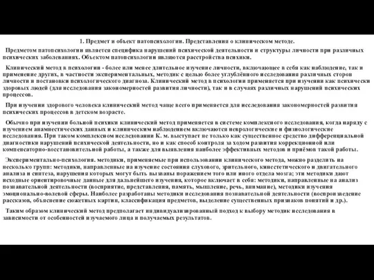 Предмет и объект патопсихологии. Представления о клиническом методе
 Использование хвойных растений при простудных заболеваниях
 Перименопауза и гормоны
 Тактика лечения и профилактика заболеваний сопровождающихся развитием судорожного синдрома
 The Tooth structure
 Морфолого-анатомическая характеристика лекарственных растений ПМР, обладающих тонизирующими свойствами
 Бронх демікпесі ұстамасында мейірбике көмегінің алгоритмі
 Реабилитация пациентов с заболеваниями сердечно-сосудистой системы
 Diagnostic diferenţial în sindromul de hipertensiune portală
 Immunobiological preparations
 Реабилитация пациентов после ЧМТ и инсультов