- Control of body temperature

Содержание
- 2. Aims To understand the body’s control of temperature and respiratory rate. To look at the anatomy
- 3. Thermoregulation The ability to keep the body temperature within its limitations even when the surrounding temperature
- 4. Temperature control Temperature control is the process of keeping the body at a constant temperature of
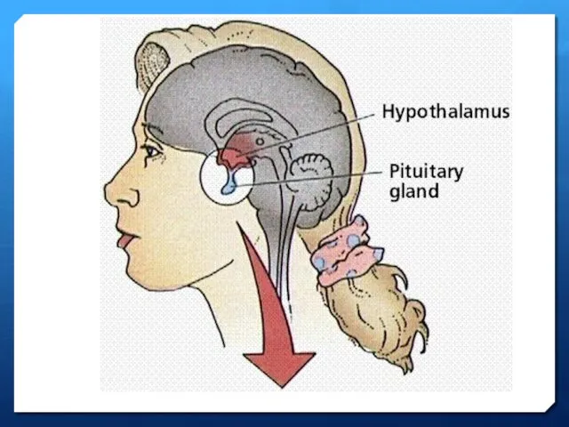
- 5. Temperature receptors in the skin detect changes in the external temperature. They pass this information to
- 7. If we are too hot or too cold, the processing centre sends nerve impulses to the
- 9. Core temperature maintained despite of environmental changes
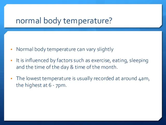
- 10. normal body temperature? Normal body temperature can vary slightly It is influenced by factors such as
- 11. normal body rhythms
- 12. The average normal body temperature taken in the mouth is 37ºC (98.6ºF), but anywhere between 36.5ºC
- 13. Causes of temperature variation Environmental Exercise Food/drink Dehydration (vomiting & diarrhoea) Drugs Infection Inflammation Disease
- 14. An increase in body temperature can occur; Infective conditions Inflammation Immunological diseases Lupus Sarcoidosis Inflammatory bowel
- 15. Considerations when taking a temperature Room temperature How is the patient dressed Temperature site The equipment
- 16. Hyperthermia (heat stroke) Occurs when the body produces or absorbs more heat the it can dissipate.
- 17. fever Fever occurs when the core temperature is set higher Usually in response to bacterial or
- 18. hypothermia Happens when the body temperature falls below 35c Hypothermia can quickly become life threatening and
- 19. Any questions?
- 20. Respiratory system Lynne Powell
- 21. Respiration is defined as; The transport of oxygen from the outside air to the cells within
- 22. Respiration v. ventilation Respiration is a chemical reaction that happens in all living cells. It is
- 24. The respiratory system The human respiratory system contains the organs that allow us to get the
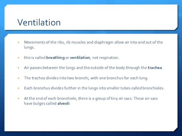
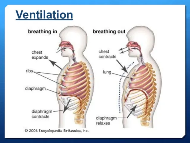
- 26. Ventilation Movements of the ribs, rib muscles and diaphragm allow air into and out of the
- 27. Ventilation
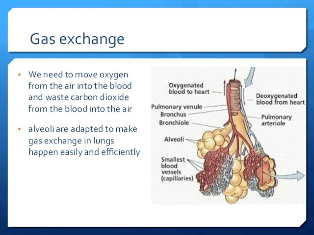
- 28. Gas exchange We need to move oxygen from the air into the blood and waste carbon
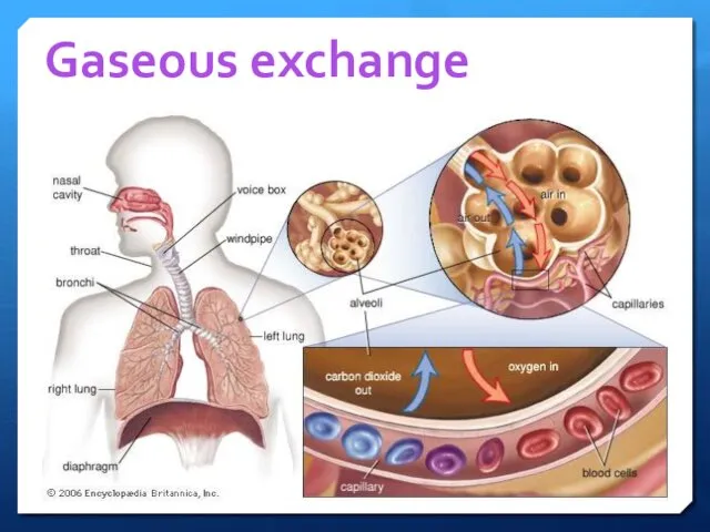
- 29. Gaseous exchange
- 30. Respirations are recorded for a number of reasons: To acquire a baseline. To monitor a patient
- 31. When measuring and recording breathing, the rate and pattern of breathing should be recorded.
- 32. The rate should be regular with equal pause between each breath. The rate can be irregular
- 33. is the patient; mouth breathing pursing the lips on expiration, using the abdominal muscles flaring the
- 34. Average respiratory rates, by age: Average adult 10-20 breaths/min Newborns: Average 44 breaths per minute Infants:
- 35. Measuring respiratory rate The human respiration rate is usually measured when a person is at rest.
- 36. PEAK FLOW MEASUREMENT Measurement of expiratory flow. Standard range peak flow meters are suitable for both
- 37. To take a peak flow reading you should: check that the pointer is at zero. preferably
- 38. Vitalograph
- 39. Infection control considerations DISPOSABLE MOUTH PIECES
- 40. ACTION PLAN FOR PATIENTS with asthma Green: 80 to 100 percent of your personal best peak
- 41. Pulse oximetry
- 42. Pulse oximetry Non-invasive method of monitoring the % of haemoglobin (Hb) saturated with oxygen.
- 43. Blood Red cells contain haemoglobin that have the ability to pick up and release oxygen under
- 44. Haemoglobin is the active oxygen carrying part of the erythrocyte (red blood cell) Blood carries oxygen
- 45. Most oxygen is carried by haemoglobin but 3 factors influence the amount of oxygen delivered to
- 46. How does a pulse oximeter work? Calibrated during manufacture. It emits two wavelengths which are partly
- 47. Limitations of pulse oximetry Not reliable in patient’s with poor circulation (e.g. peripheral vascular disease, anaemia,
- 48. Using an oximeter Resting readings should be taken for at least 5 mins The sensor is
- 49. Results of pulse oximetry 95 - 100% = Acceptable normal ranges 92% or less = Refer
- 50. Medicines & healthcare products regulatory agency
- 52. Скачать презентацию
Aims
To understand the body’s control of temperature and respiratory rate.
To look
Aims
To understand the body’s control of temperature and respiratory rate.
To look
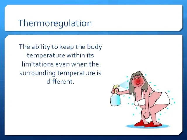
Thermoregulation
The ability to keep the body temperature within its limitations even
Thermoregulation
The ability to keep the body temperature within its limitations even
Temperature control
Temperature control is the process of keeping the body at
Temperature control
Temperature control is the process of keeping the body at
Temperature receptors in the skin detect changes in the external temperature.
Temperature receptors in the skin detect changes in the external temperature.
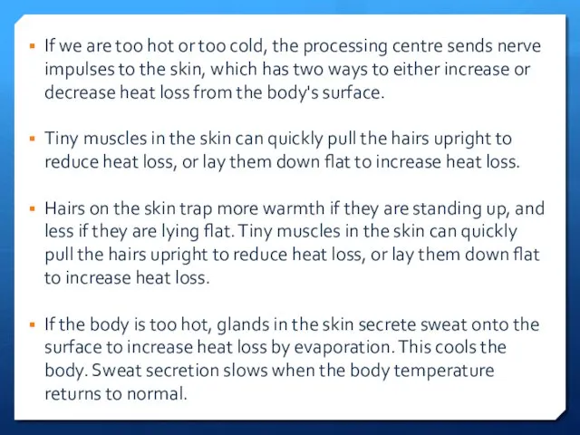
If we are too hot or too cold, the processing centre
If we are too hot or too cold, the processing centre

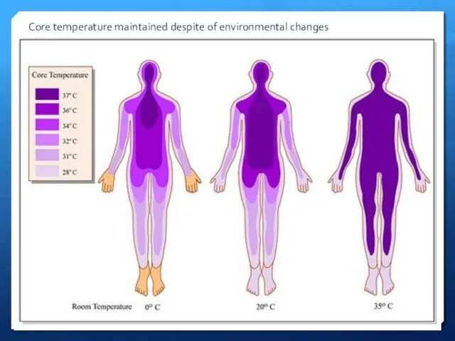
Core temperature maintained despite of environmental changes
Core temperature maintained despite of environmental changes
normal body temperature?
Normal body temperature can vary slightly
It is influenced by
normal body temperature?
Normal body temperature can vary slightly
It is influenced by
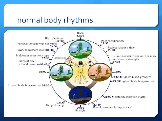
normal body rhythms
normal body rhythms
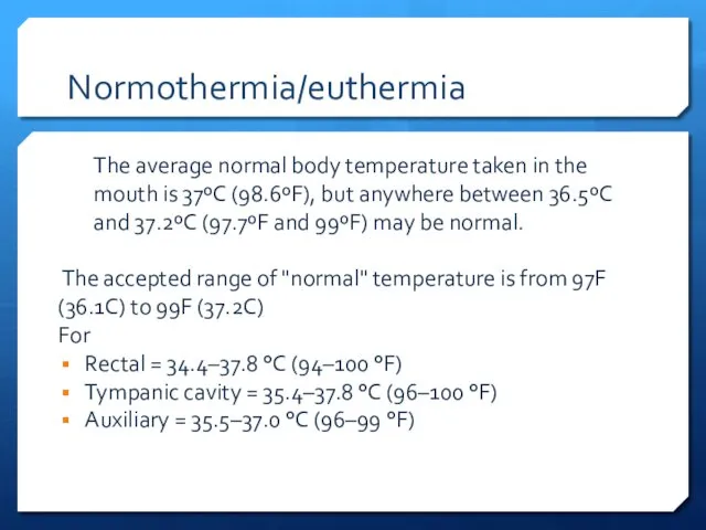
The average normal body temperature taken in the mouth is 37ºC
The average normal body temperature taken in the mouth is 37ºC
Causes of temperature variation
Environmental
Exercise
Food/drink
Dehydration (vomiting & diarrhoea)
Drugs
Infection
Inflammation
Disease
Causes of temperature variation
Environmental
Exercise
Food/drink
Dehydration (vomiting & diarrhoea)
Drugs
Infection
Inflammation
Disease
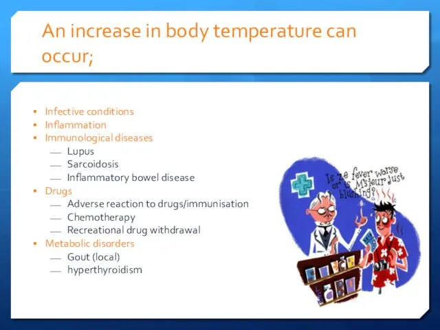
An increase in body temperature can occur;
Infective conditions
Inflammation
Immunological diseases
Lupus
Sarcoidosis
Inflammatory bowel disease
Drugs
Adverse
An increase in body temperature can occur;
Infective conditions
Inflammation
Immunological diseases
Lupus
Sarcoidosis
Inflammatory bowel disease
Drugs
Adverse
Considerations when taking a temperature
Room temperature
How is the patient dressed
Temperature site
The
Considerations when taking a temperature
Room temperature
How is the patient dressed
Temperature site
The
Hyperthermia (heat stroke)
Occurs when the body produces or absorbs more heat
Hyperthermia (heat stroke)
Occurs when the body produces or absorbs more heat
fever
Fever occurs when the core temperature is set higher
Usually in response
fever
Fever occurs when the core temperature is set higher
Usually in response
hypothermia
Happens when the body temperature falls below 35c
Hypothermia can quickly become
hypothermia
Happens when the body temperature falls below 35c
Hypothermia can quickly become

Any questions?
Any questions?
Respiratory system
Lynne Powell
Respiratory system
Lynne Powell
Respiration is defined as;
The transport of oxygen from the outside air
Respiration is defined as;
The transport of oxygen from the outside air
Respiration v. ventilation
Respiration is a chemical reaction that happens in all
Respiration v. ventilation
Respiration is a chemical reaction that happens in all

The respiratory system
The human respiratory system contains the organs that allow
The respiratory system
The human respiratory system contains the organs that allow

Ventilation
Movements of the ribs, rib muscles and diaphragm allow air into
Ventilation
Movements of the ribs, rib muscles and diaphragm allow air into
Ventilation
Ventilation
Gas exchange
We need to move oxygen from the air into the
Gas exchange
We need to move oxygen from the air into the
Gaseous exchange
Gaseous exchange
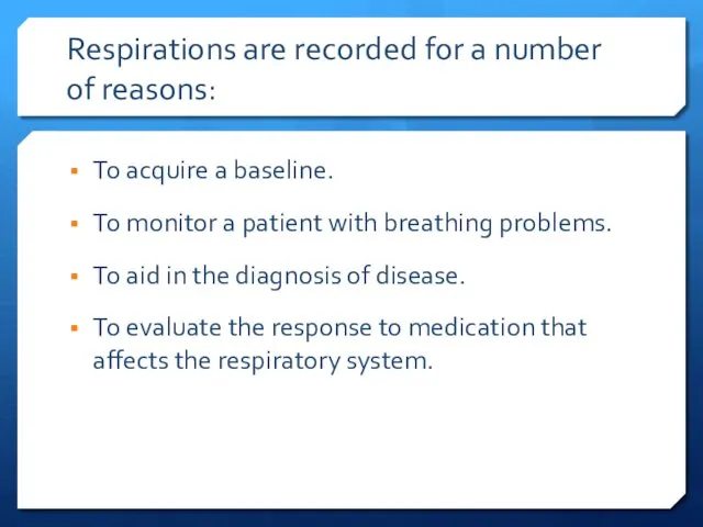
Respirations are recorded for a number of reasons:
To acquire a baseline.
To
Respirations are recorded for a number of reasons:
To acquire a baseline.
To
When measuring and recording breathing, the rate and pattern of breathing
When measuring and recording breathing, the rate and pattern of breathing
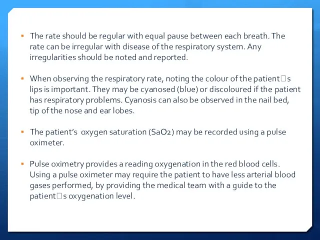
The rate should be regular with equal pause between each breath.
The rate should be regular with equal pause between each breath.
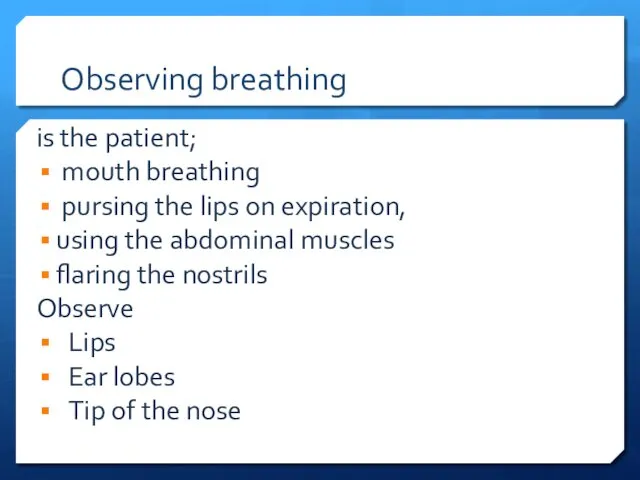
is the patient;
mouth breathing
pursing the lips on expiration,
using the
is the patient;
mouth breathing
pursing the lips on expiration,
using the
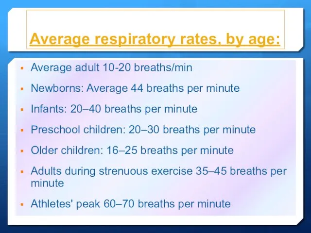
Average respiratory rates, by age:
Average adult 10-20 breaths/min
Newborns: Average 44 breaths
Average respiratory rates, by age:
Average adult 10-20 breaths/min
Newborns: Average 44 breaths
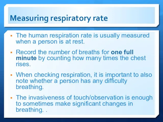
Measuring respiratory rate
The human respiration rate is usually measured when a
Measuring respiratory rate
The human respiration rate is usually measured when a
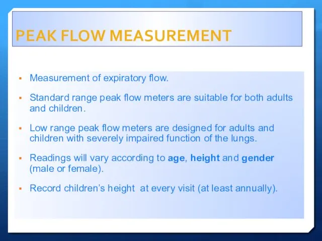
PEAK FLOW MEASUREMENT
Measurement of expiratory flow.
Standard range peak flow meters are
PEAK FLOW MEASUREMENT
Measurement of expiratory flow.
Standard range peak flow meters are
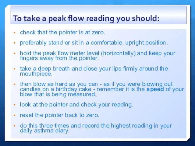
To take a peak flow reading you should:
check that the pointer
To take a peak flow reading you should:
check that the pointer
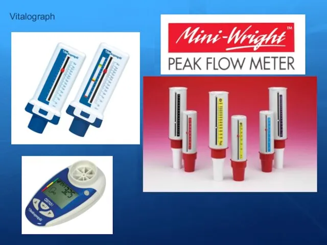
Vitalograph
Vitalograph
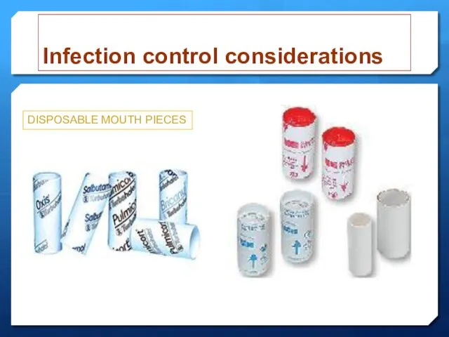
Infection control considerations
DISPOSABLE MOUTH PIECES
Infection control considerations
DISPOSABLE MOUTH PIECES
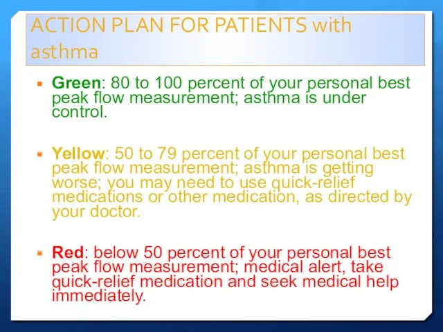
ACTION PLAN FOR PATIENTS with asthma
Green: 80 to 100 percent of
ACTION PLAN FOR PATIENTS with asthma
Green: 80 to 100 percent of

Pulse oximetry
Pulse oximetry
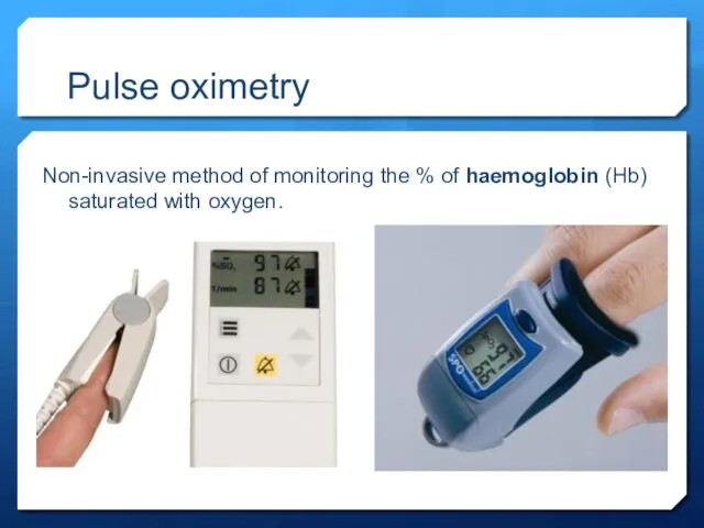
Pulse oximetry
Non-invasive method of monitoring the % of haemoglobin (Hb) saturated
Pulse oximetry
Non-invasive method of monitoring the % of haemoglobin (Hb) saturated
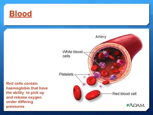
Blood
Red cells contain haemoglobin that have the ability to pick up
Blood
Red cells contain haemoglobin that have the ability to pick up
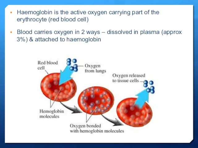
Haemoglobin is the active oxygen carrying part of the erythrocyte (red
Haemoglobin is the active oxygen carrying part of the erythrocyte (red
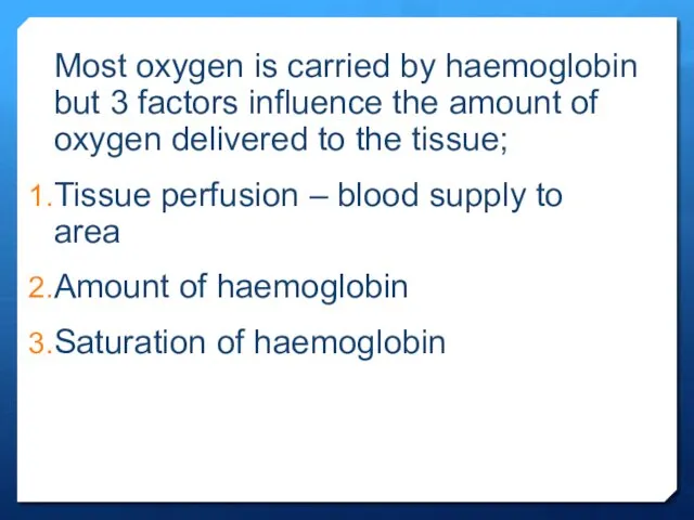
Most oxygen is carried by haemoglobin but 3 factors influence the
Most oxygen is carried by haemoglobin but 3 factors influence the
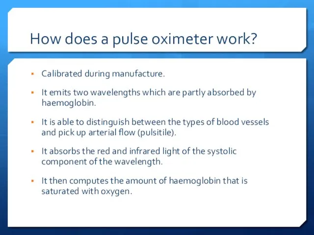
How does a pulse oximeter work?
Calibrated during manufacture.
It emits two wavelengths
How does a pulse oximeter work?
Calibrated during manufacture.
It emits two wavelengths
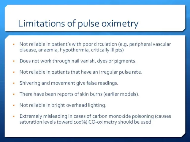
Limitations of pulse oximetry
Not reliable in patient’s with poor circulation (e.g.
Limitations of pulse oximetry
Not reliable in patient’s with poor circulation (e.g.
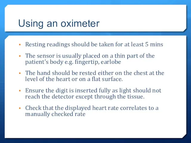
Using an oximeter
Resting readings should be taken for at least 5
Using an oximeter
Resting readings should be taken for at least 5
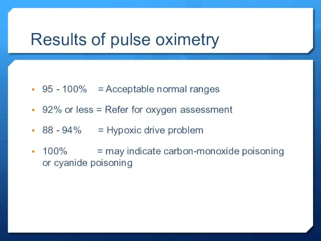
Results of pulse oximetry
95 - 100% = Acceptable normal ranges
92% or
Results of pulse oximetry
95 - 100% = Acceptable normal ranges
92% or
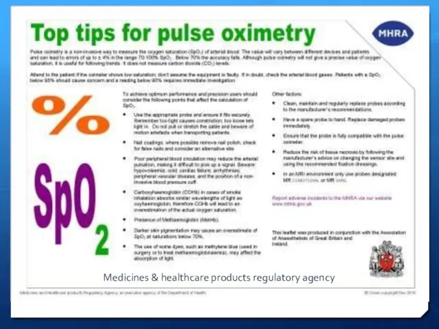
Medicines & healthcare products regulatory agency
Medicines & healthcare products regulatory agency
 Профилактика плоскостопия и нарушения осанки у учащихся начальной школы
 Рассеянный склероз, орэм. Диагностика, дифференциальная диагностика, реабилитация
 Лабораторная диагностика туберкулёзной инфекции
 Медицина в России XIX века. Развитие отечественной хирургии. (Лекция 6)
 Қол ұшы флегмоналары
 Диагностика и лечения неотложных состояний в онкологии
 Dental caries and Conditionals
 Ротавирусная инфекция
 Местная анестезия
 Заболевания новорожденных, связанные с актом родов. Родовые травмы
 Пиелонефриты
 Ауыз кілегей қабығының дерматоздар кезінде (қызыл жалпақ теміреткі, қызыл жегі, кулбіреуікше) жарақаттануы
 Внутриутробный период развития организма
 Гастроэзофагеальная рефлюксная болезнь
 Принципы лечения болезней пародонта. Составление плана комплексного лечения пародонтологического пациента
 Гипотензивные средства. Мочегонные средства. Антисклеротические средства
 Всемирный день больного — 11 февраля
 Водолечение. Гидротерапия, бальнеотерапия, талассотерапия
 Лечебная физкультура при гинекологических заболеваниях
 Шизофрения, шизотипические и бредовые расстройства
 Отёчно-асцитический синдром
 Физиологические роды
 Рентгенологические методы исследования, применяемые в кардиологии
 Коматозні стани
 Ісікке қарсы вакциналар
 Денсаулық 2020 бағдарламасы
 Влияние загрязнения атмосферного воздуха на здоровье населения и санитарные условия жизни в городах
 Гемофилия В