- Giardiasis Giardia lamblia Giardia intestinalis

Содержание
- 2. Giardiasis Most common causative agent of epidemic & endemic diarrhoea throughout the world Prevalence - 2-5%
- 3. Morphology of Giardia lamblia trophozoite Pear shaped, rounded anterior end, posterior end pointed (looks like monkey
- 4. Morphology of Giardia lamblia trophozoite ventral dorsal Scanning EM view of trophozoite surface showing the adhesive
- 5. Morphology of Giardia lamblia trophozoite Light microscope photos of trophozoites
- 6. Morphology of Giardia lamblia cyst ovoid in shape 8-12 µm long x 7-10 µm wide thick
- 7. Giardia lamblia cyst
- 8. Giardiasis Giardia intestinalis =(lamblia) Trophozoites Cysts
- 9. Infective form – mature cyst passed in feces of man Routes of transmission Feco-oral ingestion of
- 10. Life Cycle Acquire infection – ingestion of mature cysts Excystation occurs in stomach & duodenum within
- 11. Giardia – Life cycle
- 12. Giardia lamblia life cycle
- 13. Pathology Do not invade tissues Feed on mucous secretions May localise in biliary tract to avoid
- 14. Giardiasis: The Disease Asymptomatic : largest group Acute : self-limiting infection, acute watery diarrhoea, abdominal cramps,
- 15. Laboratory Diagnosis Parasitic Diagnosis Samples Stool Duodenal contents Duodenal fluid( Entero test ) Duodenal/ jejunal biopsy
- 16. Parasitic Diagnosis Microscopy Microscopy Direct Wet Mount Trophozoite with falling leaf motility in saline mount Cyst
- 17. Laboratory Diagnosis Parasitic Diagnosis Antigen detection ( Coproantigen ) ELISA Sensitivity & specificity high Culture Not
- 18. Laboratory Diagnosis Serodiagnosis ELISA Epidemiological purpose Molecular diagnosis DNA probes & PCR for research purpose
- 19. Prevention Avoid food & water that might be contaminated filtration of water (be sure filter is
- 21. Скачать презентацию
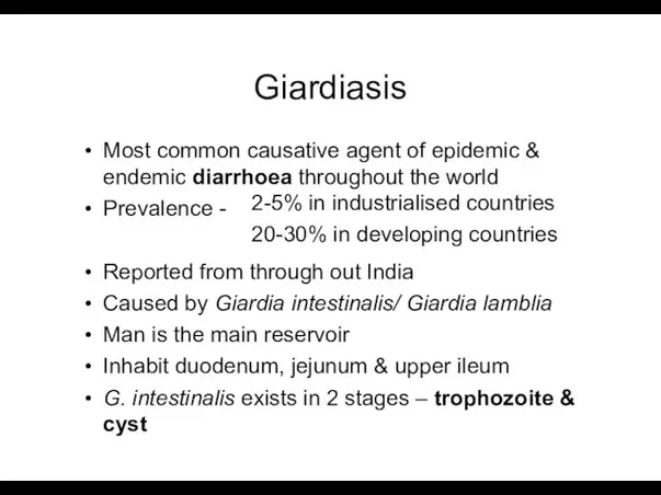
Giardiasis
Most common causative agent of epidemic & endemic diarrhoea throughout the
Giardiasis
Most common causative agent of epidemic & endemic diarrhoea throughout the
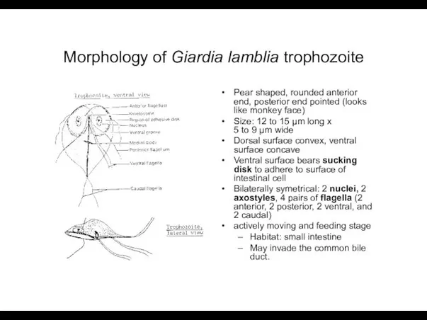
Morphology of Giardia lamblia trophozoite
Pear shaped, rounded anterior end, posterior end
Morphology of Giardia lamblia trophozoite
Pear shaped, rounded anterior end, posterior end
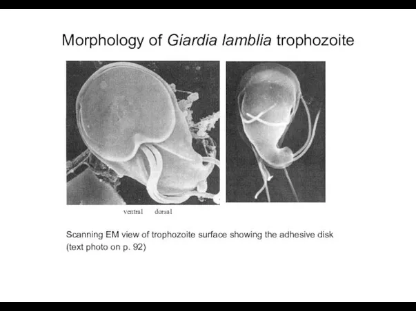
Morphology of Giardia lamblia trophozoite
ventral dorsal
Scanning EM view of trophozoite surface showing
Morphology of Giardia lamblia trophozoite
ventral dorsal
Scanning EM view of trophozoite surface showing
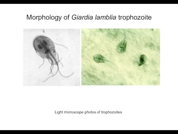
Morphology of Giardia lamblia trophozoite
Light microscope photos of trophozoites
Morphology of Giardia lamblia trophozoite
Light microscope photos of trophozoites
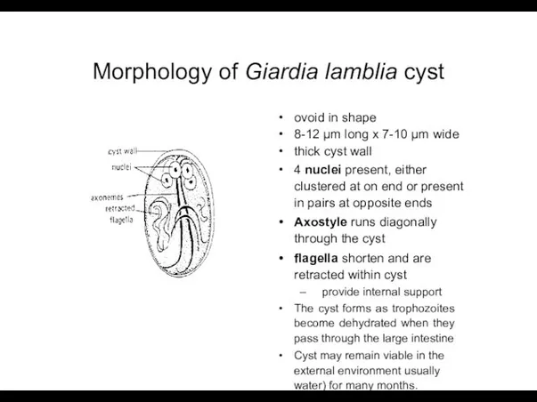
Morphology of Giardia lamblia cyst
ovoid in shape
8-12 µm long x 7-10
Morphology of Giardia lamblia cyst
ovoid in shape
8-12 µm long x 7-10
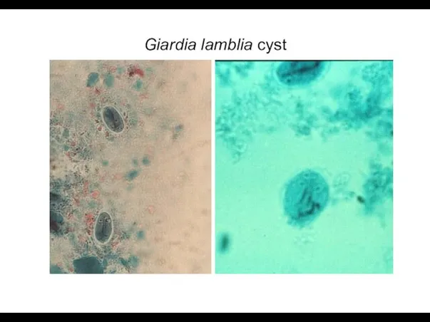
Giardia lamblia cyst
Giardia lamblia cyst
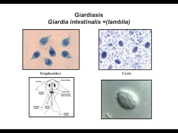
Giardiasis
Giardia intestinalis =(lamblia)
Trophozoites
Cysts
Giardiasis
Giardia intestinalis =(lamblia)
Trophozoites
Cysts
Infective form – mature cyst passed in feces of man
Routes of
Infective form – mature cyst passed in feces of man
Routes of
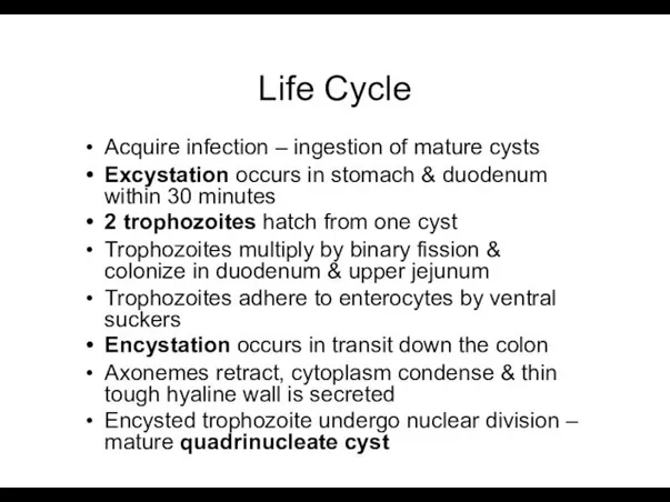
Life Cycle
Acquire infection – ingestion of mature cysts
Excystation occurs in stomach
Life Cycle
Acquire infection – ingestion of mature cysts
Excystation occurs in stomach
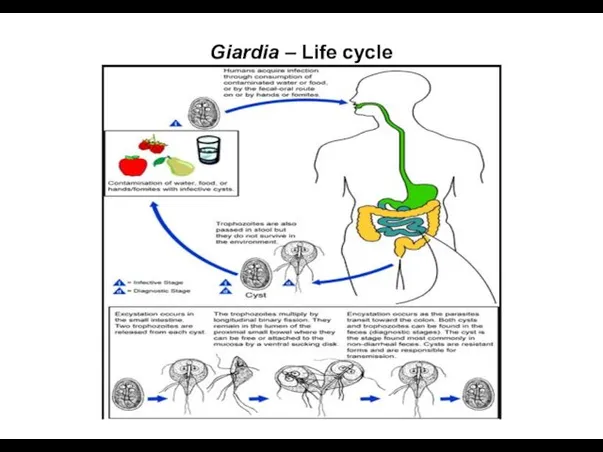
Giardia – Life cycle
Giardia – Life cycle
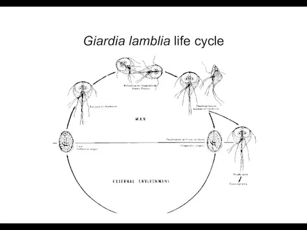
Giardia lamblia life cycle
Giardia lamblia life cycle
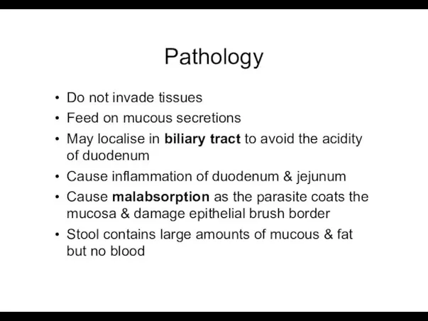
Pathology
Do not invade tissues
Feed on mucous secretions
May localise in biliary tract
Pathology
Do not invade tissues
Feed on mucous secretions
May localise in biliary tract
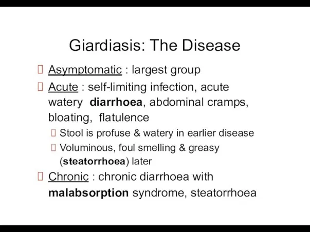
Giardiasis: The Disease
Asymptomatic : largest group
Acute : self-limiting infection, acute watery
Giardiasis: The Disease
Asymptomatic : largest group
Acute : self-limiting infection, acute watery
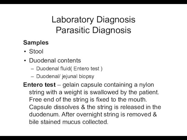
Laboratory Diagnosis Parasitic Diagnosis
Samples
Stool
Duodenal contents
Duodenal fluid( Entero test )
Duodenal/ jejunal biopsy
Entero
Laboratory Diagnosis Parasitic Diagnosis
Samples
Stool
Duodenal contents
Duodenal fluid( Entero test )
Duodenal/ jejunal biopsy
Entero
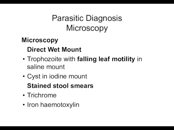
Parasitic Diagnosis Microscopy
Microscopy
Direct Wet Mount
Trophozoite with falling leaf motility in saline
Parasitic Diagnosis Microscopy
Microscopy
Direct Wet Mount
Trophozoite with falling leaf motility in saline
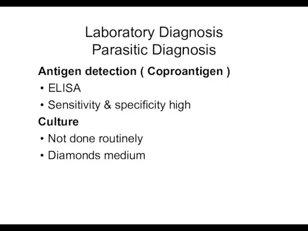
Laboratory Diagnosis Parasitic Diagnosis
Antigen detection ( Coproantigen )
ELISA
Sensitivity & specificity high
Culture
Not
Laboratory Diagnosis Parasitic Diagnosis
Antigen detection ( Coproantigen )
ELISA
Sensitivity & specificity high
Culture
Not
Laboratory Diagnosis
Serodiagnosis
ELISA
Epidemiological purpose
Molecular diagnosis
DNA probes & PCR for research purpose
Laboratory Diagnosis
Serodiagnosis
ELISA
Epidemiological purpose
Molecular diagnosis
DNA probes & PCR for research purpose
Prevention
Avoid food & water that might be contaminated
filtration of water (be
Prevention
Avoid food & water that might be contaminated
filtration of water (be
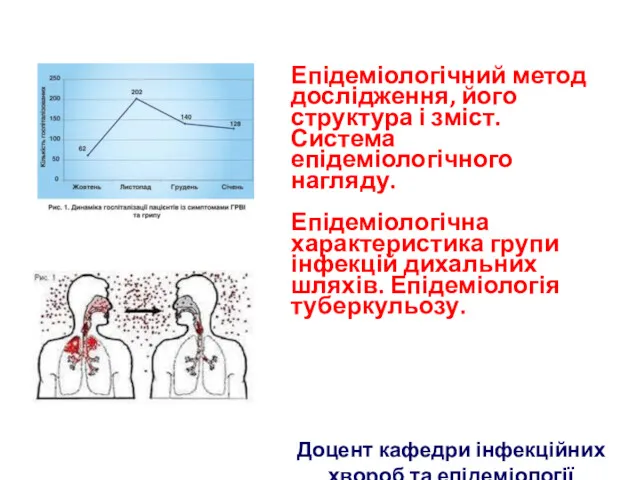 Епідеміологічний метод дослідження, його структура і зміст. Система епідеміологічного нагляду. Епідеміологія туберкульозу
 Синдром гострого запалення слизових оболонок дихальних шляхів. Грип
 Ас қорыту физиологиясы
 Физиология и методы исследования системы гемостаза
 Родовые повреждения новорожденных
 Ведение нормальных родов. Управление родовым актом
 Трансформация патологии населения. Основные социально-гигиенические проблемы современного общества
 Профилактика ВИЧ - инфекций
 Рефракция и аккомодация глаза
 Негізгі психопатологиялық синдромдар
 Риски расстройств пищевого поведения у спортсменов
 Дошкольный и преддошкольный возраст
 Беременность при туберкулезе
 Введение в иммунологию. Иммунная система
 Гіполіпідемічні лікарські засоби
 Анатомо-физиологические особенности спинального и эпидурального пространств у детей раннего возраста
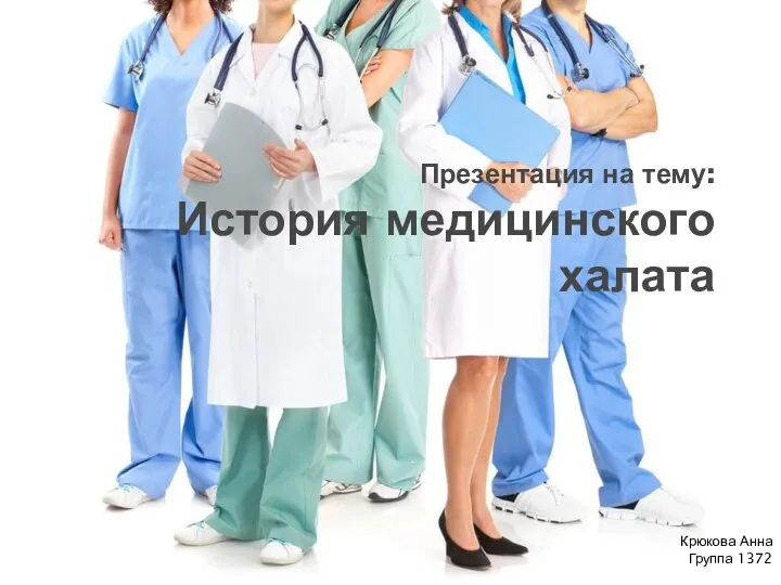 История медицинского халата
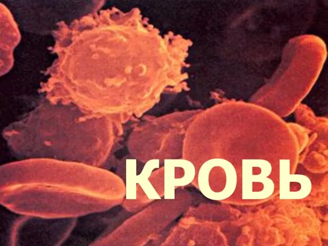 Кровь. Функции и состав крови. Группы крови
 Бронхообструктивный синдром
 ВПР мочевыводящей системы
 Пороки развития ЦНС
 Equipment and instruments of dental clinic
 Классификация острого панкреатита
 Виявлення хворих на туберкульоз. (Лекція 2)
 Ведение пациентов с болью в спине в практике терапевта и семейного врача
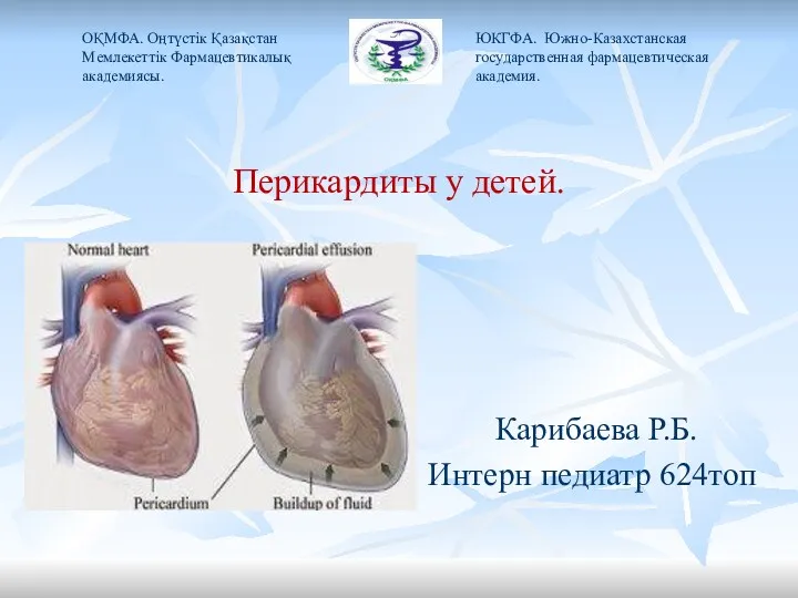 Перикардиты у детей
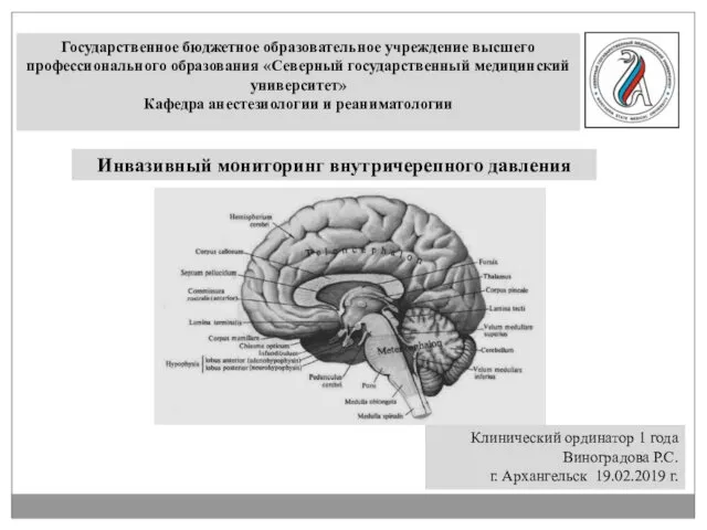 Инвазивный мониторинг внутричерепного давления
 Несеп жыныс жүйесі