- Hormones and the Endocrine System

Содержание
- 2. Overview: The Body’s Long-Distance Regulators Animal hormones are chemical signals that are secreted into the circulatory
- 3. Two systems coordinate communication throughout the body: the endocrine system and the nervous system. The endocrine
- 4. What role do hormones play in transforming a caterpillar into a butterfly?
- 5. Hormones and other signaling molecules bind to target receptors, triggering specific response pathways Chemical signals bind
- 6. Types of Secreted Signaling Molecules Secreted chemical signals include Hormones Local regulators Neurotransmitters Neurohormones Pheromones
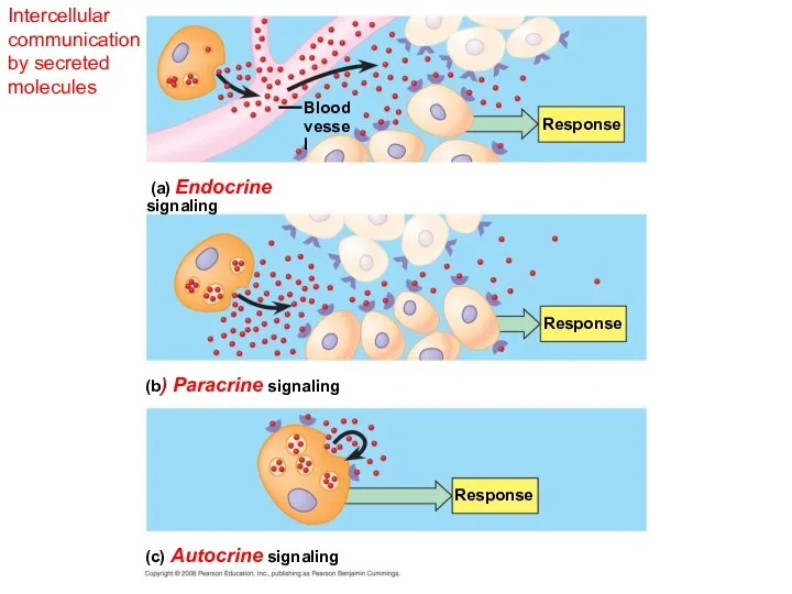
- 7. Hormones Endocrine signals (hormones) are secreted into extracellular fluids and travel via the bloodstream. Endocrine glands
- 8. Intercellular communication by secreted molecules Blood vessel Response Response Response Response (a) Endocrine signaling (b) Paracrine
- 9. Local Regulators = Short Distance Chemical Signals Local regulators are chemical signals that travel over short
- 10. Intercellular communication by secreted molecules Blood vessel Response Response Response (a) Endocrine signaling (b) Paracrine signaling
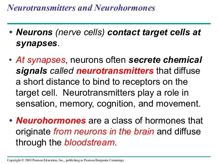
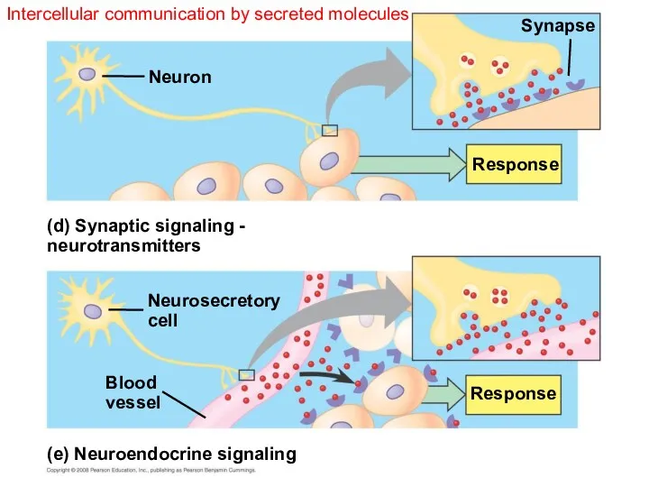
- 11. Neurotransmitters and Neurohormones Neurons (nerve cells) contact target cells at synapses. At synapses, neurons often secrete
- 12. Intercellular communication by secreted molecules Response (d) Synaptic signaling - neurotransmitters Neuron Neurosecretory cell (e) Neuroendocrine
- 13. Pheromones Pheromones are chemical signals that are released from the body and used to communicate with
- 14. Chemical Classes of Hormones Three major classes of molecules function as hormones in vertebrates: Polypeptides (proteins
- 15. Lipid-soluble hormones (steroid hormones) pass easily through cell membranes. Water-soluble hormones (polypeptides and amines) do not
- 16. Hormones differ in form and solubility Water-soluble Lipid-soluble Steroid: Cortisol Polypeptide: Insulin Amine: Epinephrine Amine: Thyroxine
- 17. Cellular Response Pathways Water and lipid soluble hormones differ in their paths through a body. Water-soluble
- 18. Signaling by any of these hormones involves three key events: Reception Signal transduction Response Binding of
- 19. Receptor location varies with hormone type NUCLEUS Signal receptor (a) (b) TARGET CELL Signal receptor Transport
- 20. Receptor location varies with hormone type Signal receptor TARGET CELL Signal receptor Transport protein Water- soluble
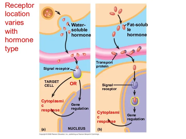
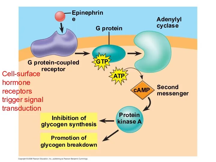
- 21. Pathway for Water-Soluble Hormones The hormone epinephrine has multiple effects in mediating the body’s response to
- 22. cAMP Second messenger Adenylyl cyclase G protein-coupled receptor ATP GTP G protein Epinephrine Inhibition of glycogen
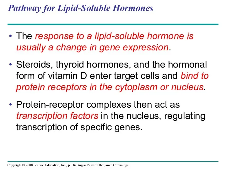
- 23. Pathway for Lipid-Soluble Hormones The response to a lipid-soluble hormone is usually a change in gene
- 24. Steroid hormone receptors are inside the cell and directly regulate gene expression Hormone (estradiol) Hormone-receptor complex
- 25. Multiple Effects of Hormones The same hormone may have different effects on target cells that have
- 26. One hormone, different effects Glycogen deposits β receptor Vessel dilates. Epinephrine (a) Liver cell Epinephrine β
- 27. Specialized role of a hormone in frog metamorphosis (a) (b)
- 28. Signaling by Local Regulators In paracrine signaling, nonhormonal chemical signals called local regulators elicit responses in
- 29. Major endocrine glands: Adrenal glands Hypothalamus Pineal gland Pituitary gland Thyroid gland Parathyroid glands Pancreas Kidney
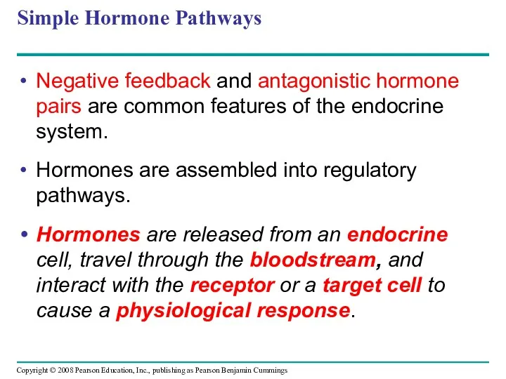
- 30. Simple Hormone Pathways Negative feedback and antagonistic hormone pairs are common features of the endocrine system.
- 31. A simple endocrine pathway Pathway Example Stimulus Low pH in duodenum S cells of duodenum secrete
- 32. A negative feedback loop inhibits a response by reducing the initial stimulus. Negative feedback reverses a
- 33. Insulin Lowers Blood Glucose Levels Homeostasis: Blood glucose level (about 90 mg/100 mL) Insulin Beta cells
- 34. Glucagon Raises Blood Glucose Levels Homeostasis: Blood glucose level (about 90 mg/100 mL) Glucagon STIMULUS: Blood
- 35. Maintenance of glucose homeostasis by insulin and glucagon Homeostasis: Blood glucose level (about 90 mg/100 mL)
- 36. Target Tissues for Insulin and Glucagon Insulin reduces blood glucose levels by Promoting the cellular uptake
- 37. Diabetes Mellitus Diabetes mellitus is an endocrine disorder caused by a deficiency of insulin or a
- 38. The endocrine and nervous systems act individually and together in regulating animal physiology Signals from the
- 39. Coordination of Endocrine and Nervous Systems in Invertebrates In insects, molting and development are controlled by
- 40. Hormonal regulation of insect development Ecdysone Brain PTTH EARLY LARVA Neurosecretory cells Corpus cardiacum Corpus allatum
- 41. Coordination of Endocrine and Nervous Systems in Vertebrates The hypothalamus receives information from the nervous system
- 42. The posterior pituitary stores and secretes hormones that are made in the hypothalamus The anterior pituitary
- 43. Endocrine glands in the human brain Spinal cord Posterior pituitary Cerebellum Pineal gland Anterior pituitary Hypothalamus
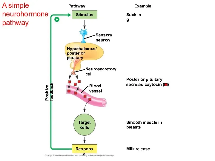
- 46. Oxytocin induces uterine contractions and the release of milk Suckling sends a message to the hypothalamus
- 47. A simple neurohormone pathway Suckling Pathway Stimulus Hypothalamus/ posterior pituitary Positive feedback Example Sensory neuron Neurosecretory
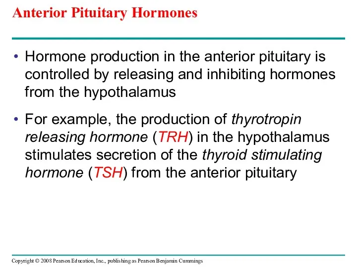
- 48. Anterior Pituitary Hormones Hormone production in the anterior pituitary is controlled by releasing and inhibiting hormones
- 49. Production and release of anterior pituitary hormones Hypothalamic releasing and inhibiting hormones Neurosecretory cells of the
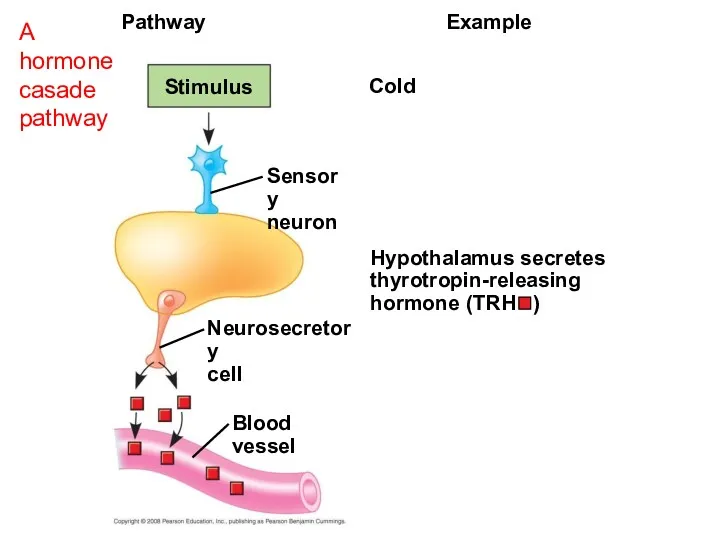
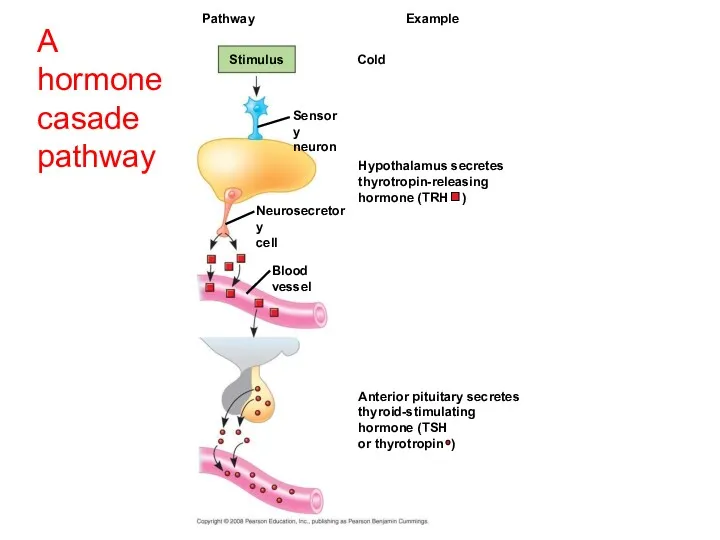
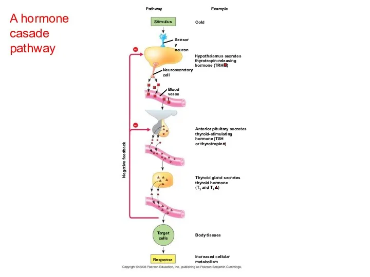
- 50. Hormone Cascade Pathways A hormone can stimulate the release of a series of other hormones, the
- 51. Cold Pathway Stimulus Blood vessel Example Sensory neuron Hypothalamus secretes thyrotropin-releasing hormone (TRH ) Neurosecretory cell
- 52. Cold Pathway Stimulus Hypothalamus secretes thyrotropin-releasing hormone (TRH ) Example Sensory neuron Neurosecretory cell Blood vessel
- 53. A hormone casade pathway Cold Pathway Stimulus Hypothalamus secretes thyrotropin-releasing hormone (TRH ) Negative feedback Example
- 54. Tropic Hormones A tropic hormone regulates the function of endocrine cells or glands. The four strictly
- 55. Nontropic Hormones - target nonendocrine tissues. Nontropic hormones produced by the anterior pituitary are: Prolactin (PRL)
- 56. Growth Hormone Growth hormone (GH) is secreted by the anterior pituitary gland and has tropic and
- 57. Endocrine signaling regulates metabolism, homeostasis, development, and behavior. Endocrine glands respond to diverse stimuli in regulating
- 58. Thyroid Hormone: Control of Metabolism and Development The thyroid gland consists of two lobes on the
- 59. Thyroid hormones stimulate metabolism and influence development and maturation. Hyperthyroidism, excessive secretion of thyroid hormones, causes
- 60. Parathyroid Hormone and Vitamin D: Control of Blood Calcium Two antagonistic hormones regulate the homeostasis of
- 61. Antagonistic Hormone Pairs control blood calcium levels PTH Parathyroid gland (behind thyroid) STIMULUS: Falling blood Ca2+
- 62. PTH increases the level of blood Ca2+ It releases Ca2+ from bone and stimulates reabsorption of
- 63. Adrenal Hormones: Response to Stress The adrenal glands are adjacent to the kidneys. Each adrenal gland
- 64. Catecholamines from the Adrenal Medulla The adrenal medulla secretes epinephrine (adrenaline) and norepinephrine (noradrenaline). These hormones
- 65. Epinephrine and norepinephrine Trigger the release of glucose and fatty acids into the blood Increase oxygen
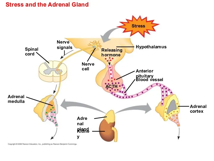
- 66. Summary: Stress and the Adrenal Gland Stress Adrenal gland Nerve cell Nerve signals Releasing hormone Hypothalamus
- 67. Stress and the Adrenal Gland Stress Adrenal gland Nerve cell Nerve signals Releasing hormone Hypothalamus Anterior
- 68. Short-term Stress and the Adrenal Gland (a) Short-term stress response Effects of epinephrine and norepinephrine: 2.
- 69. Steroid Hormones from the Adrenal Cortex The adrenal cortex releases a family of steroids called corticosteroids
- 70. Long-term Stress and the adrenal gland (b) Long-term stress response Effects of mineralocorticoids: Effects of glucocorticoids:
- 71. Glucocorticoids, such as cortisol, influence glucose metabolism and the immune system. Mineralocorticoids, such as aldosterone, affect
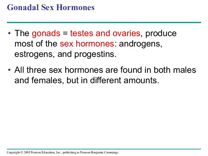
- 72. Gonadal Sex Hormones The gonads = testes and ovaries, produce most of the sex hormones: androgens,
- 73. The testes primarily synthesize androgens, mainly testosterone, which stimulate development and maintenance of the male reproductive
- 74. Estrogens, made in the ovary, most importantly estradiol, are responsible for maintenance of the female reproductive
- 75. Pineal Gland - Melatonin and Biorhythms The pineal gland, located in the brain, secretes melatonin. Light/dark
- 76. Signal Transduction Pathway Example Stimulus Low blood glucose Pancreas alpha cells secretes glucagon Endocrine cell Blood
- 77. You should now be able to: Distinguish between the following pairs of terms: hormones and local
- 79. Скачать презентацию
Overview: The Body’s Long-Distance Regulators
Animal hormones are chemical signals that are
Overview: The Body’s Long-Distance Regulators
Animal hormones are chemical signals that are
Two systems coordinate communication throughout the body: the endocrine system and
Two systems coordinate communication throughout the body: the endocrine system and
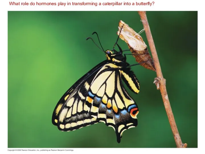
What role do hormones play in transforming a caterpillar into
What role do hormones play in transforming a caterpillar into
Hormones and other signaling molecules bind to target receptors, triggering specific
Hormones and other signaling molecules bind to target receptors, triggering specific
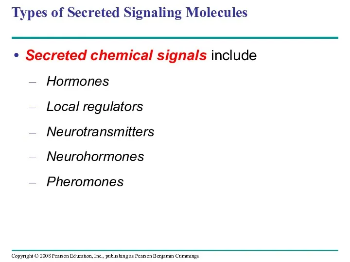
Types of Secreted Signaling Molecules
Secreted chemical signals include
Hormones
Local regulators
Neurotransmitters
Neurohormones
Pheromones
Types of Secreted Signaling Molecules
Secreted chemical signals include
Hormones
Local regulators
Neurotransmitters
Neurohormones
Pheromones
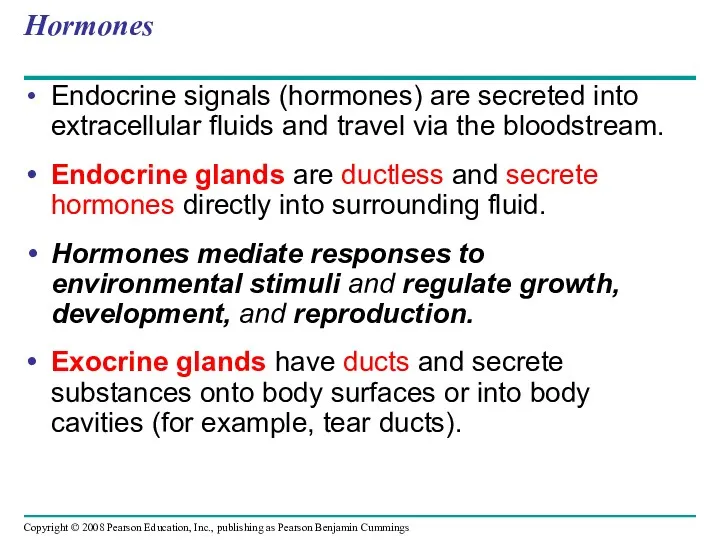
Hormones
Endocrine signals (hormones) are secreted into extracellular fluids and travel via
Hormones
Endocrine signals (hormones) are secreted into extracellular fluids and travel via
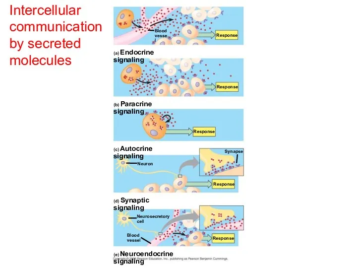
Intercellular communication by secreted molecules
Blood
vessel
Response
Response
Response
Response
(a) Endocrine signaling
(b) Paracrine signaling
(c) Autocrine signaling
(d)
Intercellular communication by secreted molecules
Blood
vessel
Response
Response
Response
Response
(a) Endocrine signaling
(b) Paracrine signaling
(c) Autocrine signaling
(d)
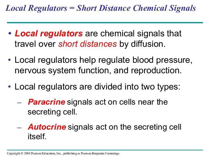
Local Regulators = Short Distance Chemical Signals
Local regulators are chemical
Local Regulators = Short Distance Chemical Signals
Local regulators are chemical
Intercellular communication
by secreted molecules
Blood
vessel
Response
Response
Response
(a) Endocrine signaling
(b) Paracrine signaling
(c) Autocrine signaling
Intercellular communication
by secreted molecules
Blood
vessel
Response
Response
Response
(a) Endocrine signaling
(b) Paracrine signaling
(c) Autocrine signaling
Neurotransmitters and Neurohormones
Neurons (nerve cells) contact target cells at synapses.
At synapses,
Neurotransmitters and Neurohormones
Neurons (nerve cells) contact target cells at synapses.
At synapses,
Intercellular communication by secreted molecules
Response
(d) Synaptic signaling - neurotransmitters
Neuron
Neurosecretory
cell
(e) Neuroendocrine signaling
Blood
vessel
Synapse
Response
Intercellular communication by secreted molecules
Response
(d) Synaptic signaling - neurotransmitters
Neuron
Neurosecretory
cell
(e) Neuroendocrine signaling
Blood
vessel
Synapse
Response
Pheromones
Pheromones are chemical signals that are released from the body and
Pheromones
Pheromones are chemical signals that are released from the body and
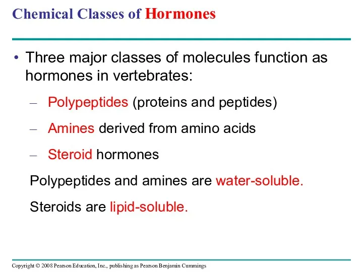
Chemical Classes of Hormones
Three major classes of molecules function as hormones
Chemical Classes of Hormones
Three major classes of molecules function as hormones
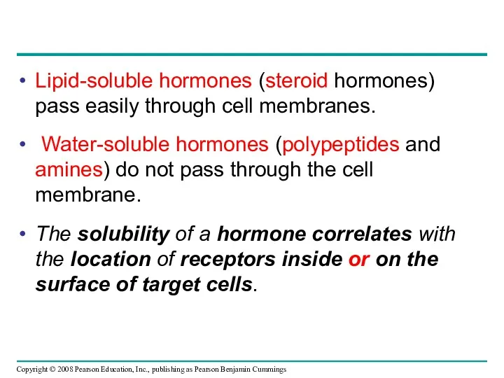
Lipid-soluble hormones (steroid hormones) pass easily through cell membranes.
Water-soluble hormones
Lipid-soluble hormones (steroid hormones) pass easily through cell membranes.
Water-soluble hormones
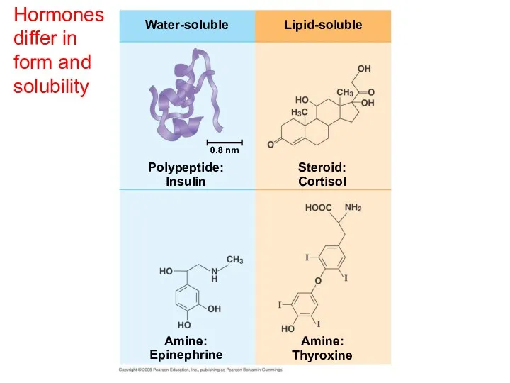
Hormones differ in form and solubility
Water-soluble
Lipid-soluble
Steroid:
Cortisol
Polypeptide:
Insulin
Amine:
Epinephrine
Amine:
Thyroxine
0.8 nm
Hormones differ in form and solubility
Water-soluble
Lipid-soluble
Steroid:
Cortisol
Polypeptide:
Insulin
Amine:
Epinephrine
Amine:
Thyroxine
0.8 nm
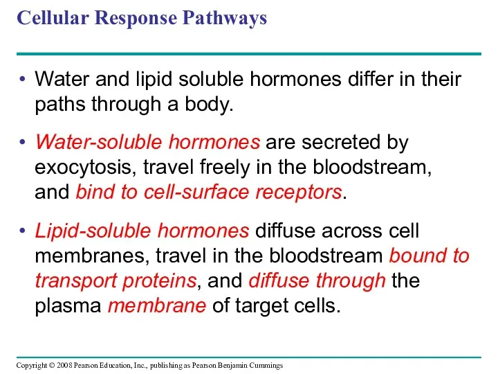
Cellular Response Pathways
Water and lipid soluble hormones differ in their paths
Cellular Response Pathways
Water and lipid soluble hormones differ in their paths
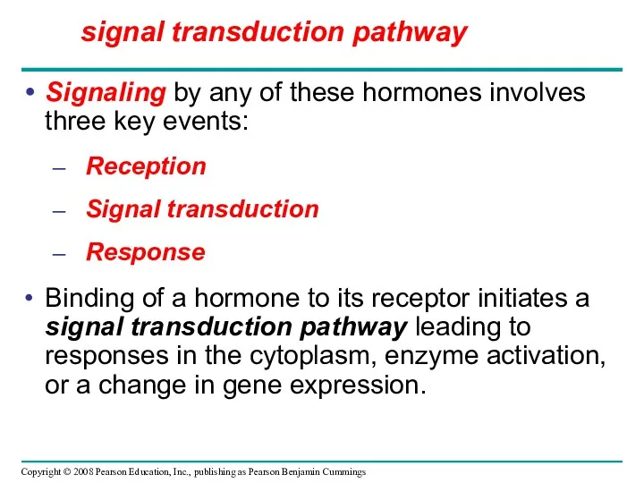
Signaling by any of these hormones involves three key events:
Reception
Signal transduction
Response
Binding
Signaling by any of these hormones involves three key events:
Reception
Signal transduction
Response
Binding
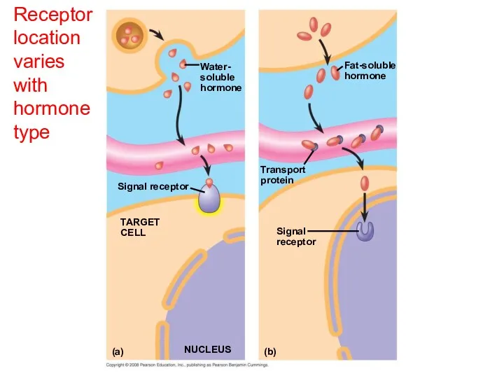
Receptor location varies with hormone type
NUCLEUS
Signal
receptor
(a)
(b)
TARGET
CELL
Signal receptor
Transport
protein
Water-
soluble
hormone
Fat-soluble
hormone
Receptor location varies with hormone type
NUCLEUS
Signal
receptor
(a)
(b)
TARGET
CELL
Signal receptor
Transport
protein
Water-
soluble
hormone
Fat-soluble
hormone
Receptor location varies with hormone type
Signal
receptor
TARGET
CELL
Signal receptor
Transport
protein
Water-
soluble
hormone
Fat-soluble
hormone
Gene
regulation
Cytoplasmic
response
Gene
regulation
Cytoplasmic
response
OR
(a)
NUCLEUS
(b)
Receptor location varies with hormone type
Signal
receptor
TARGET
CELL
Signal receptor
Transport
protein
Water-
soluble
hormone
Fat-soluble
hormone
Gene
regulation
Cytoplasmic
response
Gene
regulation
Cytoplasmic
response
OR
(a)
NUCLEUS
(b)
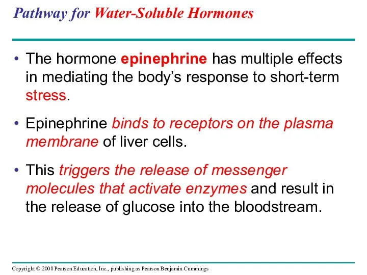
Pathway for Water-Soluble Hormones
The hormone epinephrine has multiple effects in mediating
Pathway for Water-Soluble Hormones
The hormone epinephrine has multiple effects in mediating
cAMP
Second
messenger
Adenylyl
cyclase
G protein-coupled
receptor
ATP
GTP
G protein
Epinephrine
Inhibition of
glycogen synthesis
Promotion of
glycogen breakdown
Protein
kinase A
Cell-surface hormone receptors
trigger
cAMP
Second
messenger
Adenylyl
cyclase
G protein-coupled
receptor
ATP
GTP
G protein
Epinephrine
Inhibition of
glycogen synthesis
Promotion of
glycogen breakdown
Protein
kinase A
Cell-surface hormone receptors
trigger
Pathway for Lipid-Soluble Hormones
The response to a lipid-soluble hormone is usually
Pathway for Lipid-Soluble Hormones
The response to a lipid-soluble hormone is usually
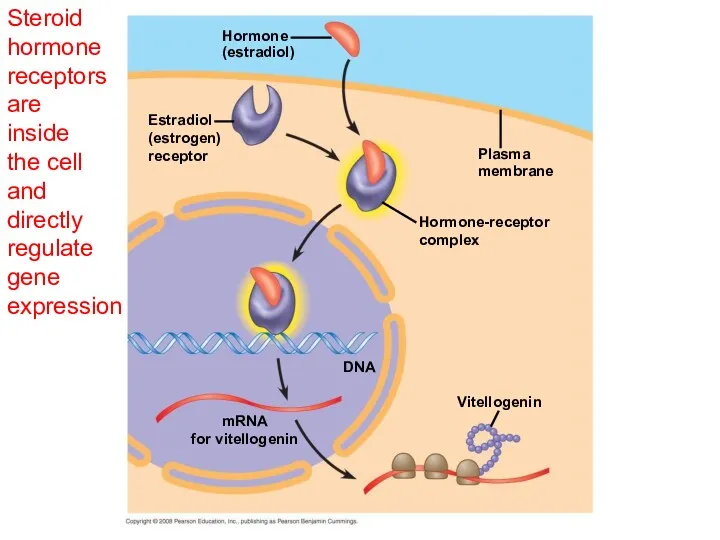
Steroid hormone receptors
are
inside
the cell
and
directly regulate
gene expression
Hormone
(estradiol)
Hormone-receptor
complex
Plasma
membrane
Estradiol
(estrogen)
receptor
DNA
Vitellogenin
mRNA
for vitellogenin
Steroid hormone receptors
are
inside
the cell
and
directly regulate
gene expression
Hormone
(estradiol)
Hormone-receptor
complex
Plasma
membrane
Estradiol
(estrogen)
receptor
DNA
Vitellogenin
mRNA
for vitellogenin
Multiple Effects of Hormones
The same hormone may have different effects on
Multiple Effects of Hormones
The same hormone may have different effects on
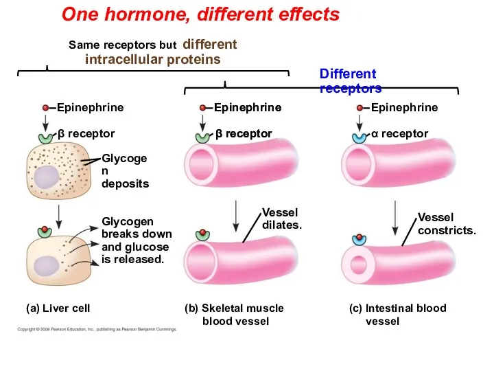
One hormone, different effects
Glycogen
deposits
β receptor
Vessel
dilates.
Epinephrine
(a) Liver cell
Epinephrine
β receptor
Glycogen
breaks down
and glucose
is
One hormone, different effects
Glycogen
deposits
β receptor
Vessel
dilates.
Epinephrine
(a) Liver cell
Epinephrine
β receptor
Glycogen
breaks down
and glucose
is
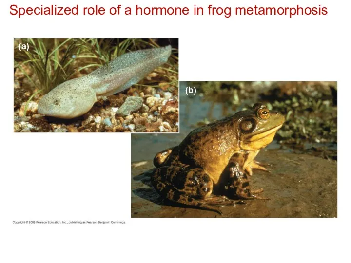
Specialized role of a hormone in frog metamorphosis
(a)
(b)
Specialized role of a hormone in frog metamorphosis
(a)
(b)
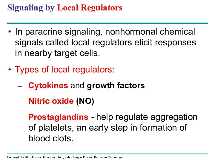
Signaling by Local Regulators
In paracrine signaling, nonhormonal chemical signals called local
Signaling by Local Regulators
In paracrine signaling, nonhormonal chemical signals called local
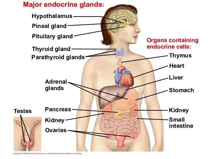
Major endocrine glands:
Adrenal
glands
Hypothalamus
Pineal gland
Pituitary gland
Thyroid gland
Parathyroid glands
Pancreas
Kidney
Ovaries
Testes
Organs containing
endocrine cells:
Thymus
Heart
Liver
Stomach
Kidney
Small
intestine
Major endocrine glands:
Adrenal
glands
Hypothalamus
Pineal gland
Pituitary gland
Thyroid gland
Parathyroid glands
Pancreas
Kidney
Ovaries
Testes
Organs containing
endocrine cells:
Thymus
Heart
Liver
Stomach
Kidney
Small
intestine
Simple Hormone Pathways
Negative feedback and antagonistic hormone pairs are common features
Simple Hormone Pathways
Negative feedback and antagonistic hormone pairs are common features
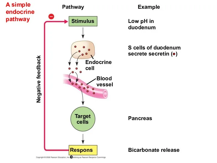
A simple endocrine pathway
Pathway
Example
Stimulus
Low pH in
duodenum
S cells of duodenum
secrete secretin (
A simple endocrine pathway
Pathway
Example
Stimulus
Low pH in
duodenum
S cells of duodenum
secrete secretin (
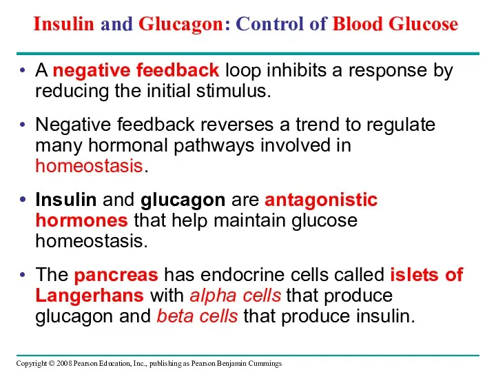
A negative feedback loop inhibits a response by reducing the initial
A negative feedback loop inhibits a response by reducing the initial
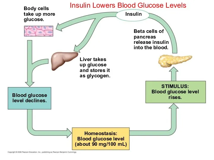
Insulin Lowers Blood Glucose Levels
Homeostasis:
Blood glucose level
(about 90 mg/100 mL)
Insulin
Beta cells
Insulin Lowers Blood Glucose Levels
Homeostasis:
Blood glucose level
(about 90 mg/100 mL)
Insulin
Beta cells
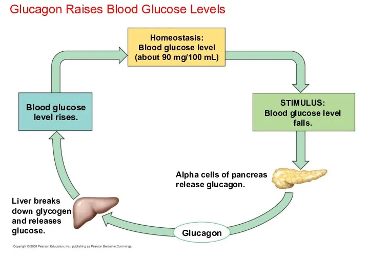
Glucagon Raises Blood Glucose Levels
Homeostasis:
Blood glucose level
(about 90 mg/100 mL)
Glucagon
STIMULUS:
Blood glucose
Glucagon Raises Blood Glucose Levels
Homeostasis:
Blood glucose level
(about 90 mg/100 mL)
Glucagon
STIMULUS:
Blood glucose
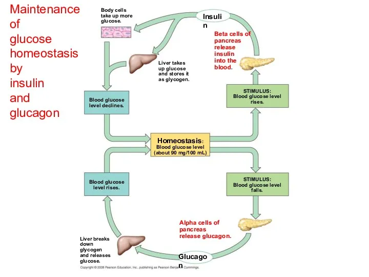
Maintenance of
glucose homeostasis by
insulin
and glucagon
Homeostasis:
Blood glucose level
(about 90
Maintenance of
glucose homeostasis by
insulin
and glucagon
Homeostasis:
Blood glucose level
(about 90
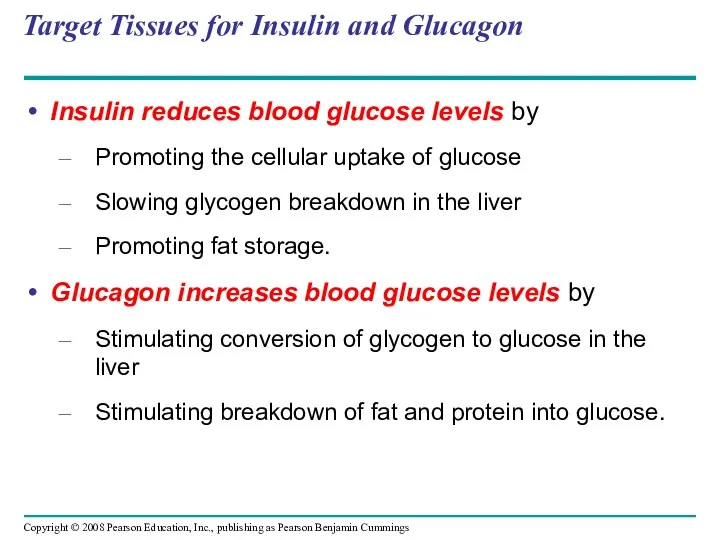
Target Tissues for Insulin and Glucagon
Insulin reduces blood glucose levels by
Promoting
Target Tissues for Insulin and Glucagon
Insulin reduces blood glucose levels by
Promoting
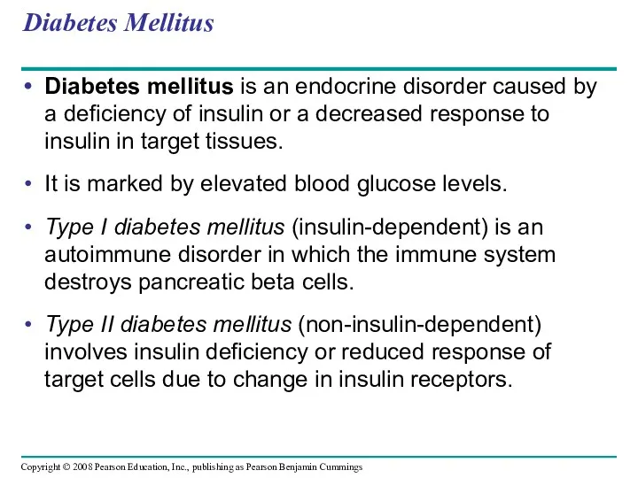
Diabetes Mellitus
Diabetes mellitus is an endocrine disorder caused by a deficiency
Diabetes Mellitus
Diabetes mellitus is an endocrine disorder caused by a deficiency
The endocrine and nervous systems act individually and together in regulating
The endocrine and nervous systems act individually and together in regulating
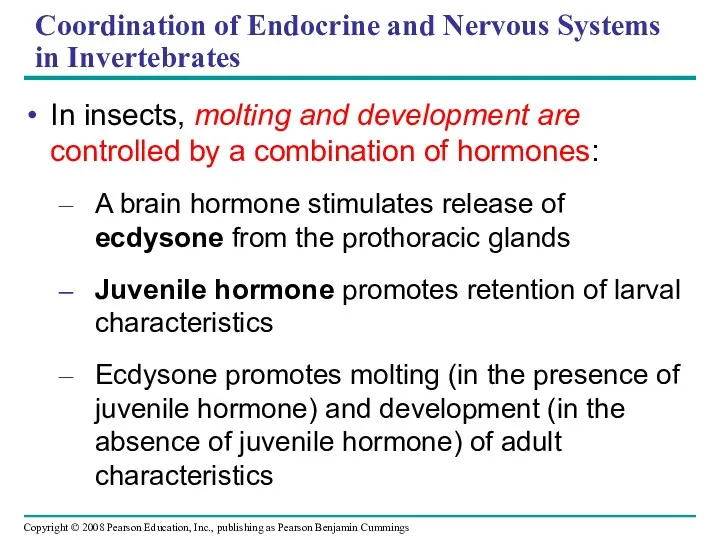
Coordination of Endocrine and Nervous Systems in Invertebrates
In insects, molting and
Coordination of Endocrine and Nervous Systems in Invertebrates
In insects, molting and
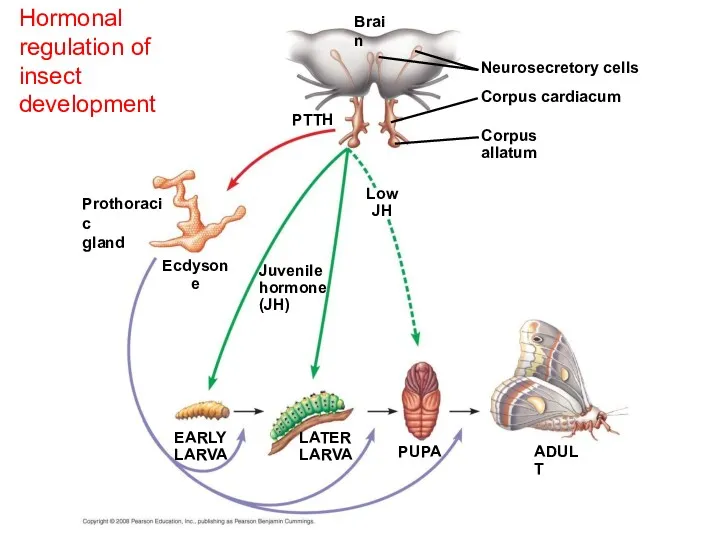
Hormonal regulation of insect development
Ecdysone
Brain
PTTH
EARLY
LARVA
Neurosecretory cells
Corpus cardiacum
Corpus allatum
LATER
LARVA
PUPA
ADULT
Low
JH
Juvenile
hormone
(JH)
Prothoracic
gland
Hormonal regulation of insect development
Ecdysone
Brain
PTTH
EARLY
LARVA
Neurosecretory cells
Corpus cardiacum
Corpus allatum
LATER
LARVA
PUPA
ADULT
Low
JH
Juvenile
hormone
(JH)
Prothoracic
gland
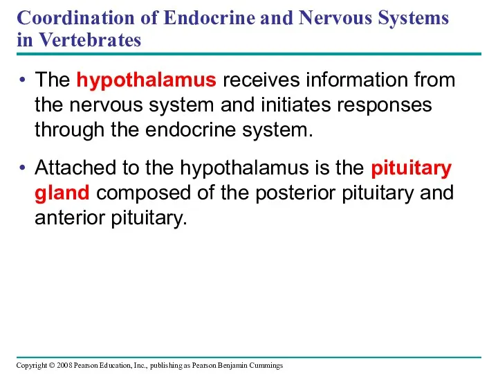
Coordination of Endocrine and Nervous Systems in Vertebrates
The hypothalamus receives information
Coordination of Endocrine and Nervous Systems in Vertebrates
The hypothalamus receives information
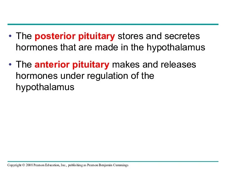
The posterior pituitary stores and secretes hormones that are made in
The posterior pituitary stores and secretes hormones that are made in
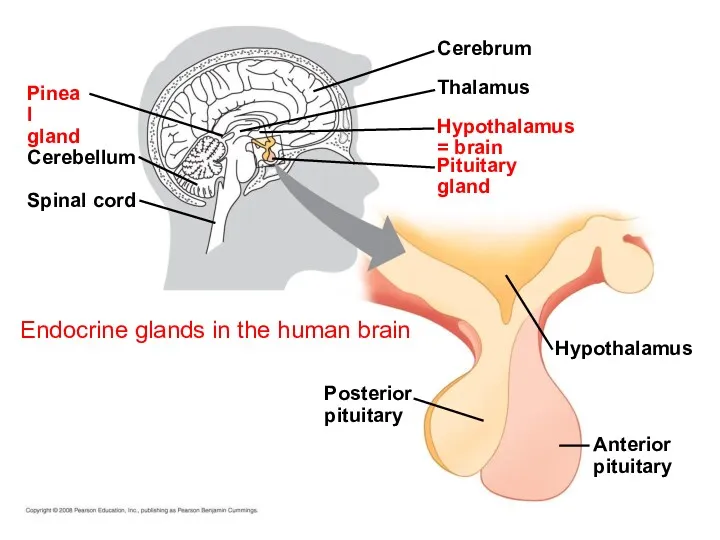
Endocrine glands in the human brain
Spinal cord
Posterior
pituitary
Cerebellum
Pineal
gland
Anterior
pituitary
Hypothalamus
Pituitary
gland
Hypothalamus = brain
Thalamus
Cerebrum
Endocrine glands in the human brain
Spinal cord
Posterior
pituitary
Cerebellum
Pineal
gland
Anterior
pituitary
Hypothalamus
Pituitary
gland
Hypothalamus = brain
Thalamus
Cerebrum

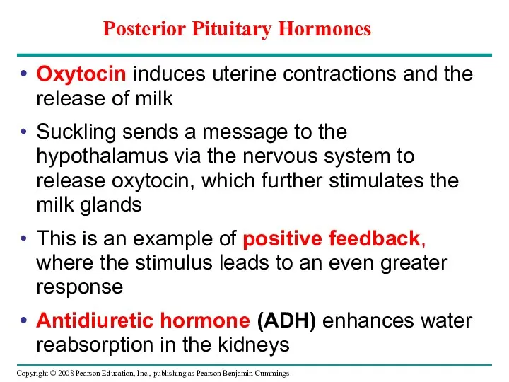
Oxytocin induces uterine contractions and the release of milk
Suckling sends a
Oxytocin induces uterine contractions and the release of milk
Suckling sends a
A simple neurohormone pathway
Suckling
Pathway
Stimulus
Hypothalamus/
posterior pituitary
Positive feedback
Example
Sensory
neuron
Neurosecretory
cell
Blood
vessel
Posterior pituitary
secretes oxytocin ( )
Target
cells
Response
Smooth muscle
A simple neurohormone pathway
Suckling
Pathway
Stimulus
Hypothalamus/
posterior pituitary
Positive feedback
Example
Sensory
neuron
Neurosecretory
cell
Blood
vessel
Posterior pituitary
secretes oxytocin ( )
Target
cells
Response
Smooth muscle
Anterior Pituitary Hormones
Hormone production in the anterior pituitary is controlled by
Anterior Pituitary Hormones
Hormone production in the anterior pituitary is controlled by
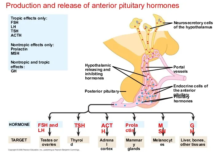
Production and release of anterior pituitary hormones
Hypothalamic
releasing and
inhibiting
hormones
Neurosecretory cells
of the hypothalamus
HORMONE
TARGET
Posterior
Production and release of anterior pituitary hormones
Hypothalamic
releasing and
inhibiting
hormones
Neurosecretory cells
of the hypothalamus
HORMONE
TARGET
Posterior
Hormone Cascade Pathways
A hormone can stimulate the release of a series
Hormone Cascade Pathways
A hormone can stimulate the release of a series
Cold
Pathway
Stimulus
Blood
vessel
Example
Sensory
neuron
Hypothalamus secretes
thyrotropin-releasing
hormone (TRH )
Neurosecretory
cell
A hormone
casade
pathway
Cold
Pathway
Stimulus
Blood
vessel
Example
Sensory
neuron
Hypothalamus secretes
thyrotropin-releasing
hormone (TRH )
Neurosecretory
cell
A hormone
casade
pathway
Cold
Pathway
Stimulus
Hypothalamus secretes
thyrotropin-releasing
hormone (TRH )
Example
Sensory
neuron
Neurosecretory
cell
Blood
vessel
+
Anterior pituitary secretes
thyroid-stimulating
hormone (TSH
or thyrotropin )
A hormone
casade
pathway
Cold
Pathway
Stimulus
Hypothalamus secretes
thyrotropin-releasing
hormone (TRH )
Example
Sensory
neuron
Neurosecretory
cell
Blood
vessel
+
Anterior pituitary secretes
thyroid-stimulating
hormone (TSH
or thyrotropin )
A hormone
casade
pathway
A hormone
casade
pathway
Cold
Pathway
Stimulus
Hypothalamus secretes
thyrotropin-releasing
hormone (TRH )
Negative feedback
Example
Sensory
neuron
Neurosecretory
cell
Blood
vessel
Anterior pituitary secretes
thyroid-stimulating
hormone (TSH
or thyrotropin
A hormone
casade
pathway
Cold
Pathway
Stimulus
Hypothalamus secretes
thyrotropin-releasing
hormone (TRH )
Negative feedback
Example
Sensory
neuron
Neurosecretory
cell
Blood
vessel
Anterior pituitary secretes thyroid-stimulating hormone (TSH or thyrotropin
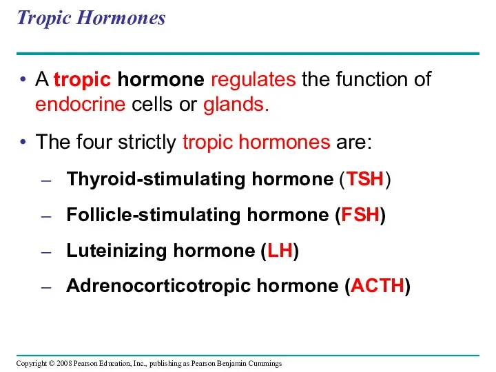
Tropic Hormones
A tropic hormone regulates the function of endocrine cells or
Tropic Hormones
A tropic hormone regulates the function of endocrine cells or
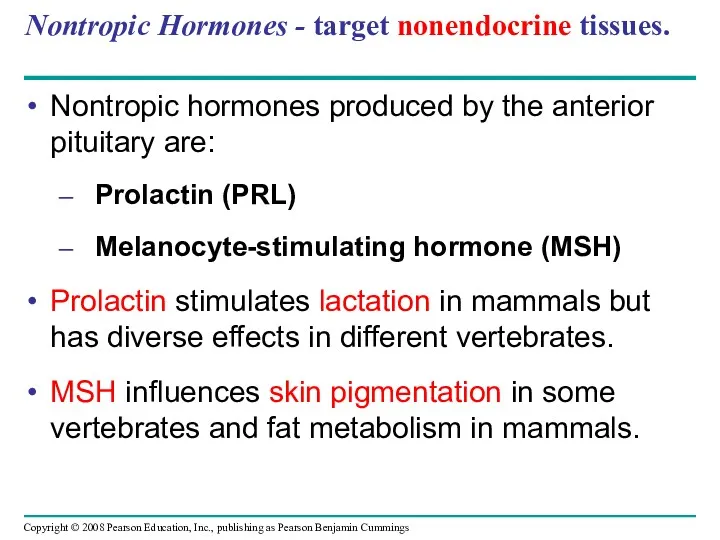
Nontropic Hormones - target nonendocrine tissues.
Nontropic hormones produced by the anterior
Nontropic Hormones - target nonendocrine tissues.
Nontropic hormones produced by the anterior
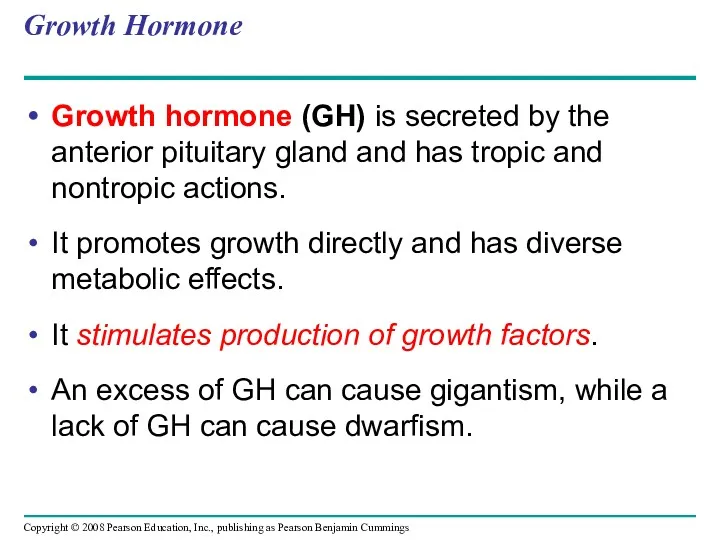
Growth Hormone
Growth hormone (GH) is secreted by the anterior pituitary gland
Growth Hormone
Growth hormone (GH) is secreted by the anterior pituitary gland
Endocrine signaling regulates metabolism, homeostasis, development, and behavior.
Endocrine glands respond to
Endocrine signaling regulates metabolism, homeostasis, development, and behavior.
Endocrine glands respond to
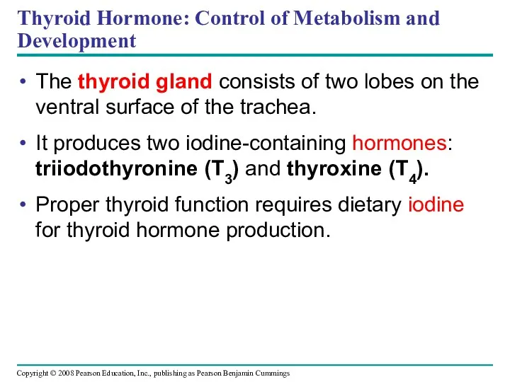
Thyroid Hormone: Control of Metabolism and Development
The thyroid gland consists of
Thyroid Hormone: Control of Metabolism and Development
The thyroid gland consists of
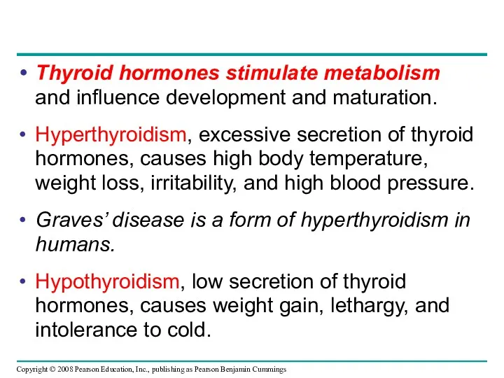
Thyroid hormones stimulate metabolism and influence development and maturation.
Hyperthyroidism, excessive secretion
Thyroid hormones stimulate metabolism and influence development and maturation.
Hyperthyroidism, excessive secretion
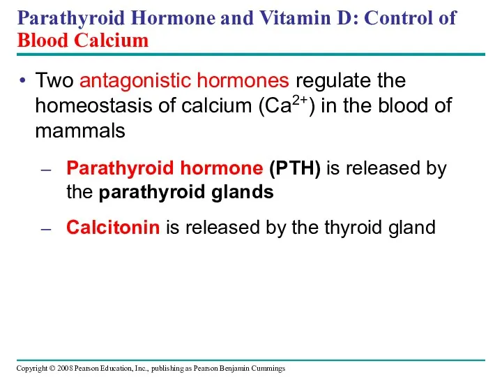
Parathyroid Hormone and Vitamin D: Control of Blood Calcium
Two antagonistic hormones
Parathyroid Hormone and Vitamin D: Control of Blood Calcium
Two antagonistic hormones
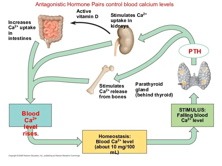
Antagonistic Hormone Pairs control blood calcium levels
PTH
Parathyroid gland
(behind thyroid)
STIMULUS:
Falling blood
Ca2+ level
Homeostasis:
Blood
Antagonistic Hormone Pairs control blood calcium levels
PTH
Parathyroid gland
(behind thyroid)
STIMULUS:
Falling blood
Ca2+ level
Homeostasis:
Blood
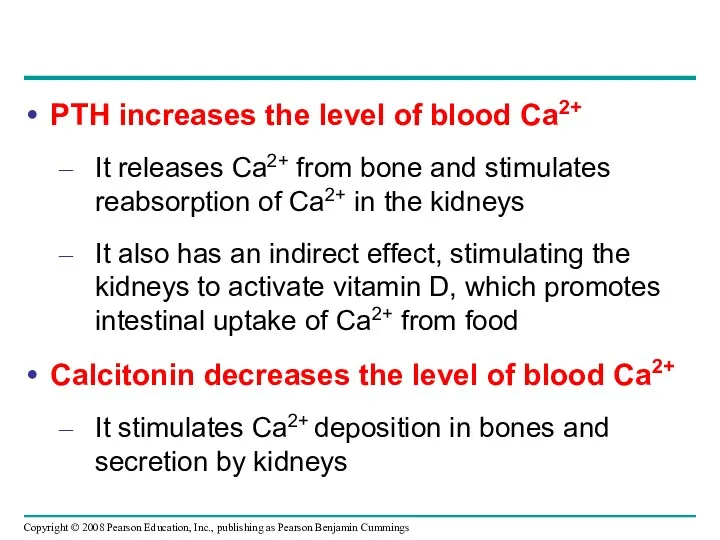
PTH increases the level of blood Ca2+
It releases Ca2+ from bone
PTH increases the level of blood Ca2+
It releases Ca2+ from bone
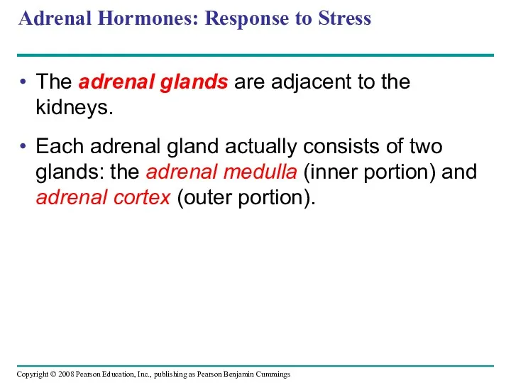
Adrenal Hormones: Response to Stress
The adrenal glands are adjacent to the
Adrenal Hormones: Response to Stress
The adrenal glands are adjacent to the
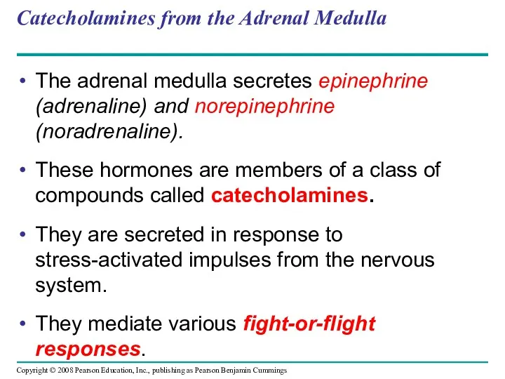
Catecholamines from the Adrenal Medulla
The adrenal medulla secretes epinephrine (adrenaline) and
Catecholamines from the Adrenal Medulla
The adrenal medulla secretes epinephrine (adrenaline) and
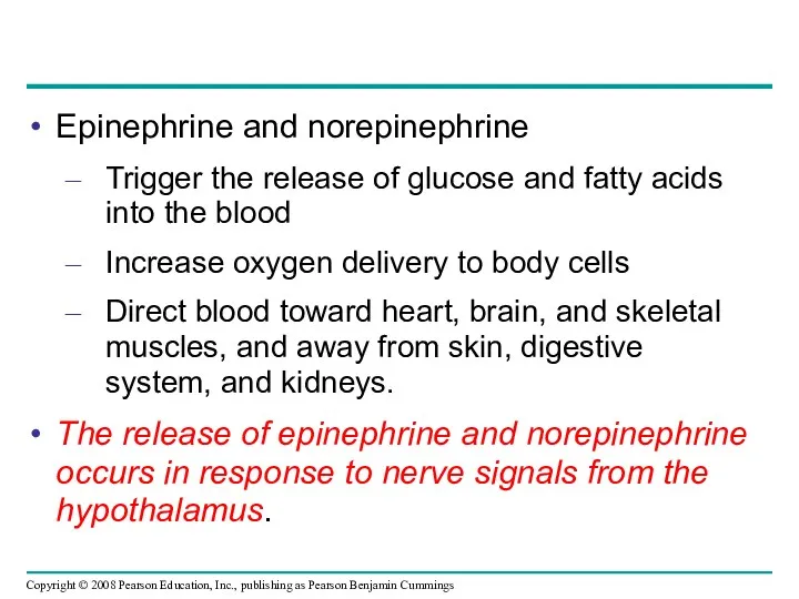
Epinephrine and norepinephrine
Trigger the release of glucose and fatty acids into
Epinephrine and norepinephrine
Trigger the release of glucose and fatty acids into
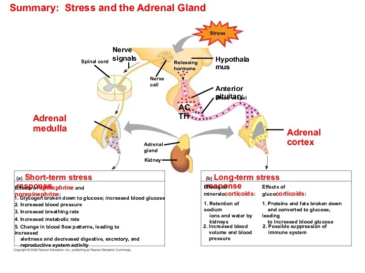
Summary: Stress and the Adrenal Gland
Stress
Adrenal
gland
Nerve
cell
Nerve
signals
Releasing
hormone
Hypothalamus
Anterior pituitary
Blood vessel
ACTH
Adrenal cortex
Spinal cord
Adrenal
Summary: Stress and the Adrenal Gland
Stress
Adrenal
gland
Nerve
cell
Nerve
signals
Releasing
hormone
Hypothalamus
Anterior pituitary
Blood vessel
ACTH
Adrenal cortex
Spinal cord
Adrenal
Stress and the Adrenal Gland
Stress
Adrenal
gland
Nerve
cell
Nerve
signals
Releasing
hormone
Hypothalamus
Anterior pituitary
Blood vessel
ACTH
Adrenal
cortex
Spinal cord
Adrenal
medulla
Kidney
Stress and the Adrenal Gland
Stress
Adrenal
gland
Nerve
cell
Nerve
signals
Releasing
hormone
Hypothalamus
Anterior pituitary
Blood vessel
ACTH
Adrenal
cortex
Spinal cord
Adrenal
medulla
Kidney
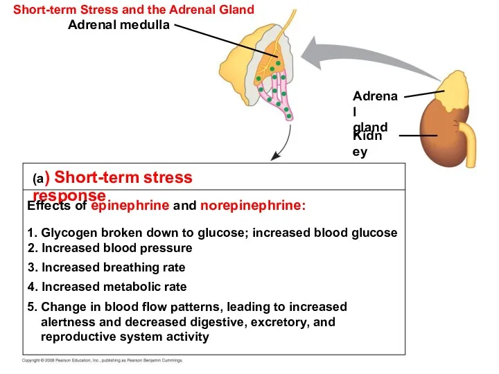
Short-term Stress and the Adrenal Gland
(a) Short-term stress response
Effects of epinephrine
Short-term Stress and the Adrenal Gland
(a) Short-term stress response
Effects of epinephrine
Steroid Hormones from the Adrenal Cortex
The adrenal cortex releases a family
Steroid Hormones from the Adrenal Cortex
The adrenal cortex releases a family
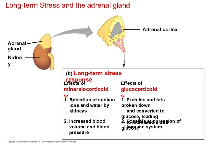
Long-term Stress and the adrenal gland
(b) Long-term stress response
Effects of
mineralocorticoids:
Effects of
glucocorticoids:
1.
Long-term Stress and the adrenal gland
(b) Long-term stress response
Effects of
mineralocorticoids:
Effects of
glucocorticoids:
1.
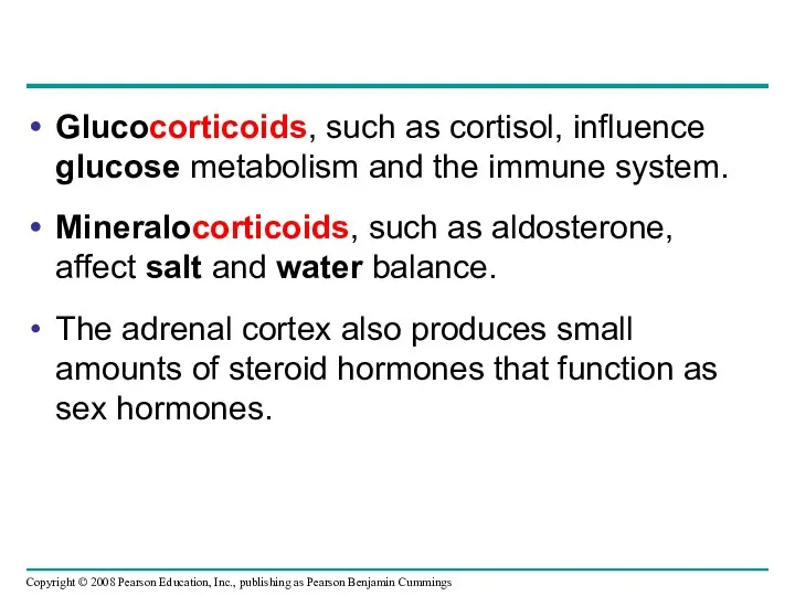
Glucocorticoids, such as cortisol, influence glucose metabolism and the immune system.
Mineralocorticoids,
Glucocorticoids, such as cortisol, influence glucose metabolism and the immune system.
Mineralocorticoids,
Gonadal Sex Hormones
The gonads = testes and ovaries, produce most of
Gonadal Sex Hormones
The gonads = testes and ovaries, produce most of
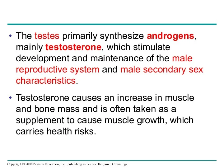
The testes primarily synthesize androgens, mainly testosterone, which stimulate development and
The testes primarily synthesize androgens, mainly testosterone, which stimulate development and
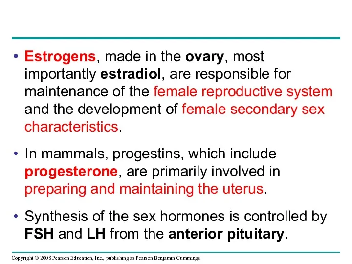
Estrogens, made in the ovary, most importantly estradiol, are responsible for
Estrogens, made in the ovary, most importantly estradiol, are responsible for
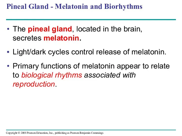
Pineal Gland - Melatonin and Biorhythms
The pineal gland, located in the
Pineal Gland - Melatonin and Biorhythms
The pineal gland, located in the
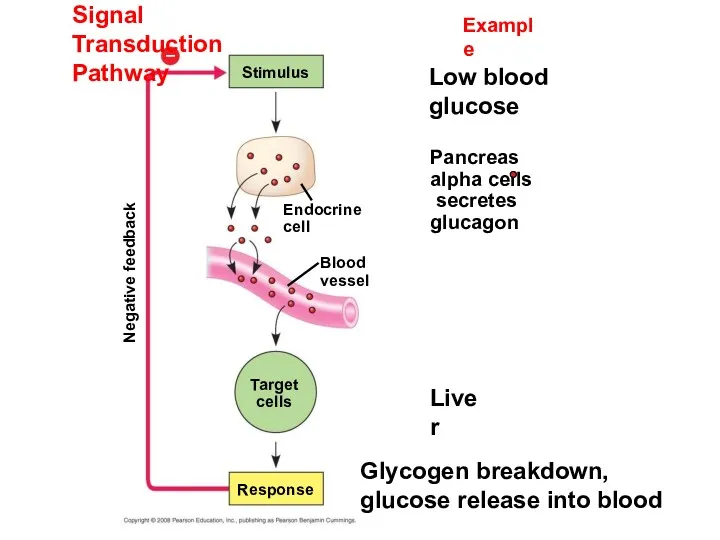
Signal Transduction Pathway
Example
Stimulus
Low blood glucose
Pancreas alpha cells
secretes
glucagon
Endocrine
cell
Blood
vessel
Liver
Target
cells
Response
Glycogen breakdown,
glucose
Signal Transduction Pathway
Example
Stimulus
Low blood glucose
Pancreas alpha cells
secretes
glucagon
Endocrine
cell
Blood
vessel
Liver
Target
cells
Response
Glycogen breakdown, glucose
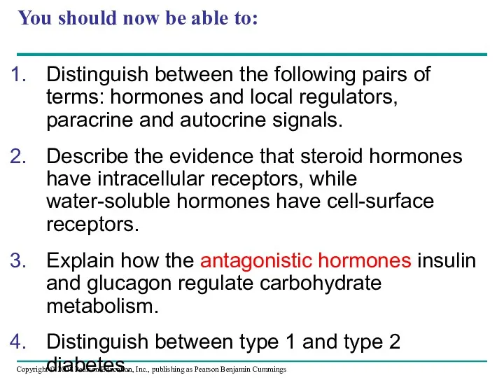
You should now be able to:
Distinguish between the following pairs of
You should now be able to:
Distinguish between the following pairs of
 Внутриутробные инфекции у новорожденных детей
 Пациент және оның жақындарының қауіпсіз қоршаған ортасы
 Тағамдық токсикоинфекциялар
 Лечение при аллергозах
 Методическое пособие Партограмма как интегральный метод контроля за клиническим течением родов
 Human excretory system
 Свойства растворов электролитов. (Лекция 4)
 Әр түрлі қан тамырлық бассейндердің зақымдану кезіндегі клиникалық синдромдары
 Аритмии - нарушения ритма сердца. Лекция
 Қоршаған орта мен тұрғындар денсаулығы үшін Қазақстан аймақтарындағы көп жылдық ядролық сынақтар салдары
 Основы кардиохирургии. Введение
 Патология слюнных желез
 Раны. Анатомия ран
 Особенности детей ОВЗ с умственной отсталостью. Обучение и воспитание
 Травмы мягких тканей
 Материнская смертность
 Макулодистрофия. Патогенетические аспекты. Модифицированная клиническая классификация. Лечение глаз
 Критерии выбора дезинфицирующих средств при организации дезинфекционно-стерилизационного режима
 5_6_Средства_на_дыхательную_систему
 Развитие репродуктивной системы женщины. Нейроэндокринная регуляция менструального цикла
 Сердечно-легочная реанимация
 Эндокринді бездердің қатерлі және қатесіз ісіктері
 Ортопедическое лечения ВНЧС перед протезированием
 Инфекционно-токсический шок при менингококковой инфекции. Мероприятия при ИТШ
 Влияние болезней кровообращения на течение беременности
 Чрезвычайные ситуации инфекционных заболеваний людей и животных
 Санаторно-курортное обслуживание населения в СССР
 Ортопедические и комплексные методы лечения деформации зубных рядов