- Immunophysilogy of lung

Содержание
- 6. For more than 70 years, surfactant was perceived to be a soap-like substance that reduced surface
- 7. Augmented production of SP-A by the maturing fetal lung at term provides a key hormonal stimulus
- 8. Surfactant protein A (SP-A) has been shown to inhibit the maturation of dendritic cells (DCs), whereas
- 9. A consequence of apoptotic-body uptake by a phagocyte is induction of an anti-inflammatory response by the
- 10. The roles of macrophages in clearing apoptotic cells and cellular debris in health and disease are
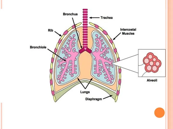
- 11. The alveolar membrane is the largest surface of the body in contact with the outside environment.
- 12. Alveolar macrophages are long-lived, with a turnover rate of only approximately 40% in 1 year. By
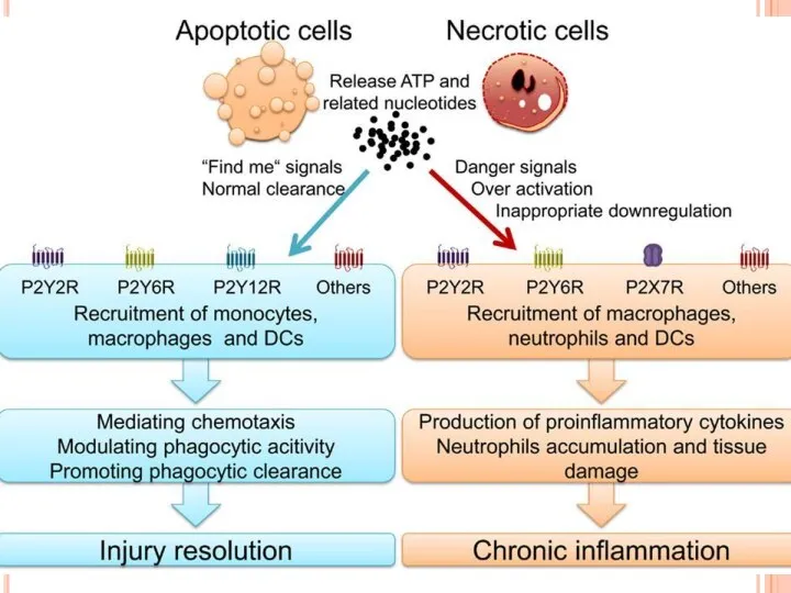
- 13. The non-inflammatory clearance of apoptotic cells (a) compared with the pro-inflammatory ingestion of necrotic cells and
- 18. In healthy individuals, the airspaces are replete with mechanisms that prevent an inflammatory response from occurring.
- 19. Table 1 | The specific phenotype of mouse macrophages from different sites Surface Marker Peritoneal macrophage
- 20. Innate immune functions of alveolar macrophages. As the resident innate immune cell of the pulmonary airspace,
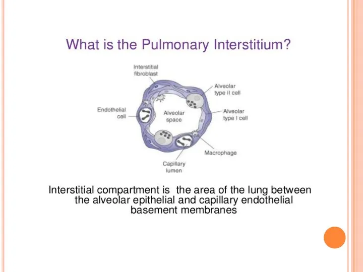
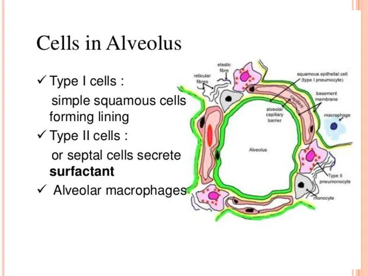
- 21. Alveolar macrophages reside in the airspaces juxtaposed with type I alveolar epithelial cells or with type
- 23. Скачать презентацию
For more than 70 years, surfactant was perceived to be a
For more than 70 years, surfactant was perceived to be a
Augmented production of SP-A by the maturing fetal lung at term
Augmented production of SP-A by the maturing fetal lung at term
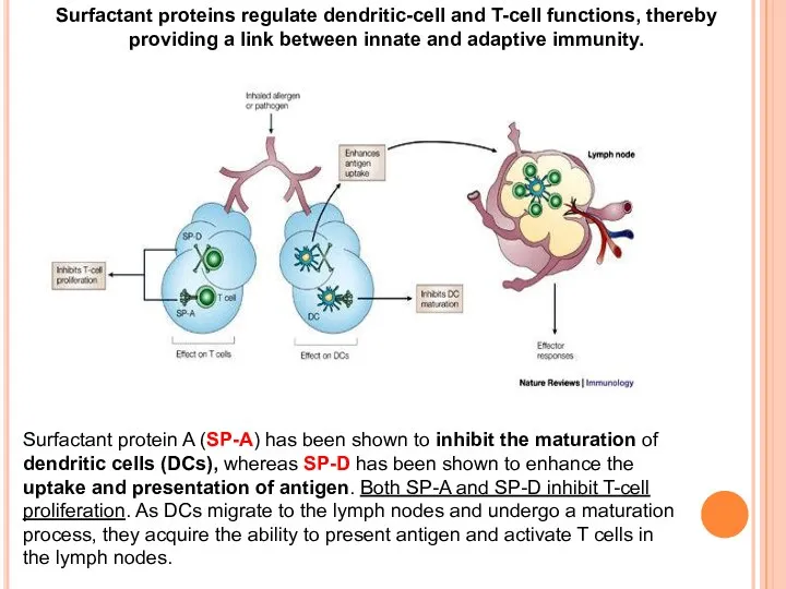
Surfactant protein A (SP-A) has been shown to inhibit the maturation
Surfactant protein A (SP-A) has been shown to inhibit the maturation
A consequence of apoptotic-body uptake by a phagocyte is induction of
A consequence of apoptotic-body uptake by a phagocyte is induction of
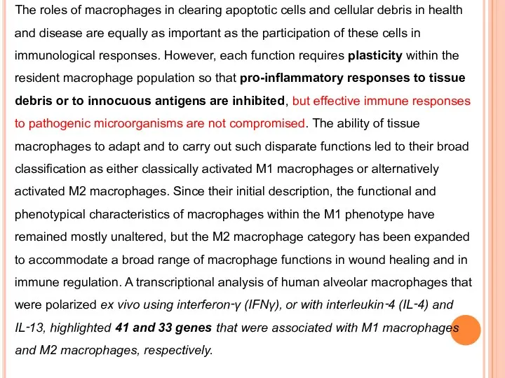
The roles of macrophages in clearing apoptotic cells and cellular debris
The roles of macrophages in clearing apoptotic cells and cellular debris
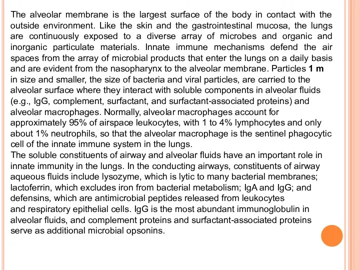
The alveolar membrane is the largest surface of the body in
The alveolar membrane is the largest surface of the body in
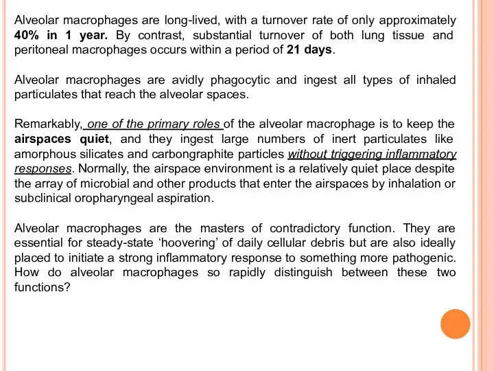
Alveolar macrophages are long-lived, with a turnover rate of only approximately
Alveolar macrophages are long-lived, with a turnover rate of only approximately
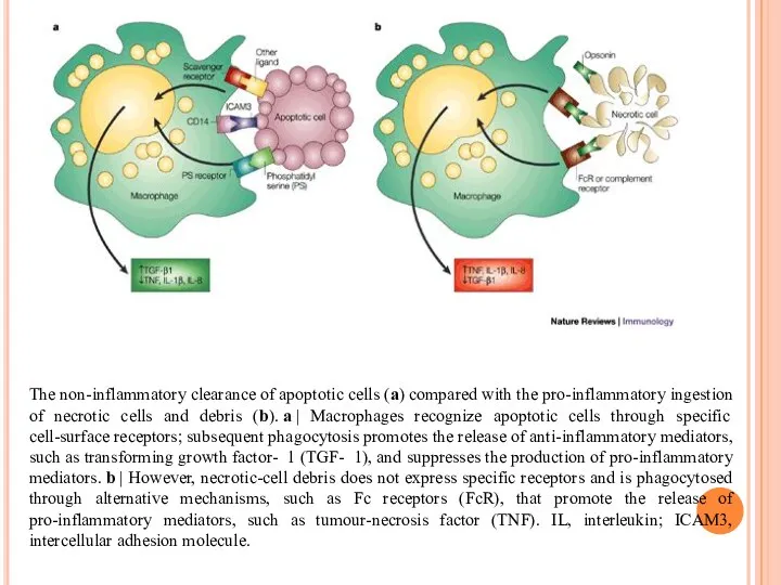
The non-inflammatory clearance of apoptotic cells (a) compared with the pro-inflammatory
The non-inflammatory clearance of apoptotic cells (a) compared with the pro-inflammatory
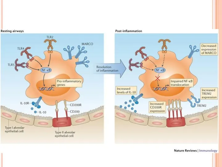
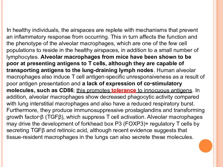
In healthy individuals, the airspaces are replete with mechanisms that prevent
In healthy individuals, the airspaces are replete with mechanisms that prevent
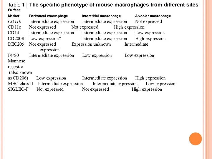
Table 1 | The specific phenotype of mouse macrophages from different
Table 1 | The specific phenotype of mouse macrophages from different
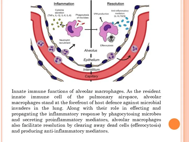
Innate immune functions of alveolar macrophages. As the resident innate immune
Innate immune functions of alveolar macrophages. As the resident innate immune
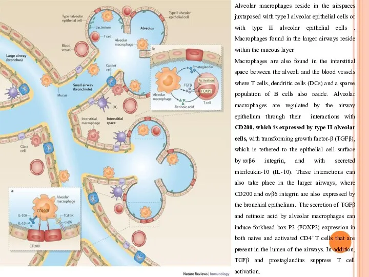
Alveolar macrophages reside in the airspaces juxtaposed with type I alveolar
Alveolar macrophages reside in the airspaces juxtaposed with type I alveolar
 Таргетная терапия немелкоклеточного рака лёгких
 Емхананың профилактикалық бөлмесінде тексеру жүргізу. Тұрғын халықтың диспансеризациясы
 Сестринский процесс при гастритах
 Нарушение равновесия жидких сред, расстройства крово- и лимфообращения
 Врожденные пороки развития желудочно-кишечного тракта
 Остеохондроз и методы его лечения
 Вирусты инфекциялардың химиотерапиясының артықшылықтары
 Новообразование орбиты
 Острые лейкозы у детей
 Лечение желчнокаменной болезни
 Сахарный диабет. Диагностика СД и других нарушений углеводного обмена
 Маточные кровотечения. Новые подходы к известной проблеме
 Организация акушерско-гинекологической помощи. Репродуктивное здоровье населения в России
 Химиоэмболизация в онкологии
 Кровь. Лейкоциты. Иммунитет
 Злоякісні пухлини голови та шиї
 Психофармкотерапия психических и поведенческих расстройств в процессе медицинской реабилитации у детей
 Көпіршікті дерматоздар: Ұшық тәрізді Дюринг дерматозы
 Местная анестезия. Виды и методы проведения. Принципы сердечно-легочной реанимации
 Синдром сердечной недостаточности
 Ветряная оспа
 Доброякісні пухлини ж.с.о. Фонові та передракові захворювання ж.с.о
 Основы наложения кожных швов
 Послеоперационное обезболивание
 Қылилық
 Соединительно-тканный массаж. Меридианы
 Патофизиология - наука о природе заболеваний
 Неврит. Виды неврита