- Introduction into morphology of tumors tumors from epithelium

Содержание
- 2. Neoplasia (tumor, neoplasm) - it is the process of new uncontrolled growth, characterized by violation of
- 3. There are 3 types of tumors according to prognosis: 1. benign tumors – slowly growing, not
- 4. All tumors, benign and malignant, have two basic components: 1) proliferating neoplastic cells that constitute their
- 5. I. In general, benign tumors are designated by attaching the suffix -oma to the cell type
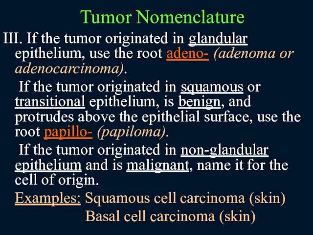
- 6. III. If the tumor originated in glandular epithelium, use the root adeno- (adenoma or adenocarcinoma). If
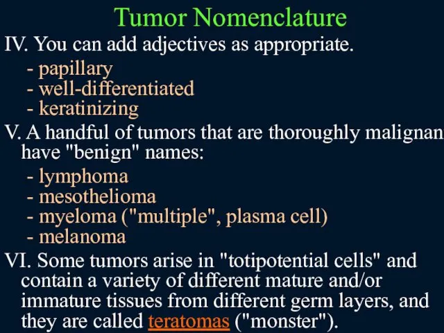
- 7. IV. You can add adjectives as appropriate. - papillary - well-differentiated - keratinizing V. A handful
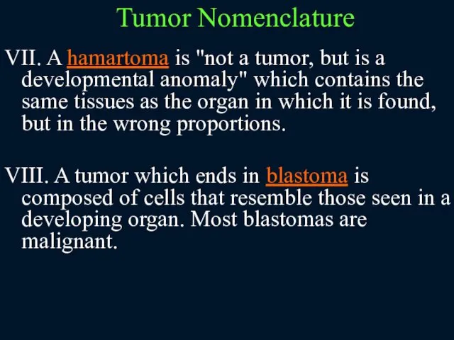
- 8. VII. A hamartoma is "not a tumor, but is a developmental anomaly" which contains the same
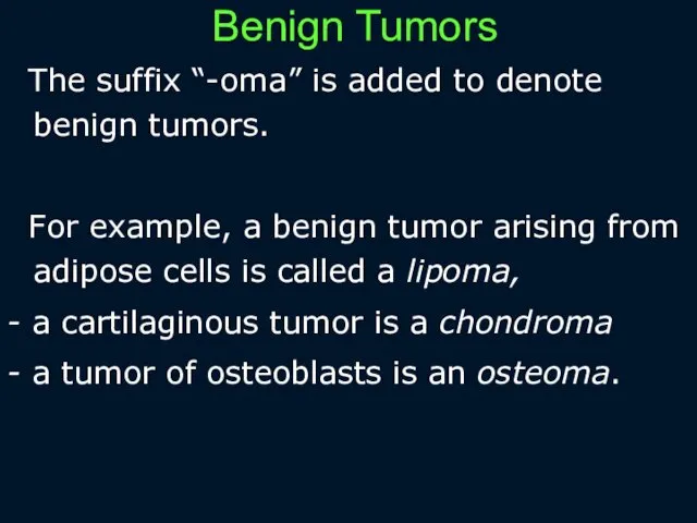
- 9. Benign Tumors The suffix “-oma” is added to denote benign tumors. For example, a benign tumor
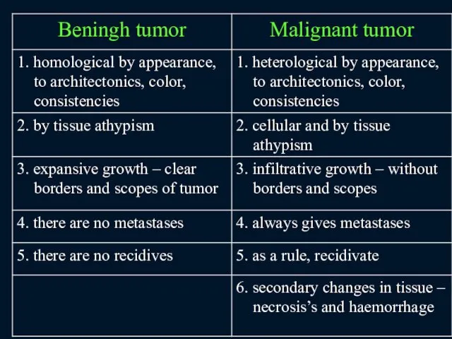
- 10. Characteristics of benign tumors: 1. Cells resemble normal cells and tumor architecture resembles that of the
- 11. Characteristics of malignant tumors 1. Malignant tumors generally grow more rapidly than benign tumors. 2. Cells
- 13. Stages of malignant tumor 1. The before tumor changes of tissue – is dysplasia (duration of
- 14. DYSPLASIA ("atypia", "pre-cancer"): It is abnormal epithelium with "loss of uniformity of the individual cells, as
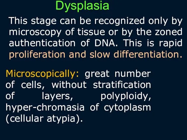
- 15. Dysplasia This stage can be recognized only by microscopy of tissue or by the zoned authentication
- 16. Dysplasia really can reflect: - initial stage of development of cancer - the process of reparation
- 17. Dysplasia – it is facultative pre-cancer in: - Breаst - cervix of uterus - endometrium -
- 18. The stage of formation of tumor rudiment (non-invasive stage) It is appearance of different clones of
- 19. Microscopically: 1. intensive proliferation of cells with the presence of pathological mitosis’s 2. structural atypism (cellular
- 20. The Invasive stage – migration of tumor cells into surrounding tissues. Tumor cells connects with fibronectin
- 21. Metastatical stage – tumor cells must grow up to the vessels. Cancer cells have the tropism
- 22. Metastatic spread: There are four routes: 1) Spreading by serous surfaces 2) Mechanical transplantation (rare, typically
- 23. Features of metastatic stage 1. the first tumor cells are destroyed by macrophages 2. there is
- 24. The Anatomy-histological classification According to the appearance: - cellular infiltrate in tissue - nodules - polypus
- 25. According to the type of growth: - unicentrical – from one tumor rudiment - policentrical (in
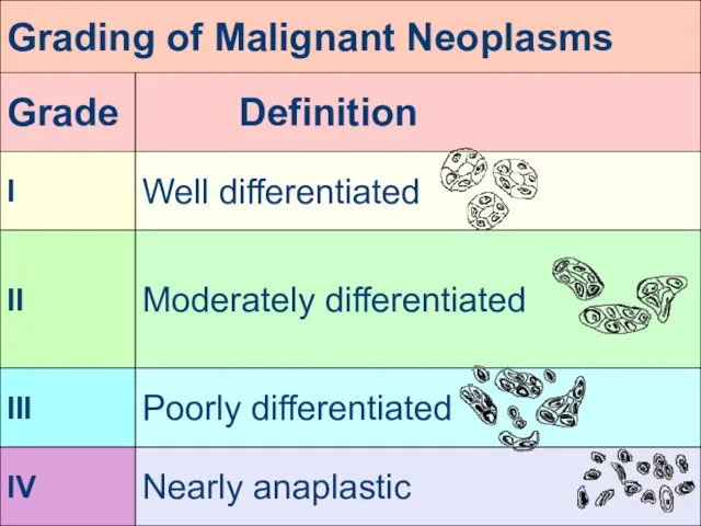
- 26. According to the degree of maturity tumors are devided: - differentiate – characterized by the slow
- 27. Grade and Stage: Tumor grade: assigned by the pathologist to reflect the cancer's degree of differentiation.
- 28. Grade and Stage: Tumor stage: assigned by the clinician on the basis of all available information
- 30. Alternative system: TNM "T" for tumor: T1 might mean primary tumor is smaller than 1 cm
- 31. Generally, tumors of high grade present at high stage, while tumors of low grade present at
- 32. Benign epithelial tumors Benign epithelial tumors are subdivided according to their origin from different types of
- 33. Papillomas It is broad-based superficial tumor of branching villous vascular stroma covered by neoplastic epithelium. Bening
- 34. Adenomas Benign epithelial tumor from the epithelium of the glands and glandular organs. More often they
- 35. Squamous cell carcinomas These arise anywhere there is a stratified squamous epithelium, either healthy (skin, esophagus,
- 36. Adenocarcinomas These arise anywhere there are glands, even single-celled glands (i.e., goblet cells) Look for any
- 37. I. Maligant neoplasms arising from tissue embryologically derived from ectoderm or endoderm are usually carcinomas. Examples
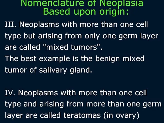
- 38. III. Neoplasms with more than one cell type but arising from only one germ layer are
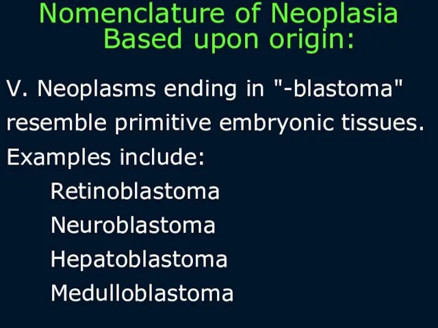
- 39. V. Neoplasms ending in "-blastoma" resemble primitive embryonic tissues. Examples include: Retinoblastoma Neuroblastoma Hepatoblastoma Medulloblastoma Nomenclature
- 41. Скачать презентацию
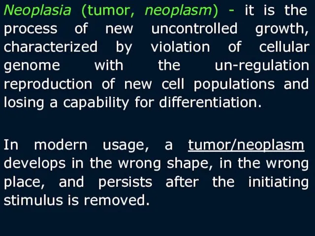
Neoplasia (tumor, neoplasm) - it is the process of new uncontrolled
Neoplasia (tumor, neoplasm) - it is the process of new uncontrolled
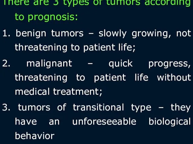
There are 3 types of tumors according to prognosis:
1. benign tumors
There are 3 types of tumors according to prognosis:
1. benign tumors
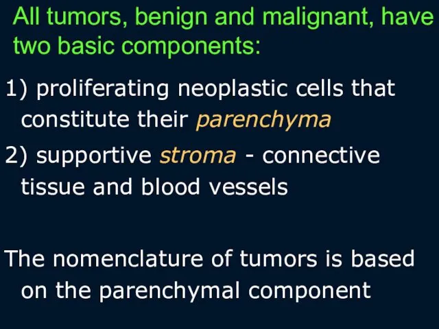
All tumors, benign and malignant, have two basic components:
1) proliferating
All tumors, benign and malignant, have two basic components:
1) proliferating
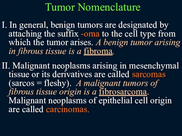
I. In general, benign tumors are designated by attaching the suffix
I. In general, benign tumors are designated by attaching the suffix
III. If the tumor originated in glandular epithelium, use the root
III. If the tumor originated in glandular epithelium, use the root
IV. You can add adjectives as appropriate.
- papillary
- well-differentiated
IV. You can add adjectives as appropriate.
- papillary
- well-differentiated
VII. A hamartoma is "not a tumor, but is a developmental
Benign Tumors
The suffix “-oma” is added to denote benign tumors.
For
Benign Tumors
The suffix “-oma” is added to denote benign tumors.
For
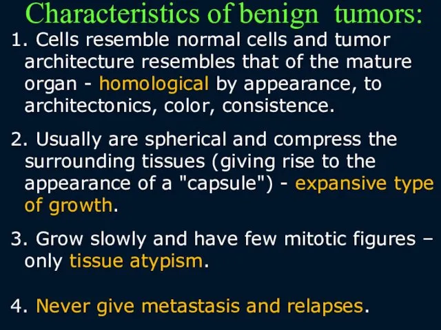
Characteristics of benign tumors:
1. Cells resemble normal cells and tumor architecture
Characteristics of benign tumors:
1. Cells resemble normal cells and tumor architecture
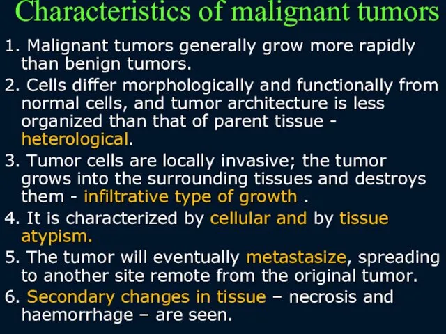
Characteristics of malignant tumors
1. Malignant tumors generally grow more rapidly
Characteristics of malignant tumors
1. Malignant tumors generally grow more rapidly
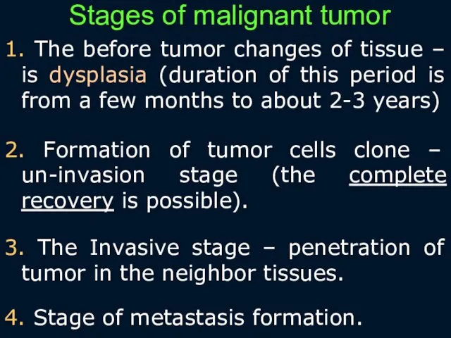
Stages of malignant tumor
1. The before tumor changes of tissue –
Stages of malignant tumor
1. The before tumor changes of tissue –
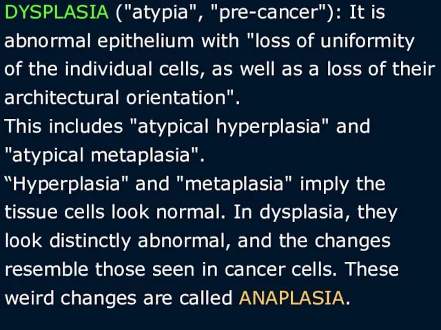
DYSPLASIA ("atypia", "pre-cancer"): It is abnormal epithelium with "loss of uniformity
DYSPLASIA ("atypia", "pre-cancer"): It is abnormal epithelium with "loss of uniformity
Dysplasia
This stage can be recognized only by microscopy of tissue
Dysplasia
This stage can be recognized only by microscopy of tissue
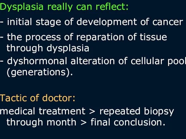
Dysplasia really can reflect:
- initial stage of development of cancer
- the
Dysplasia really can reflect:
- initial stage of development of cancer
- the
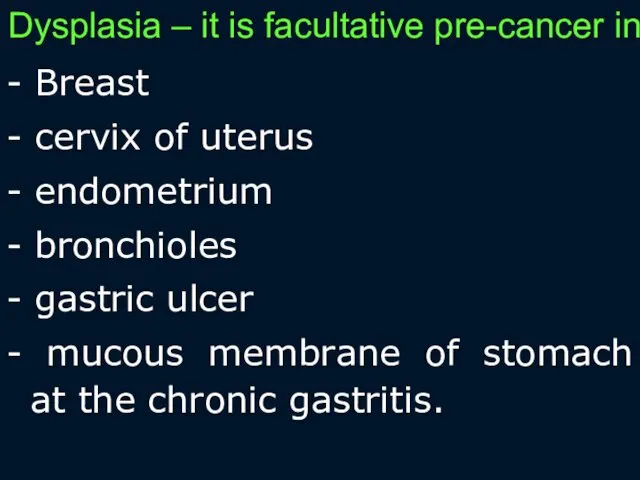
Dysplasia – it is facultative pre-cancer in:
- Breаst
- cervix of uterus
-
Dysplasia – it is facultative pre-cancer in:
- Breаst
- cervix of uterus
-
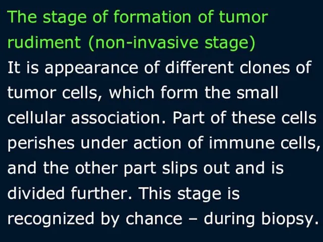
The stage of formation of tumor rudiment (non-invasive stage)
It is
The stage of formation of tumor rudiment (non-invasive stage) It is
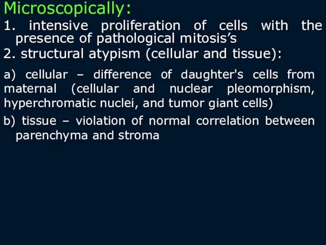
Microscopically:
1. intensive proliferation of cells with the presence of pathological
Microscopically:
1. intensive proliferation of cells with the presence of pathological
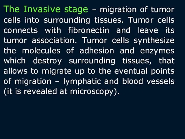
The Invasive stage – migration of tumor cells into surrounding tissues.
The Invasive stage – migration of tumor cells into surrounding tissues.
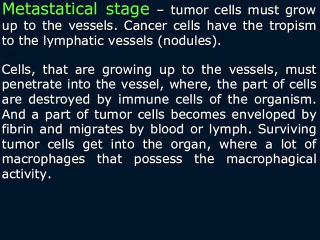
Metastatical stage – tumor cells must grow up to the vessels.
Metastatical stage – tumor cells must grow up to the vessels.
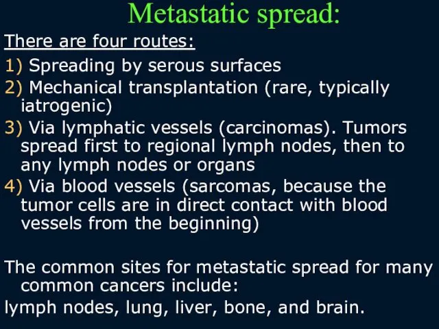
Metastatic spread:
There are four routes:
1) Spreading by serous surfaces
2)
Metastatic spread:
There are four routes:
1) Spreading by serous surfaces
2)
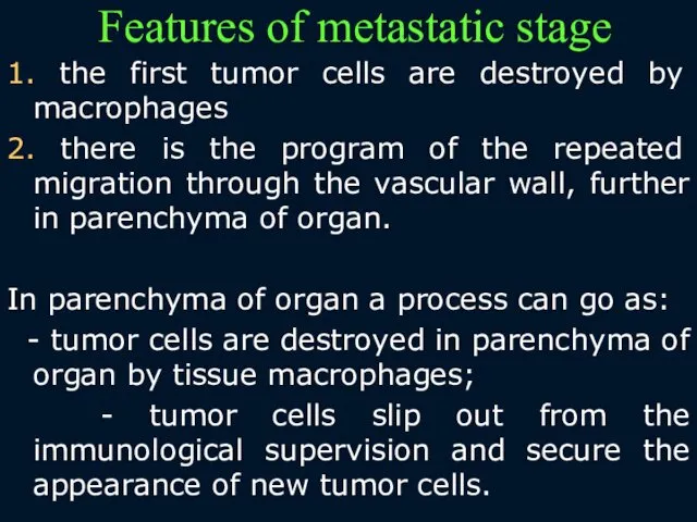
Features of metastatic stage
1. the first tumor cells are destroyed by
Features of metastatic stage
1. the first tumor cells are destroyed by
The Anatomy-histological classification
According to the appearance:
- cellular infiltrate in tissue
-
The Anatomy-histological classification
According to the appearance:
- cellular infiltrate in tissue
-
According to the type of growth:
- unicentrical – from one tumor
According to the type of growth:
- unicentrical – from one tumor
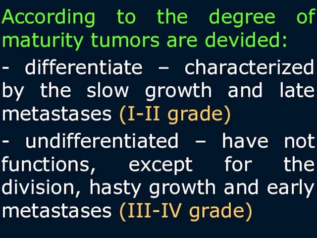
According to the degree of maturity tumors are devided:
- differentiate –
According to the degree of maturity tumors are devided:
- differentiate –
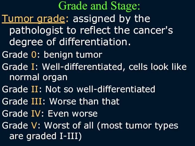
Grade and Stage:
Tumor grade: assigned by the pathologist to reflect
Grade and Stage:
Tumor grade: assigned by the pathologist to reflect
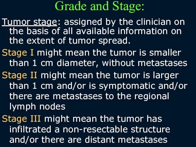
Grade and Stage:
Tumor stage: assigned by the clinician on the basis
Grade and Stage:
Tumor stage: assigned by the clinician on the basis
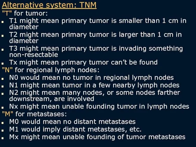
Alternative system: TNM
"T" for tumor:
T1 might mean primary tumor
Alternative system: TNM
"T" for tumor:
T1 might mean primary tumor
Generally, tumors of high grade present at high stage, while tumors
Generally, tumors of high grade present at high stage, while tumors
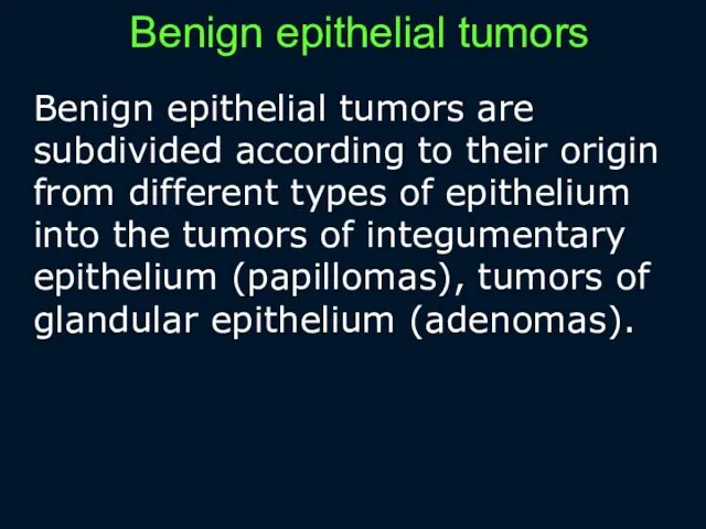
Benign epithelial tumors
Benign epithelial tumors are subdivided according to their origin
Benign epithelial tumors
Benign epithelial tumors are subdivided according to their origin
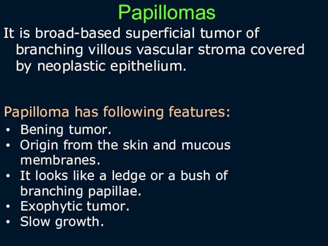
Papillomas
It is broad-based superficial tumor of branching villous vascular stroma covered
Papillomas
It is broad-based superficial tumor of branching villous vascular stroma covered
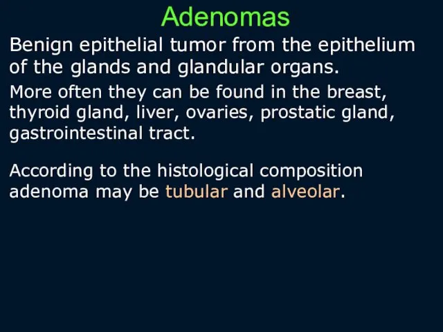
Adenomas
Benign epithelial tumor from the epithelium of the glands and glandular
Adenomas
Benign epithelial tumor from the epithelium of the glands and glandular
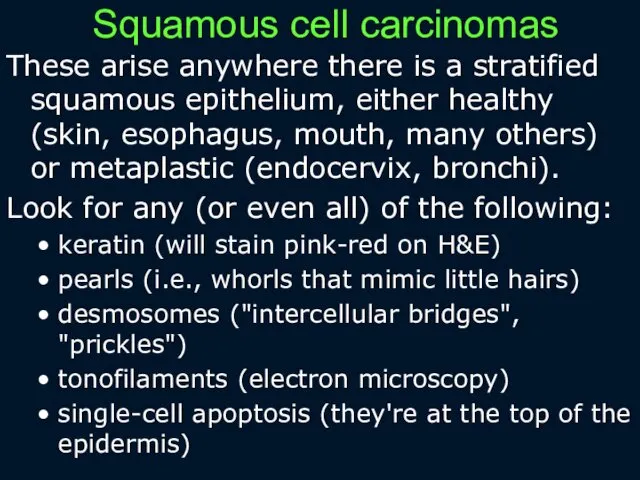
Squamous cell carcinomas
These arise anywhere there is a stratified squamous epithelium,
Squamous cell carcinomas
These arise anywhere there is a stratified squamous epithelium,
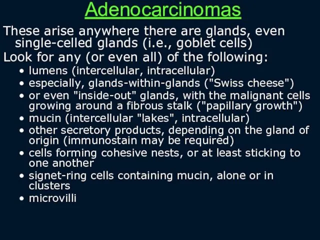
Adenocarcinomas
These arise anywhere there are glands, even single-celled glands (i.e., goblet
Adenocarcinomas
These arise anywhere there are glands, even single-celled glands (i.e., goblet
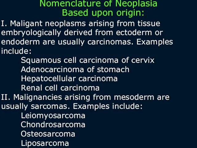
I. Maligant neoplasms arising from tissue embryologically derived from ectoderm or
I. Maligant neoplasms arising from tissue embryologically derived from ectoderm or
III. Neoplasms with more than one cell type but arising from
III. Neoplasms with more than one cell type but arising from
V. Neoplasms ending in "-blastoma" resemble primitive embryonic tissues.
Examples include:
Retinoblastoma
V. Neoplasms ending in "-blastoma" resemble primitive embryonic tissues.
Examples include:
Retinoblastoma
 Иммунопатогенез аутоиммунных заболеваний. Принципы лечения иммунозависимой патологии
 Иерсинии - псевдотуберкулезный микроб. Микробиология
 Нарушения голоса: виды и причины
 Сложное дополнение
 Chronic Lymphocytic Leukemia
 Созылмалы тубулоинтерстициалды нефрит
 Психические заболевания позднего возраста. Инволюционная депрессия. Болезнь Альцгеймера. Болезнь Пика
 Принципы нейрореабилитации
 Лямблии
 Дәрігердің көшбасшылық қасиеттері
 Сосудистая жесткость, сосудистый возраст или сосудистое старение. Эффективное использование маркера в практике частной клиники
 Підвищення точності ультразвукового зондування медико-біологічних об‘єктів, багаточастотним фазовим методом далекометрії
 Тұрғындарды індеттен қорғау жүйесінде иммундық профилактиканың алатын орны
 Анестезия у детей
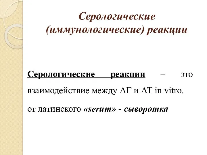 Серологические (иммунологические) реакции
 Внутрибольничная инфекция
 Виды нарушения осанки: лордоз, кифоз, сколиоз и другие, их влияние на здоровье и самочувствие детей
 Инфекционный эндокардит
 Клинико-фармакологические аспекты терапии хронической боли
 Рациональное питание. Болезни, связанные с характером питания
 Роль фельдшера в реабилитации пациентов после инфаркта миокарда
 Изменения СОПР при дерматозах
 Защита миокарда при проведении искусственного кровообращения
 Medicine in Ancient Rome
 Острый ринит
 Роль медицинской сестры при уходе за пациентами с сахарным диабетом II типа. Участие в лечебно-диагностических процессах
 Асқорыту органдарының қатерсіз және қатерлі ісіктері
 Понятие о системе крови. Физико-химические свойства крови. Физиология плазмы и эритроцитов