- Lab 2 Esophagus & Stomach

Содержание
- 2. K34 Esophagus - Upper As with all the tubular portion of the GI tract, the esophagus
- 3. K35 Esophagus - Upper Esophagus - Upper The mucosa itself is composed of three layers. 1.
- 4. K36 Esophagus - Upper The esophageal submucosa is a wide layer of varying densities. Larger blood
- 5. K37 Esophagus - Upper The ducts from the submucosal or esophageal glands course through the layers
- 6. K38 Esophagus - Upper Both layers of the muscularis externa are composed of skeletal muscle.
- 7. K39 Esophagus - Upper Skeletal muscle of the muscularis externa
- 8. K40 Esophagus - Upper Skeletal muscle of the muscularis externa, longitudinal section.
- 9. K41 Esophagus - Upper Skeletal muscle of the muscularis externa, cross section.
- 10. K42 Esophagus - Middle Distinguish the four major layers: 1. Mucosa = epithelium, stratified squamous, mucous
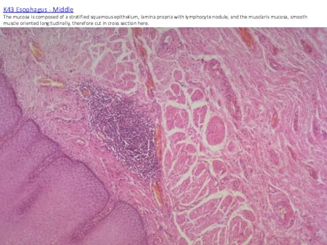
- 11. K43 Esophagus - Middle The mucosa is composed of a stratified squamous epithelium, lamina propria with
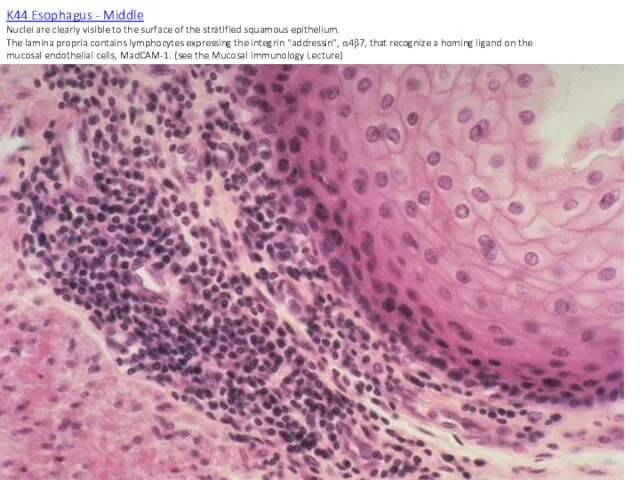
- 12. K44 Esophagus - Middle Nuclei are clearly visible to the surface of the stratified squamous epithelium.
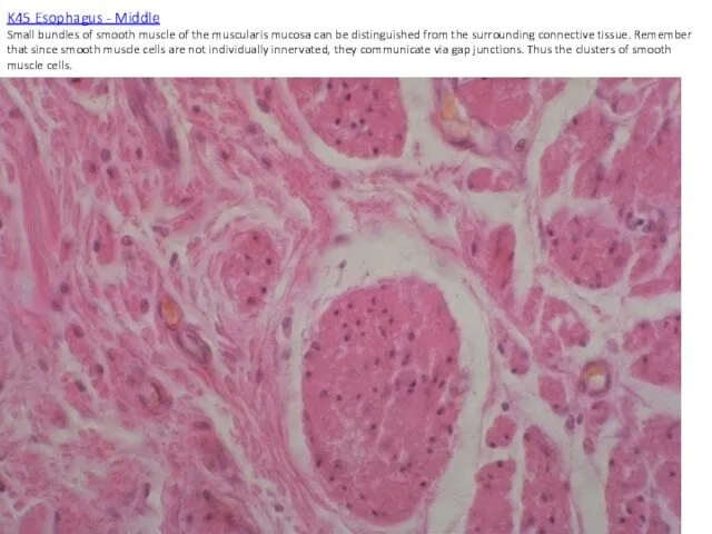
- 13. K45 Esophagus - Middle Small bundles of smooth muscle of the muscularis mucosa can be distinguished
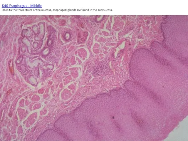
- 14. K46 Esophagus - Middle Deep to the three strata of the mucosa, esophageal glands are found
- 15. K47 Esophagus - Middle Ducts from the submucosal esophageal glands penetrate the strata of the mucosa
- 16. K48 Esophagus - Middle The muscularis externa is a blend of both skeletal and smooth muscle.
- 17. K49 Esophagus - Middle Muscle fibers of the inner circular layer are cut in longitudinal orientation.
- 18. K50 Esophagus - Middle Muscularis externa - longitudinal section through both smooth and skeletal muscle.
- 19. K51 Esophagus - Middle Muscularis externa - cross section through both smooth and skeletal muscle.
- 20. K52 Esophagus - Lower Distinguish the four major layers: 1. Mucosa = epithelium, lamina propria, muscularis
- 21. K53 Esophagus Lower
- 22. K54 Esophagus Lower The lower esophagus is unique for the muscularis externa built of only smoth
- 23. K55 Esophagus Lower Higher power view of smooth muscle cut in longitudinal section.
- 24. K56 Esophagus Lower Both the circular and longitudinal layers of the muscularis externa are smooth muscle
- 25. K57 Esophagus Lower Smooth muscle cut in cross section.
- 26. K58 Espohageal-Stomach Junction There is an abrupt transition from the histology of the esophagus to the
- 27. K59 Esophagus-Stomach Junction The transition between esophagus and stomach is abrupt and reflects the different functions
- 28. K60 Esophagus-Stomach Junction Occasionally, at the junction of the cardiac stomach and the esophagus there is
- 29. K61 Stomach - Cardiac The cardiac stomach displays pits and glands with a 1:1 ratio in
- 30. K62 Stomach - Cardiac The cardiac stomach is characterized by surface pits into which drain 2-4
- 31. K63 Stomach - Cardiac The round morphology of the parietal cells is due to the massive
- 32. K64 Stomach - Body This is the histological appearance of the stomach in the fundus, body
- 33. K65 Stomach - Body The pits are lined by surface mucous cells (note pale pink cytoplasm).
- 34. K66 Stomach - Body The base of the glands are slightly basophilic due to the stored
- 35. K67 Stomach - Body Surface mucous cells of the gastric glands. These secrete mucous in response
- 36. K68 Stomach - Body The surface pits penetrate the lamina propria. Lymphocytes can be seen throughout
- 37. K69 Stomach - Body Cross section from the base of the gastric glands displaying chief cells.
- 38. K70 Stomach - body The slide shows the mid-region or body of the gastric glands. The
- 39. K71 Stomach - Body The rugae of the stomach are large folds that contain a core
- 40. K72 Stomach - Pylorus In the pyloric stomach the ratio of the lengths of pits to
- 41. K73 Stomach - Plyorus Pits with surface mucous cells and glands with neutral mucous cells form
- 42. K74 Stoamch - Pylorus The glands of the pylorus contain mainly neutral mucous secreting cells. The
- 43. K75 Stomach - Muscularis Externa The smooth muscle of the muscularis externa is innervated by the
- 45. Скачать презентацию
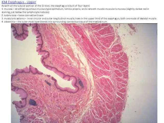
K34 Esophagus - Upper
As with all the tubular portion of the
K34 Esophagus - Upper As with all the tubular portion of the
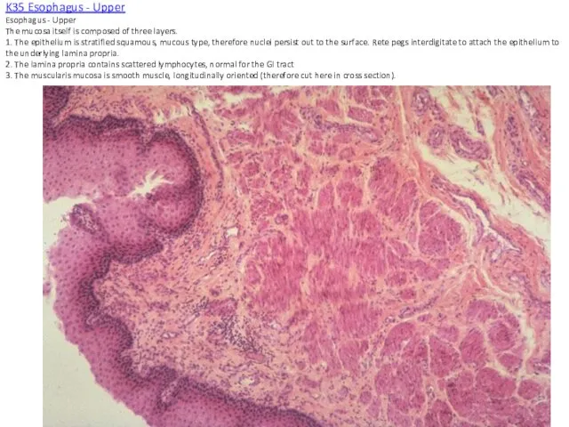
K35 Esophagus - Upper
Esophagus - Upper
The mucosa itself is composed of
K35 Esophagus - Upper Esophagus - Upper The mucosa itself is composed of
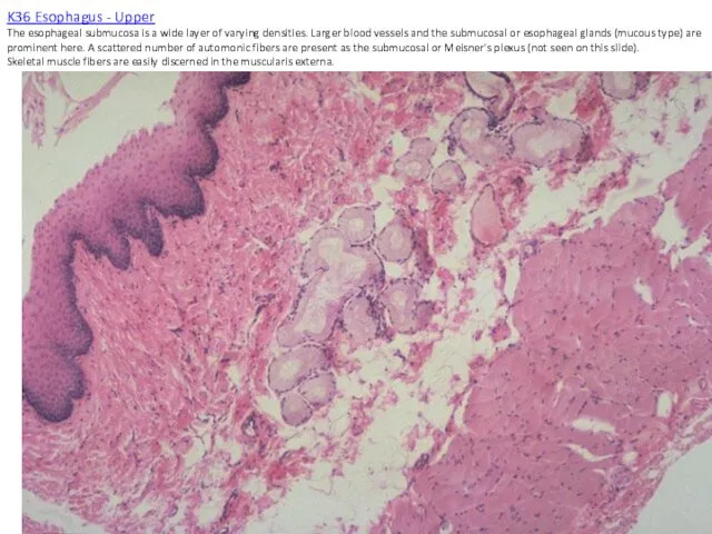
K36 Esophagus - Upper
The esophageal submucosa is a wide layer of
K36 Esophagus - Upper The esophageal submucosa is a wide layer of
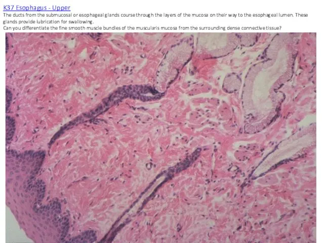
K37 Esophagus - Upper
The ducts from the submucosal or esophageal glands
K37 Esophagus - Upper The ducts from the submucosal or esophageal glands
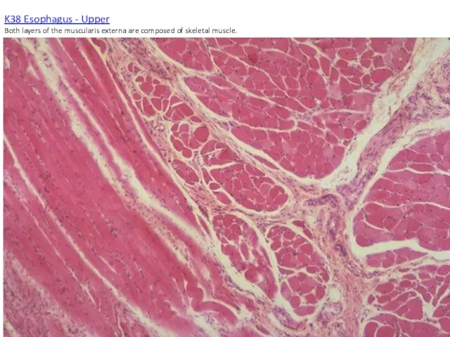
K38 Esophagus - Upper
Both layers of the muscularis externa are composed
K38 Esophagus - Upper Both layers of the muscularis externa are composed
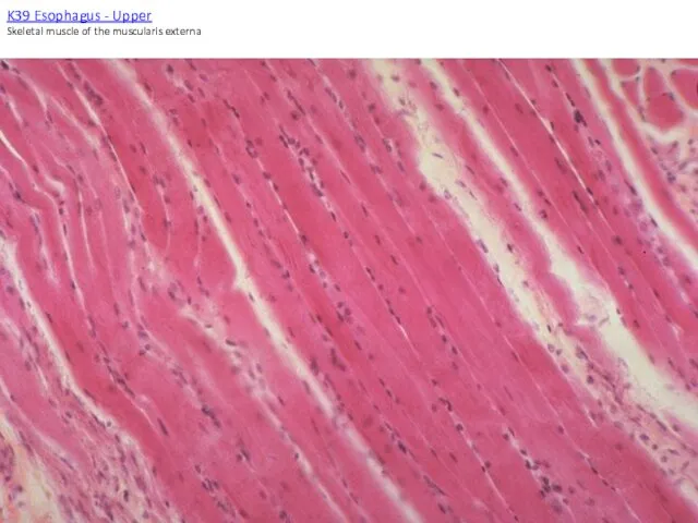
K39 Esophagus - Upper
Skeletal muscle of the muscularis externa
K39 Esophagus - Upper
Skeletal muscle of the muscularis externa

K40 Esophagus - Upper
Skeletal muscle of the muscularis externa, longitudinal section.
K40 Esophagus - Upper
Skeletal muscle of the muscularis externa, longitudinal section.
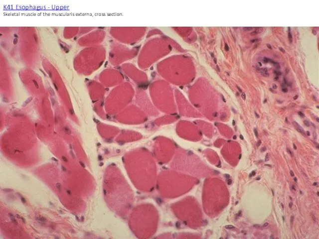
K41 Esophagus - Upper
Skeletal muscle of the muscularis externa, cross section.
K41 Esophagus - Upper
Skeletal muscle of the muscularis externa, cross section.
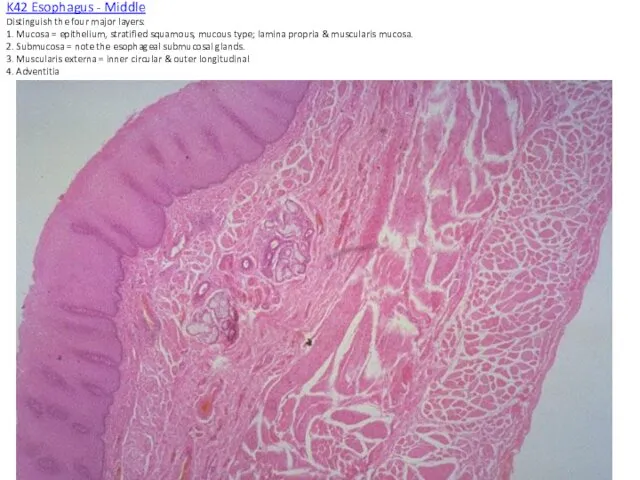
K42 Esophagus - Middle
Distinguish the four major layers:
1. Mucosa = epithelium,
K42 Esophagus - Middle Distinguish the four major layers: 1. Mucosa = epithelium,
K43 Esophagus - Middle
The mucosa is composed of a stratified squamous
K43 Esophagus - Middle The mucosa is composed of a stratified squamous
K44 Esophagus - Middle
Nuclei are clearly visible to the surface of
K44 Esophagus - Middle Nuclei are clearly visible to the surface of
K45 Esophagus - Middle
Small bundles of smooth muscle of the muscularis
K45 Esophagus - Middle Small bundles of smooth muscle of the muscularis
K46 Esophagus - Middle
Deep to the three strata of the mucosa,
K46 Esophagus - Middle Deep to the three strata of the mucosa,
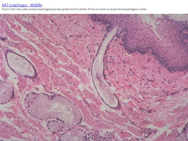
K47 Esophagus - Middle
Ducts from the submucosal esophageal glands penetrate
K47 Esophagus - Middle Ducts from the submucosal esophageal glands penetrate
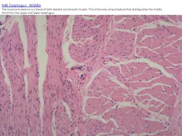
K48 Esophagus - Middle
The muscularis externa is a blend of both
K48 Esophagus - Middle The muscularis externa is a blend of both
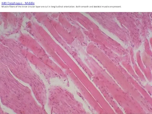
K49 Esophagus - Middle
Muscle fibers of the inner circular layer are
K49 Esophagus - Middle Muscle fibers of the inner circular layer are
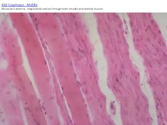
K50 Esophagus - Middle
Muscularis externa - longitudinal section through both smooth
K50 Esophagus - Middle Muscularis externa - longitudinal section through both smooth
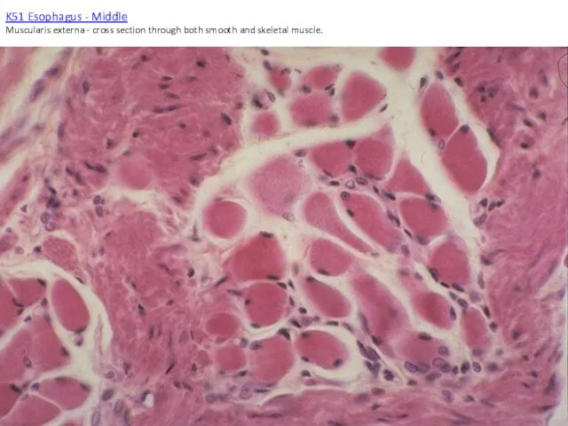
K51 Esophagus - Middle
Muscularis externa - cross section through both smooth
K51 Esophagus - Middle Muscularis externa - cross section through both smooth
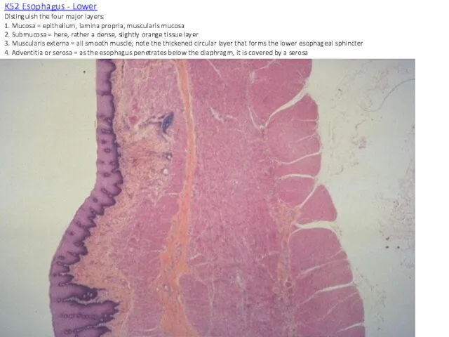
K52 Esophagus - Lower
Distinguish the four major layers:
1. Mucosa = epithelium,
K52 Esophagus - Lower Distinguish the four major layers: 1. Mucosa = epithelium,
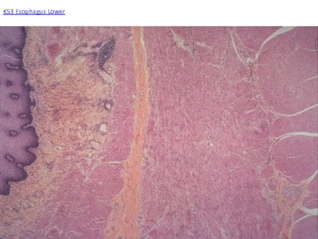
K53 Esophagus Lower
K53 Esophagus Lower
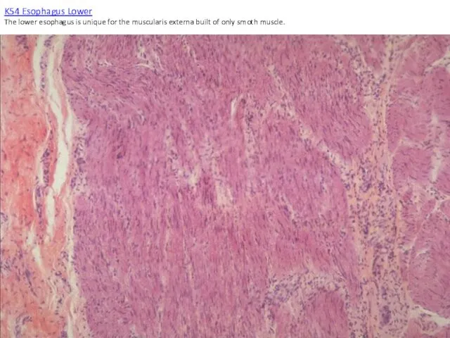
K54 Esophagus Lower
The lower esophagus is unique for the muscularis externa
K54 Esophagus Lower The lower esophagus is unique for the muscularis externa
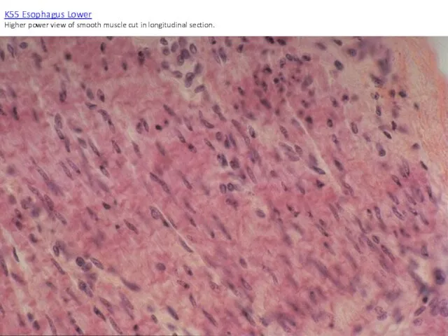
K55 Esophagus Lower
Higher power view of smooth muscle cut in longitudinal
K55 Esophagus Lower Higher power view of smooth muscle cut in longitudinal
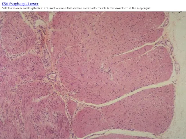
K56 Esophagus Lower
Both the circular and longitudinal layers of the muscularis
K56 Esophagus Lower Both the circular and longitudinal layers of the muscularis
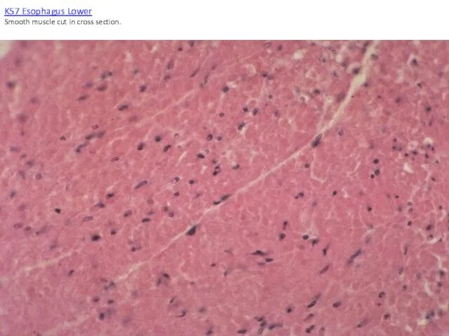
K57 Esophagus Lower
Smooth muscle cut in cross section.
K57 Esophagus Lower
Smooth muscle cut in cross section.
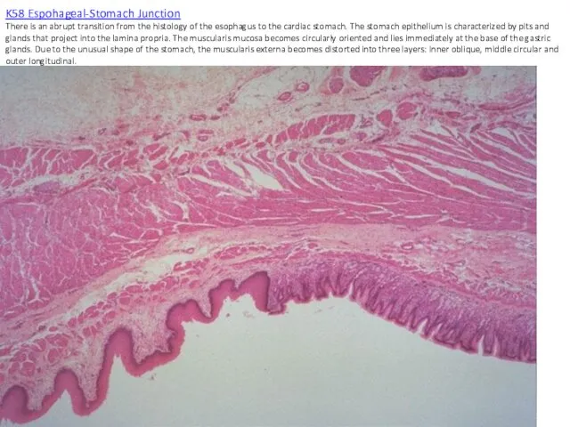
K58 Espohageal-Stomach Junction
There is an abrupt transition from the histology of
K58 Espohageal-Stomach Junction There is an abrupt transition from the histology of
K59 Esophagus-Stomach Junction
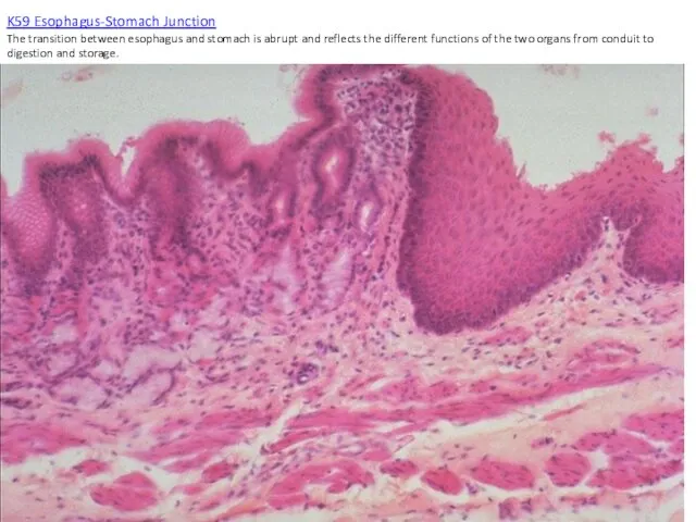
The transition between esophagus and stomach is abrupt and
K59 Esophagus-Stomach Junction The transition between esophagus and stomach is abrupt and
K60 Esophagus-Stomach Junction
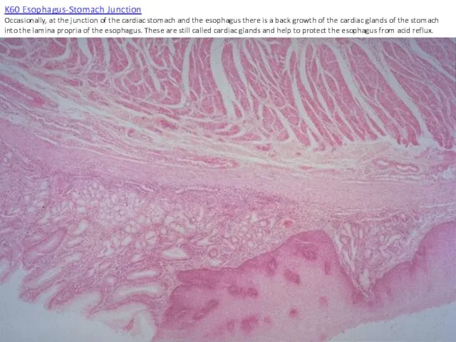
Occasionally, at the junction of the cardiac stomach and
K60 Esophagus-Stomach Junction Occasionally, at the junction of the cardiac stomach and
K61 Stomach - Cardiac
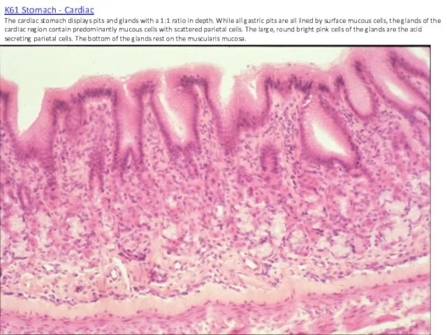
The cardiac stomach displays pits and glands with
K61 Stomach - Cardiac The cardiac stomach displays pits and glands with
K62 Stomach - Cardiac
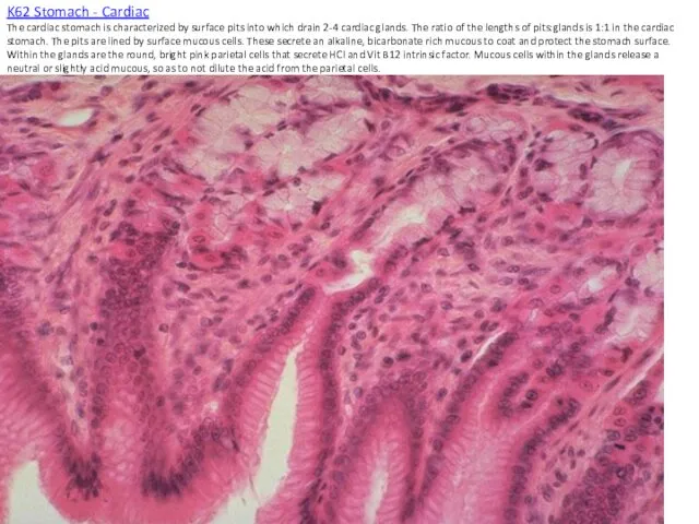
The cardiac stomach is characterized by surface pits
K62 Stomach - Cardiac The cardiac stomach is characterized by surface pits
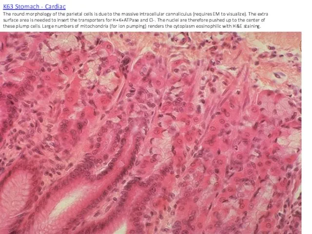
K63 Stomach - Cardiac
The round morphology of the parietal cells is
K63 Stomach - Cardiac The round morphology of the parietal cells is
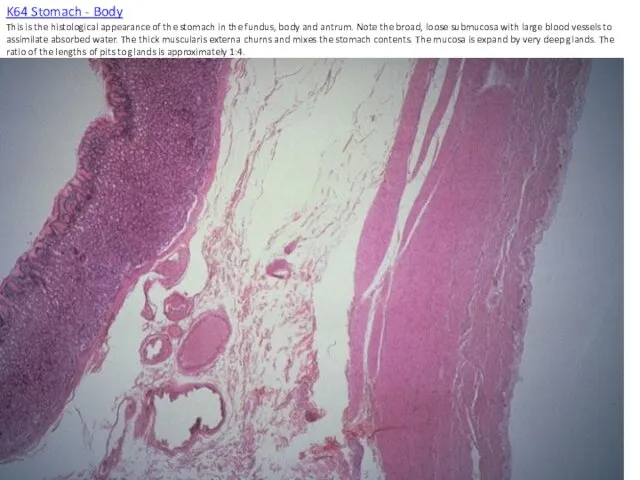
K64 Stomach - Body
This is the histological appearance of the stomach
K64 Stomach - Body This is the histological appearance of the stomach
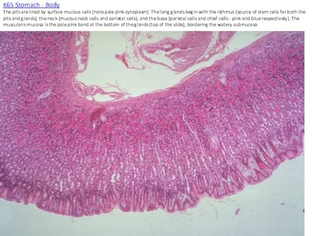
K65 Stomach - Body
The pits are lined by surface mucous cells
K65 Stomach - Body The pits are lined by surface mucous cells
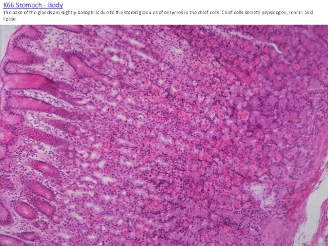
K66 Stomach - Body
The base of the glands are slightly basophilic
K66 Stomach - Body The base of the glands are slightly basophilic
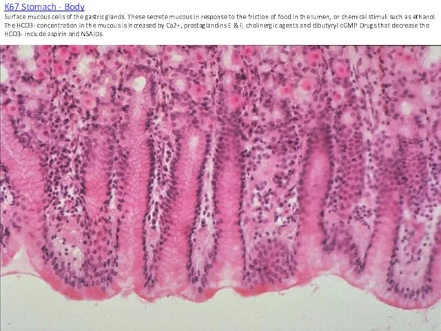
K67 Stomach - Body
Surface mucous cells of the gastric glands. These
K67 Stomach - Body Surface mucous cells of the gastric glands. These
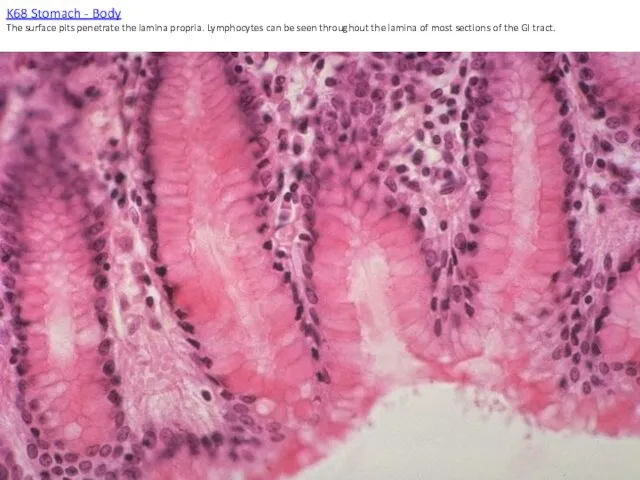
K68 Stomach - Body
The surface pits penetrate the lamina propria. Lymphocytes
K68 Stomach - Body The surface pits penetrate the lamina propria. Lymphocytes
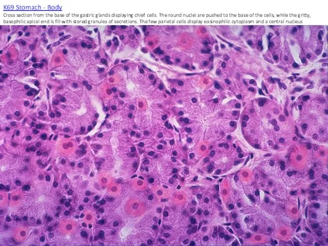
K69 Stomach - Body
Cross section from the base of the gastric
K69 Stomach - Body Cross section from the base of the gastric
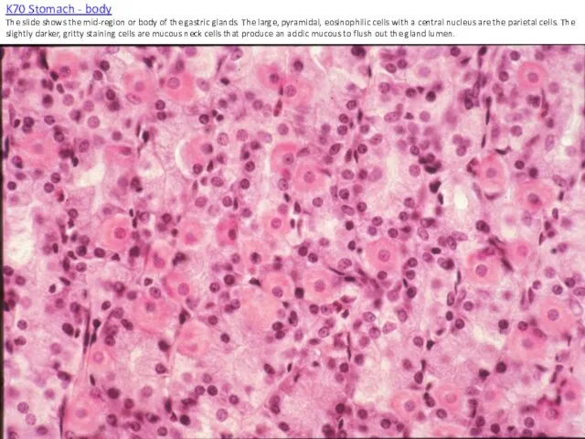
K70 Stomach - body
The slide shows the mid-region or body of
K70 Stomach - body The slide shows the mid-region or body of
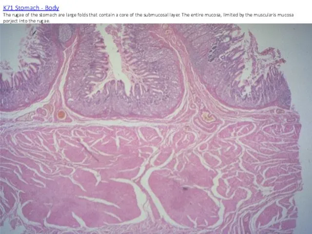
K71 Stomach - Body
The rugae of the stomach are large folds
K71 Stomach - Body The rugae of the stomach are large folds
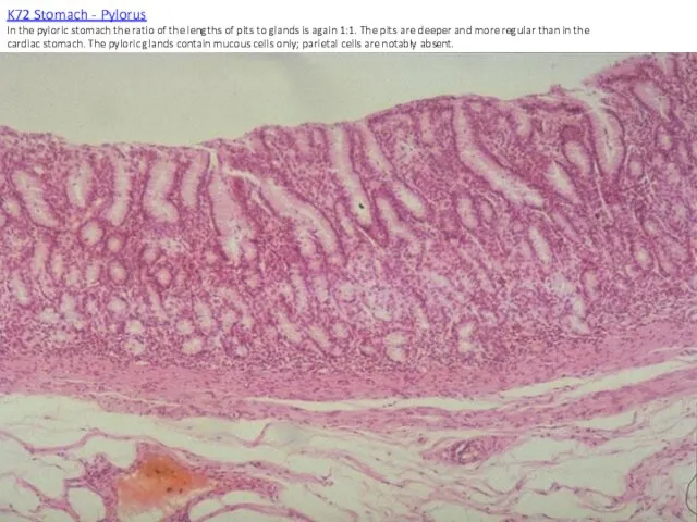
K72 Stomach - Pylorus
In the pyloric stomach the ratio of the
K72 Stomach - Pylorus In the pyloric stomach the ratio of the
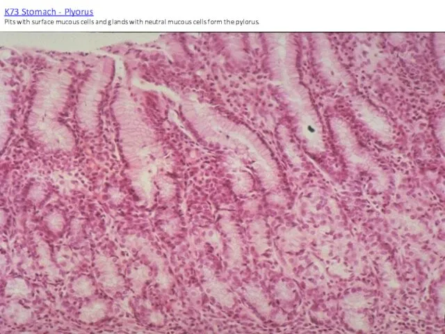
K73 Stomach - Plyorus
Pits with surface mucous cells and glands with
K73 Stomach - Plyorus Pits with surface mucous cells and glands with
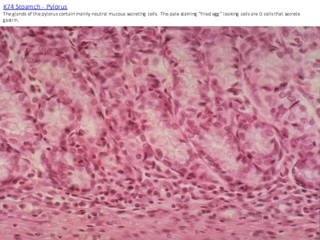
K74 Stoamch - Pylorus
The glands of the pylorus contain mainly neutral
K74 Stoamch - Pylorus The glands of the pylorus contain mainly neutral
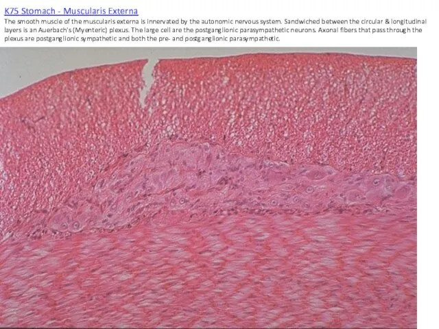
K75 Stomach - Muscularis Externa
The smooth muscle of the muscularis externa
K75 Stomach - Muscularis Externa The smooth muscle of the muscularis externa
 Первая помощь при ушибах, растяжениях, вывихах и переломах
 Unsatisfactory progress of labor (parturition)
 Система государственных учреждений, обеспечивающих контроль качества лекарственных средств
 Оказание первой помощи при отсутствии сознания, остановке дыхания и кровообращения
 Mac-анестезия в эндоскопии и малоинвазивной хирургии
 Лифома Ходжкина
 Введение в оперативную гинекологию
 Бауыр циррозы
 Профилактика нарушения осанки детей
 Клиническая фармакология антигипертензивных ЛС. Фармакотерапия артериальной гипертензии
 Эхокардиография
 Гемостаз
 Острая пневмония
 Научные статьи в электронных базах данных о факторе риска крови в развитии лихорадки Эбола
 Ожоги глаз. Классификация
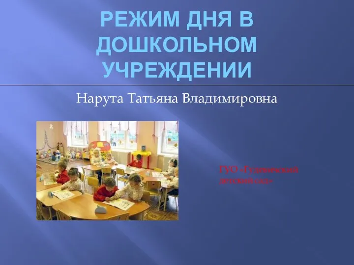 Режим дня в дошкольном учреждении
 Диадинамотерапия: показания и противопоказания
 Красная волчанка
 Проблема бессонницы в структуре соматических заболеваний
 Искусственный интеллект в медицине
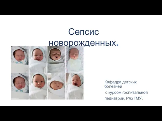 Сепсис новорожденных
 Ультразвуковое исследование голеностопного сустава
 Дәрігер мен науқастың тиімді қарым-қатынасына кедергі келтіретін бөгеттер
 Нарушения аффективной сферы
 Здоровое питание в школе и дома
 Хирургические лазеры в оториноларингологии
 Проблемы невынашивания. Современные принципы лечения
 Постхолецистэктомический синдром