- Medical biology cytogenetic methods

Содержание
- 2. GROUP- 1 Members: Prajval Deshmukh Sukanya Mondal Shahzad Kareekunnan
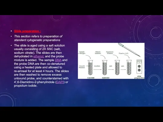
- 3. Slide preparation : This section refers to preparation of standard cytogenetic preparations The slide is aged
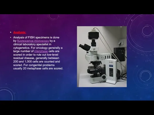
- 4. Analysis: Analysis of FISH specimens is done by fluorescence microscopy by a clinical laboratory specialist in
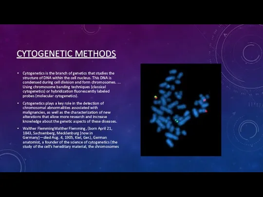
- 5. CYTOGENETIC METHODS Cytogenetics is the branch of genetics that studies the structure of DNA within the
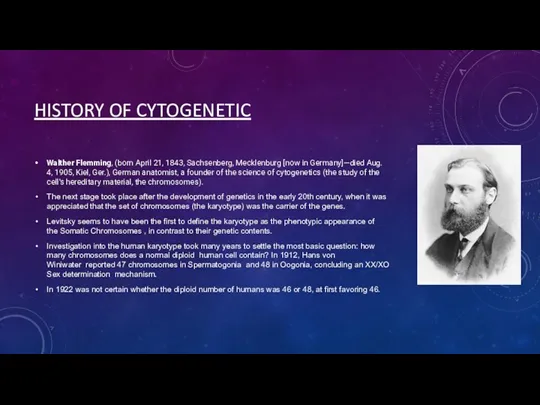
- 6. HISTORY OF CYTOGENETIC Walther Flemming, (born April 21, 1843, Sachsenberg, Mecklenburg [now in Germany]—died Aug. 4,
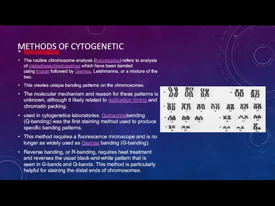
- 7. METHODS OF CYTOGENETIC Karyotyping The routine chromosome analysis (Karyotyping) refers to analysis of metaphasechromosomes which have
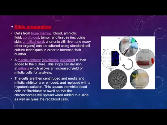
- 8. Slide preparation Cells from bone marrow, blood, amniotic fluid, cord blood, tumor, and tissues (including skin,
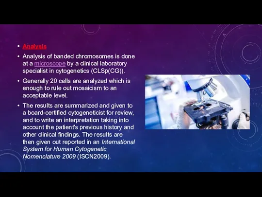
- 9. Analysis Analysis of banded chromosomes is done at a microscope by a clinical laboratory specialist in
- 10. Fluorescent in situ hybridization Fluorescent in situ hybridization (FISH) refers to using fluorescently labeled probe to
- 11. FUTURE OF CYTOGENETICS Advances now focus on molecular cytogenetics including automated systems for counting the results
- 12. QUESTIONS FOR OTHER MEMBERS: Cytogenetic method ? 2.Genealogical diagnostic methods ? 3. Biochemical method. PCR and
- 14. Скачать презентацию
GROUP- 1
Members:
Prajval Deshmukh
Sukanya Mondal
Shahzad Kareekunnan
GROUP- 1
Members:
Prajval Deshmukh
Sukanya Mondal
Shahzad Kareekunnan
Slide preparation :
This section refers to preparation of standard cytogenetic preparations
The
Slide preparation :
This section refers to preparation of standard cytogenetic preparations
The
Analysis:
Analysis of FISH specimens is done by fluorescence microscopy by a clinical laboratory
Analysis:
Analysis of FISH specimens is done by fluorescence microscopy by a clinical laboratory
CYTOGENETIC METHODS
Cytogenetics is the branch of genetics that studies the structure
CYTOGENETIC METHODS
Cytogenetics is the branch of genetics that studies the structure
HISTORY OF CYTOGENETIC
Walther Flemming, (born April 21, 1843, Sachsenberg, Mecklenburg
HISTORY OF CYTOGENETIC
Walther Flemming, (born April 21, 1843, Sachsenberg, Mecklenburg
METHODS OF CYTOGENETIC
Karyotyping
The routine chromosome analysis (Karyotyping) refers to analysis of metaphasechromosomes which
METHODS OF CYTOGENETIC
Karyotyping
The routine chromosome analysis (Karyotyping) refers to analysis of metaphasechromosomes which
Slide preparation
Cells from bone marrow, blood, amniotic fluid, cord blood, tumor, and tissues
Slide preparation
Cells from bone marrow, blood, amniotic fluid, cord blood, tumor, and tissues
Analysis
Analysis of banded chromosomes is done at a microscope by a clinical laboratory
Analysis
Analysis of banded chromosomes is done at a microscope by a clinical laboratory
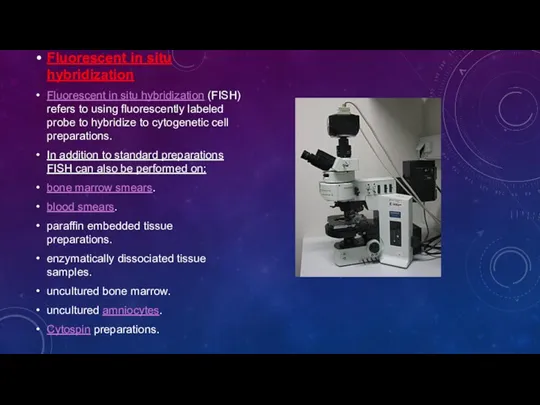
Fluorescent in situ hybridization
Fluorescent in situ hybridization (FISH) refers to using fluorescently
Fluorescent in situ hybridization
Fluorescent in situ hybridization (FISH) refers to using fluorescently

FUTURE OF CYTOGENETICS
Advances now focus on molecular cytogenetics including automated systems for counting
FUTURE OF CYTOGENETICS
Advances now focus on molecular cytogenetics including automated systems for counting
QUESTIONS FOR OTHER MEMBERS:
Cytogenetic method ?
2.Genealogical diagnostic methods ?
3. Biochemical
QUESTIONS FOR OTHER MEMBERS:
Cytogenetic method ?
2.Genealogical diagnostic methods ?
3. Biochemical
 Дезинфекция
 Лапароскопические методы лечения заболеваний пищевода и желчного пузыря
 Применение крестора (резувастатина) при лечении больных с мультифокальным атеросклерозом
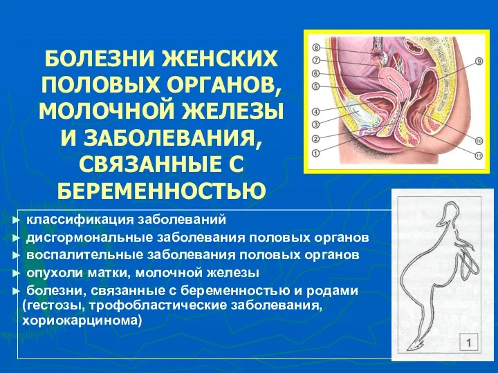 Болезни женских половых органов, молочной железы и заболевания, связанные с беременностью
 Наследственные и врожденные заболевания и заболевания передаваемые половым путем. 8 класс
 Базовые знания о меридианах
 Никотиновая зависимость и как от нее избавится
 Мышцы областей тела. Мышцы спины поверхностный слой
 Природне вигодовування дітей раннього віку
 Issues Affecting ART Success: Adherence, ARV Toxicity, Drug Interactions
 Синдром Марфана
 Медико-санитарное обеспечение населения при ликвидации последствий ЧС химической природы
 Интервенционная кардиология .Виды стендов
 Металлические яды, изоляция минерализацией. Применение металлов и их соединений в клинической фармации
 Оказание первой помощи
 Эндогенные психические заболевания
 Дифференциальная диагностика и лечение одышки
 ХСН: причины, классификация, консервативное лечение
 Ведение медицинской документации
 Предоперационный период. Хирургическая операция. Послеоперационный период
 Проведение врачебных назначений. Десмургия
 Естественное вскармливание ребенка первого года жизни
 СРС: Транспортировка больных и пострадавших в машине скорой помощи
 Депрессивные расстройства в неврологии
 Эбола, Марбург қызбаларының және жапон энцефалитінің эпидемиологиялық сипаттамасы. Алдын алу және эпидемияға қарсы шаралардың
 Постизометрическая релаксация мышц
 Особенности ухода за детьми грудного возраста
 Патопсихология