- Medical protozoology

Содержание
- 2. A NOTE ON TAXONOMY Adl et al. 2012 The revised classification of eukaryotes. The Journal of
- 3. PROTIST VS. PROTOZOA Kingdom Protista (single-celled eukaryotic organisms) Protozoa (heterotrophic protists) was considered as phylum within
- 4. IMPORTANT TERMS Trophozoite – actively motile feeding stage. Cyst – resistant, infective stage.
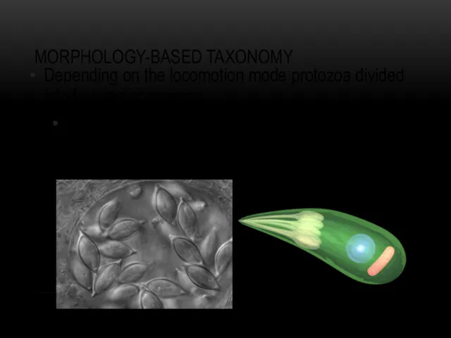
- 5. MORPHOLOGY-BASED TAXONOMY Depending on the locomotion mode protozoa divided into four major groups: Phylum Flagellates (move
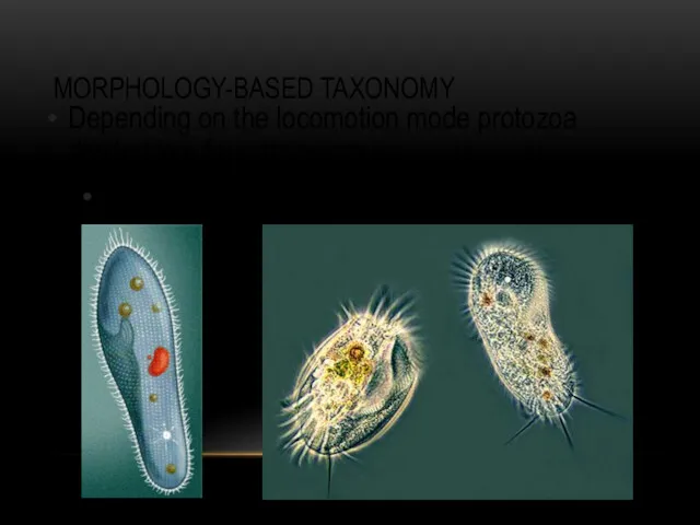
- 6. MORPHOLOGY-BASED TAXONOMY Depending on the locomotion mode protozoa divided into four major groups: Phylum Amoebae (by
- 7. MORPHOLOGY-BASED TAXONOMY Depending on the locomotion mode protozoa divided into four major groups: Phylum Ciliates (by
- 8. MORPHOLOGY-BASED TAXONOMY Depending on the locomotion mode protozoa divided into four major groups: Phylum Sporozoa (lacking
- 9. PARASITIC AMOEBAE Six species of amoebae are common in humans in most parts of the world
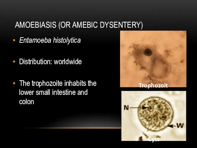
- 10. AMOEBIASIS (OR AMEBIC DYSENTERY) Entamoeba histolytica Distribution: worldwide The trophozoite inhabits the lower small intestine and
- 11. LIFE CYCLE OF E. HISTOLYTICA Infective stage: cyst Mode of infection: ingestion cysts-contaminated food or water
- 12. DIAGNOSIS AND PREVENTION OF AMOEBIASIS Diagnosis: Microscopical determination of cysts in fecal samples, serology. Prevention: Avoidance
- 13. NON-INTESTINE PARASITIC AMOEBAE … human infection is not an obligate part of their life cycle. Naegleria
- 14. PARASITIC CILIATES The only ciliates known to infect human is Balantidium coli – Balantidiosis (dysentery) Distribution:
- 15. LIFE CYCLE OF B. COLI Infective stage: cyst Mode of infection: ingestion cysts-contaminated food or water
- 16. PARASITIC FLAGELLATES Depending on the infected organs, parasitic flagellates might be classified in two broad groups.
- 17. LAMBLIASIS (GIARDIASIS) Giardia duodenalis (a.k.a G. lamblia, G. intestinalis or Lamblia intestinalis) Distribution: Worldwide The parasite
- 18. LIFE CYCLE OF G. DUODENALIS Infective stage: cyst Mode of infection: ingestion cysts-contaminated food or water
- 19. DIAGNOSIS AND PREVENTION OF LAMBLIASIS Diagnosis: Microscopic determination of trophozoites and cysts in faecal samples. Prevention:
- 20. HUMAN TRICHOMONIASIS Trichomonas vaginalis Distribution: Worldwide Trophozoites live in the vagina and prostate gland.
- 21. LIFE CYCLE OF T. VAGINALIS Infective stage: trophozoites Mode of infection: through sexual or genital contact
- 22. DIAGNOSIS AND PREVENTION OF TRICHOMONIASIS Diagnosis: Microscopic detection of trophozoites in mucus samples. Prophylaxis: Avoid unprotected
- 23. MORPHOLOGICAL STAGES OF HAEMOFLAGELLATES
- 24. TRYPANOSOMIASIS OR TRYPANOSOMOSIS ... several diseases in vertebrates caused by parasitic trypanosomes T. cruzi (Chagas disease)
- 25. CHAGAS DISEASE (OR AMERICAN TRYPANOSOMIASIS) Distribution: Mainly in Latin America Infection mode: Invasion through bite wound
- 26. LIFE CYCLE OF T. CRUZI
- 27. CLINICAL SYMPTOMS, DIAGNOSIS AND PREVENTION Main clinical symptoms: Chagom at bite site, fever, hepatosplenomegaly, cardiomegaly, aneurisms.
- 28. SLEEPING SICKNESS OR AFRICAN TRYPANOSOMIASIS Distribution: 36 countries of sub Saharan Africa Infective stage: Metacyclic trypanosome
- 29. LIFE CYCLE OF T. BRUCEI
- 30. CLINICAL SYMPTOMS, DIAGNOSIS AND PREVENTION Main clinical symptoms: Fever, local edema, possibly polyadenitis, neural complications, death.
- 31. LEISHMANIASIS More than 20 species of Leishmania infect humans L. donovani (Visceral leishmaniasis or Kala-azar) L.
- 32. LIFE CYCLE OF LEISHMANIA Infective stage: Promastigote Mode of infection: Sand fly (Lutzomyia and Phlebotomus)
- 33. CLINICAL SYMPTOMS, DIAGNOSIS AND PREVENTION Cutaneous Leishmaniasis (CL) Main clinical symptoms: Skin nodules, ulceration, necrosis. Diagnosis:
- 34. CLINICAL SYMPTOMS, DIAGNOSIS AND PREVENTION Cutaneous Leishmaniasis (CL) Main clinical symptoms: Skin nodules, ulceration, necrosis. Diagnosis:
- 35. DIAGNOSIS AND PREVENTION OF BALANTIDIASIS Diagnosis: Microscopic determination of cysts and trophozoites in fecal smears. Prevention:
- 37. Скачать презентацию
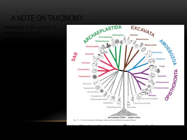
A NOTE ON TAXONOMY
Adl et al. 2012 The revised classification of
A NOTE ON TAXONOMY
Adl et al. 2012 The revised classification of
PROTIST VS. PROTOZOA
Kingdom Protista (single-celled eukaryotic organisms)
Protozoa (heterotrophic protists) was considered
PROTIST VS. PROTOZOA
Kingdom Protista (single-celled eukaryotic organisms)
Protozoa (heterotrophic protists) was considered

IMPORTANT TERMS
Trophozoite – actively motile feeding stage.
Cyst – resistant, infective stage.
IMPORTANT TERMS
Trophozoite – actively motile feeding stage.
Cyst – resistant, infective stage.
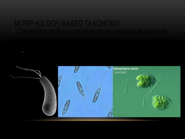
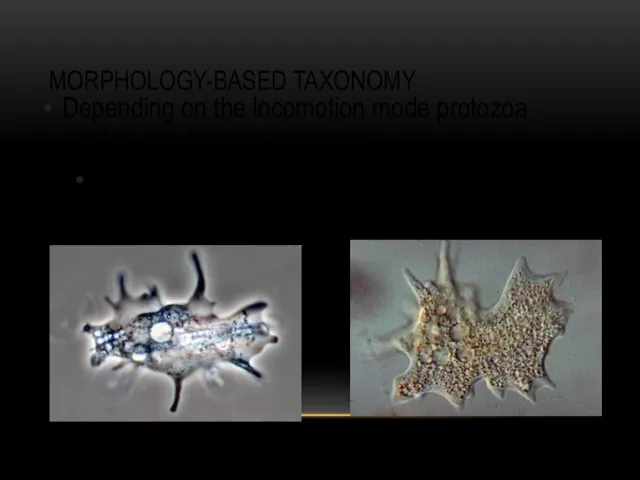
MORPHOLOGY-BASED TAXONOMY
Depending on the locomotion mode protozoa divided into four major
MORPHOLOGY-BASED TAXONOMY
Depending on the locomotion mode protozoa divided into four major
MORPHOLOGY-BASED TAXONOMY
Depending on the locomotion mode protozoa divided into four major
MORPHOLOGY-BASED TAXONOMY
Depending on the locomotion mode protozoa divided into four major
MORPHOLOGY-BASED TAXONOMY
Depending on the locomotion mode protozoa divided into four major
MORPHOLOGY-BASED TAXONOMY
Depending on the locomotion mode protozoa divided into four major
MORPHOLOGY-BASED TAXONOMY
Depending on the locomotion mode protozoa divided into four major
MORPHOLOGY-BASED TAXONOMY
Depending on the locomotion mode protozoa divided into four major
PARASITIC AMOEBAE
Six species of amoebae are common in humans in most
PARASITIC AMOEBAE
Six species of amoebae are common in humans in most
AMOEBIASIS (OR AMEBIC DYSENTERY)
Entamoeba histolytica
Distribution: worldwide
The trophozoite inhabits the lower small
AMOEBIASIS (OR AMEBIC DYSENTERY)
Entamoeba histolytica
Distribution: worldwide
The trophozoite inhabits the lower small
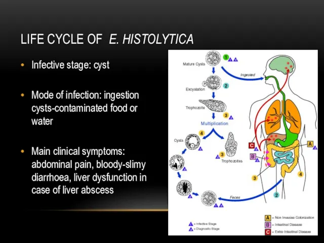
LIFE CYCLE OF E. HISTOLYTICA
Infective stage: cyst
Mode of infection: ingestion cysts-contaminated
LIFE CYCLE OF E. HISTOLYTICA
Infective stage: cyst
Mode of infection: ingestion cysts-contaminated
DIAGNOSIS AND PREVENTION OF AMOEBIASIS
Diagnosis: Microscopical determination of cysts in fecal
DIAGNOSIS AND PREVENTION OF AMOEBIASIS
Diagnosis: Microscopical determination of cysts in fecal
NON-INTESTINE PARASITIC AMOEBAE
… human infection is not an obligate part of
NON-INTESTINE PARASITIC AMOEBAE
… human infection is not an obligate part of
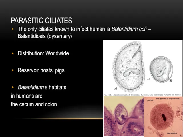
PARASITIC CILIATES
The only ciliates known to infect human is Balantidium coli
PARASITIC CILIATES
The only ciliates known to infect human is Balantidium coli
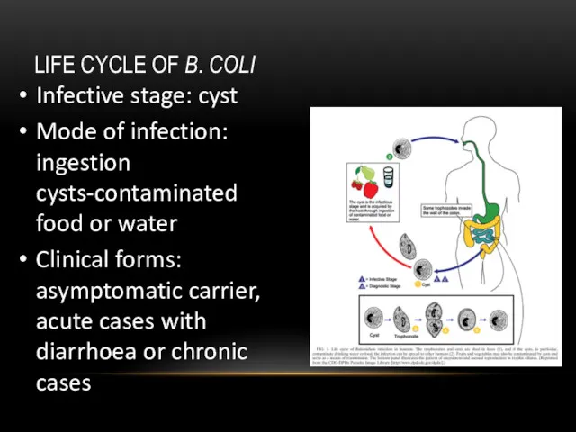
LIFE CYCLE OF B. COLI
Infective stage: cyst
Mode of infection: ingestion
LIFE CYCLE OF B. COLI
Infective stage: cyst
Mode of infection: ingestion
PARASITIC FLAGELLATES
Depending on the infected organs, parasitic flagellates might be classified
PARASITIC FLAGELLATES
Depending on the infected organs, parasitic flagellates might be classified
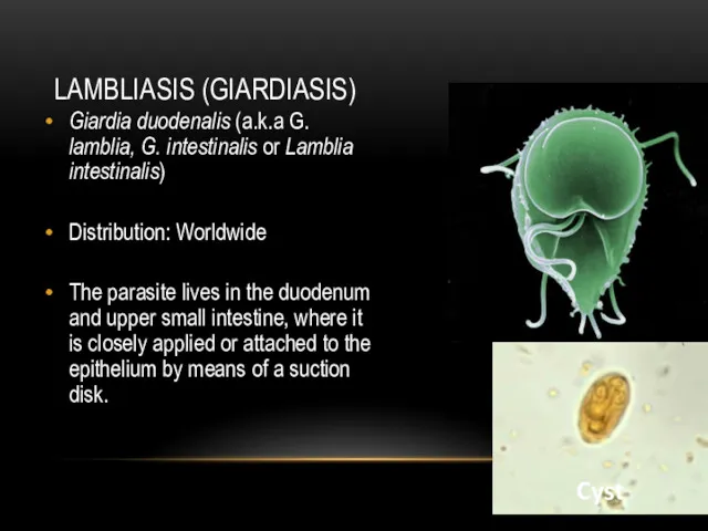
LAMBLIASIS (GIARDIASIS)
Giardia duodenalis (a.k.a G. lamblia, G. intestinalis or Lamblia intestinalis)
Distribution:
LAMBLIASIS (GIARDIASIS)
Giardia duodenalis (a.k.a G. lamblia, G. intestinalis or Lamblia intestinalis)
Distribution:
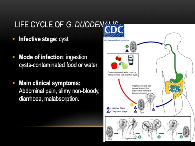
LIFE CYCLE OF G. DUODENALIS
Infective stage: cyst
Mode of infection: ingestion
LIFE CYCLE OF G. DUODENALIS
Infective stage: cyst
Mode of infection: ingestion
DIAGNOSIS AND PREVENTION OF LAMBLIASIS
Diagnosis: Microscopic determination of trophozoites and
DIAGNOSIS AND PREVENTION OF LAMBLIASIS
Diagnosis: Microscopic determination of trophozoites and
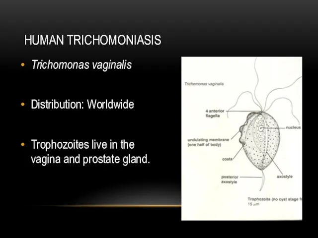
HUMAN TRICHOMONIASIS
Trichomonas vaginalis
Distribution: Worldwide
Trophozoites live in the vagina and prostate gland.
HUMAN TRICHOMONIASIS
Trichomonas vaginalis
Distribution: Worldwide
Trophozoites live in the vagina and prostate gland.
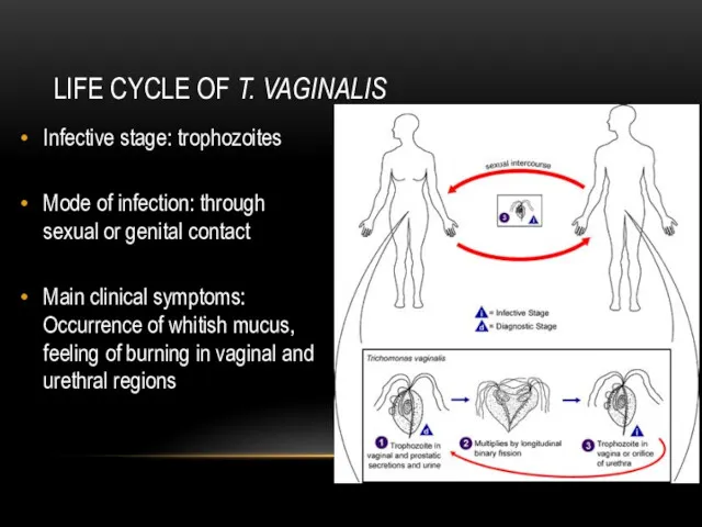
LIFE CYCLE OF T. VAGINALIS
Infective stage: trophozoites
Mode of infection: through
LIFE CYCLE OF T. VAGINALIS
Infective stage: trophozoites
Mode of infection: through
DIAGNOSIS AND PREVENTION OF TRICHOMONIASIS
Diagnosis: Microscopic detection of trophozoites in
DIAGNOSIS AND PREVENTION OF TRICHOMONIASIS
Diagnosis: Microscopic detection of trophozoites in
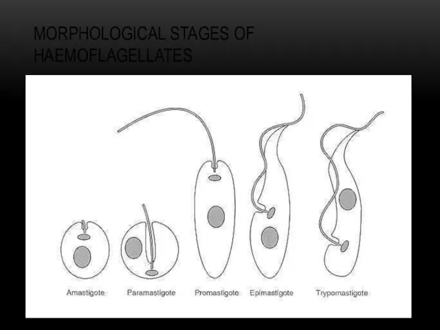
MORPHOLOGICAL STAGES OF HAEMOFLAGELLATES
MORPHOLOGICAL STAGES OF HAEMOFLAGELLATES
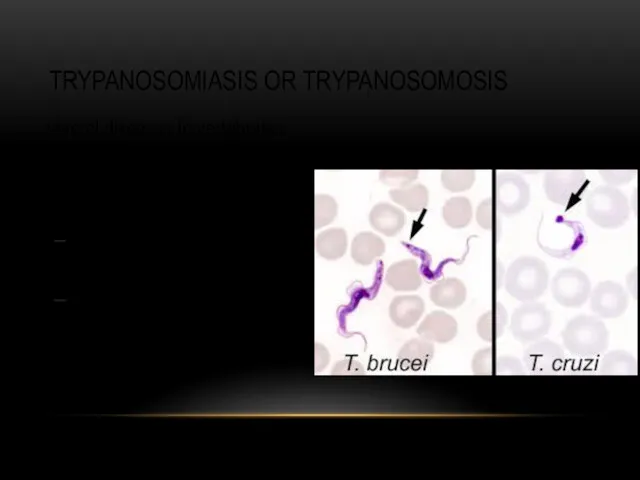
TRYPANOSOMIASIS OR TRYPANOSOMOSIS
... several diseases in vertebrates caused by parasitic trypanosomes
T. cruzi (Chagas
TRYPANOSOMIASIS OR TRYPANOSOMOSIS
... several diseases in vertebrates caused by parasitic trypanosomes
T. cruzi (Chagas
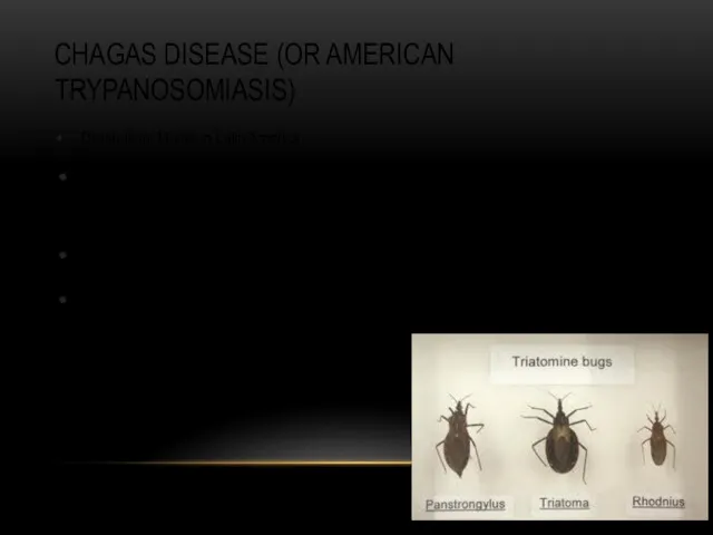
CHAGAS DISEASE (OR AMERICAN TRYPANOSOMIASIS)
Distribution: Mainly in Latin America
Infection mode: Invasion
CHAGAS DISEASE (OR AMERICAN TRYPANOSOMIASIS)
Distribution: Mainly in Latin America
Infection mode: Invasion
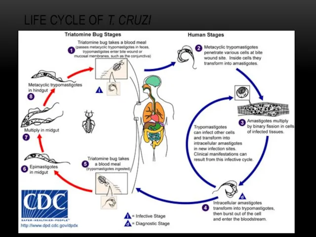
LIFE CYCLE OF T. CRUZI
LIFE CYCLE OF T. CRUZI
CLINICAL SYMPTOMS, DIAGNOSIS AND PREVENTION
Main clinical symptoms: Chagom at bite site,
CLINICAL SYMPTOMS, DIAGNOSIS AND PREVENTION
Main clinical symptoms: Chagom at bite site,
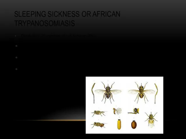
SLEEPING SICKNESS OR AFRICAN TRYPANOSOMIASIS
Distribution: 36 countries of sub Saharan Africa
Infective
SLEEPING SICKNESS OR AFRICAN TRYPANOSOMIASIS
Distribution: 36 countries of sub Saharan Africa
Infective
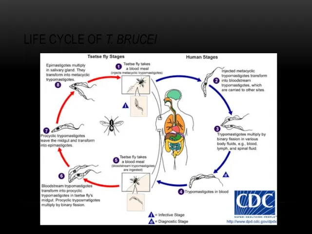
LIFE CYCLE OF T. BRUCEI
LIFE CYCLE OF T. BRUCEI
CLINICAL SYMPTOMS, DIAGNOSIS AND PREVENTION
Main clinical symptoms: Fever, local edema, possibly
CLINICAL SYMPTOMS, DIAGNOSIS AND PREVENTION
Main clinical symptoms: Fever, local edema, possibly
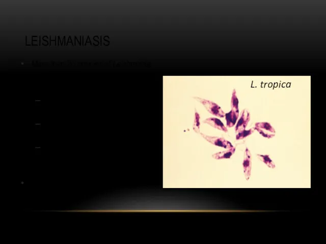
LEISHMANIASIS
More than 20 species of Leishmania infect humans
L. donovani (Visceral leishmaniasis or
LEISHMANIASIS
More than 20 species of Leishmania infect humans
L. donovani (Visceral leishmaniasis or
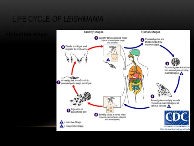
LIFE CYCLE OF LEISHMANIA
Infective stage: Promastigote
Mode of infection: Sand fly (Lutzomyia
LIFE CYCLE OF LEISHMANIA
Infective stage: Promastigote
Mode of infection: Sand fly (Lutzomyia
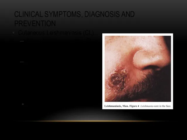
CLINICAL SYMPTOMS, DIAGNOSIS AND PREVENTION
Cutaneous Leishmaniasis (CL)
Main clinical symptoms: Skin nodules,
CLINICAL SYMPTOMS, DIAGNOSIS AND PREVENTION
Cutaneous Leishmaniasis (CL)
Main clinical symptoms: Skin nodules,
CLINICAL SYMPTOMS, DIAGNOSIS AND PREVENTION
Cutaneous Leishmaniasis (CL)
Main clinical symptoms: Skin nodules,
CLINICAL SYMPTOMS, DIAGNOSIS AND PREVENTION
Cutaneous Leishmaniasis (CL)
Main clinical symptoms: Skin nodules,
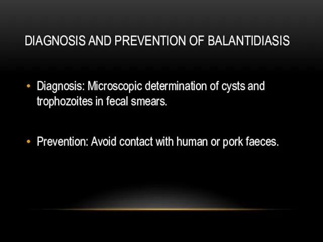
DIAGNOSIS AND PREVENTION OF BALANTIDIASIS
Diagnosis: Microscopic determination of cysts and
DIAGNOSIS AND PREVENTION OF BALANTIDIASIS
Diagnosis: Microscopic determination of cysts and
 Бүйрек аурулары
 Нейропсихологические синдромы нарушения памяти. Методы нейропсихологического обследования. Восстановительная работа
 Гостра серцева недостатність
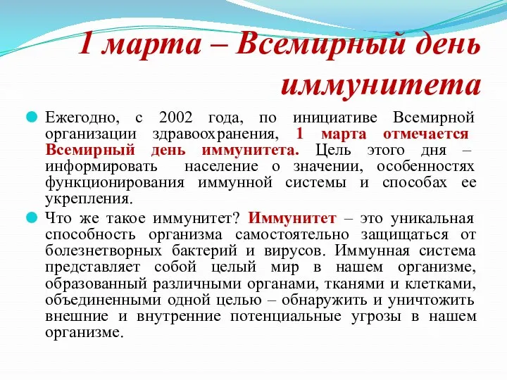 Всемирный день иммунитета
 The final impression
 Рак шейки и тела матки
 Симптоматология острого и хронического гломерулонефрита, пиелонефрита, почечной недостаточности
 Анатомия пәні және мақсаты. Тіндерге, ағзаларға және ағзалар жүйесіне жалпы сипаттама. Адам эмбриогенезінің алғашқы сатысы
 Лекарственные препараты
 Eating Disorders
 ОРВИ и грипп
 Twins diagnostic methods
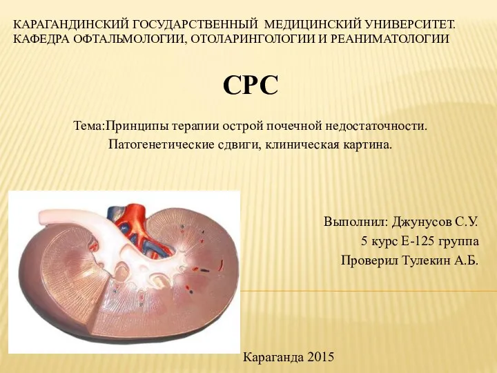 Принципы терапии острой почечной недостаточности. Патогенетические сдвиги, клиническая картина
 Вывихи и переломы зубов, альвеолярного отростка. Методы лечения
 Жүктілік және Туберкулез
 Основные аспекты содержания и методики АФК. Адаптивная физическая культура
 Воспаление. Этиология. Медиаторы. Виды. Острое воспаление
 Павел Захарович Кондоиди (1710-1760)
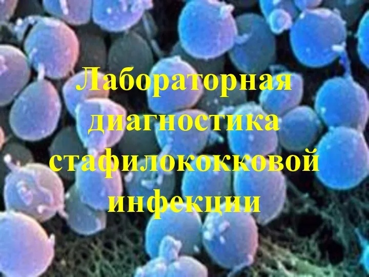 Лабораторная диагностика стафилококковой инфекции
 Анафилактикалық және гиповолемиялық шок
 Клиническая фармакология лекарственных средств, влияющих на гемостаз
 First aid
 Жүктілік кезіндегі жатыр мойнының рагі
 Патофизиология опухолевого роста
 СП при раке желудка
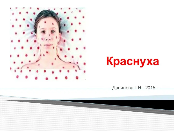 Краснуха
 Возрождение, медицина
 Лечение огнестрельных ранений живота