- Nematodes (round worms)

Содержание
- 2. All the important human parasites of the Phylum Nemathelminthes (Aschelminthes) belong to the Class Nematoda. They
- 3. The parasitic nematodes are divided into: 1. Intestinal nematodes 1.1. Intestinal nematodes with tissue stage A.
- 4. INTESTINAL NEMATODES WITH TISSUE STAGE
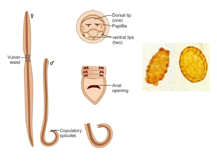
- 5. Ascaris lumbricoides These are common roundworms infecting more than 700 million people worldwide. Morphology Male adult
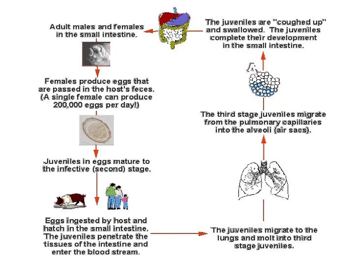
- 7. Life cycle Life cycle of Ascaris involves only 1 host. Natural host: Man. There is no
- 9. Symptoms Fever, urticaria, angioneurotic edema, wheezing, and conjunctivitis. Acute biliary obstruction or pancreatitis, obstructive appendicitis. Spoliative
- 10. Diagnosis
- 11. Treatment pyrantel pamoate 11 mg/kg once; maximum 1 g, albendazole 400 mg once, mebendazole 100 g
- 12. HOOK WORMS
- 13. HOOK WORMS There are two species of hookworm: 1. Ancylostoma duodenale 2. Necator americanus The adults
- 14. Ancylostoma duodenale Grayish-white in color. The body is slightly ventrally curved. The anterior end follows the
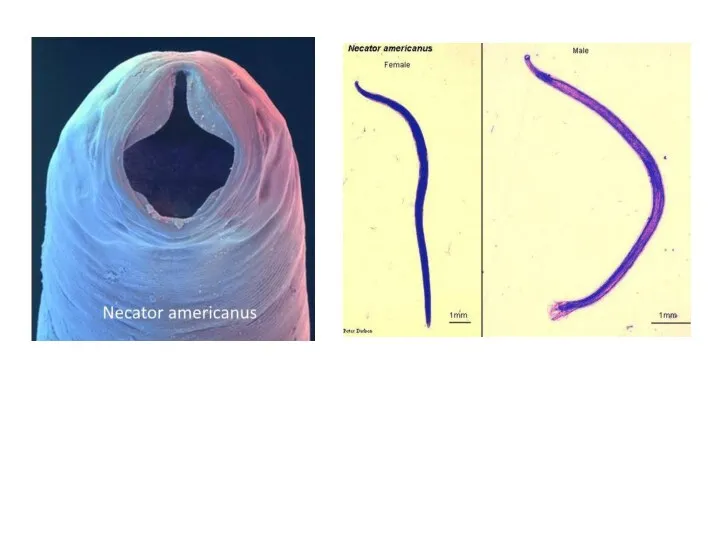
- 16. Necator americanus This species, so called American hookworm, is found in predominantly the tropics. The anterior
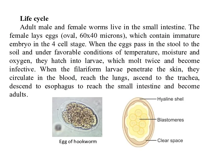
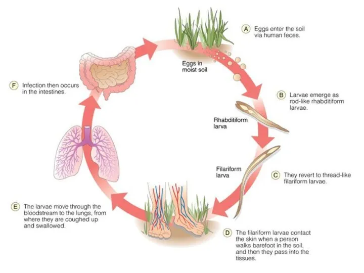
- 18. Life cycle Adult male and female worms live in the small intestine. The female lays eggs
- 20. Symptoms Adult worms in the intestine feed on blood causing iron deficiency anemia. The larvae may
- 21. LARVA MIGRANS
- 22. There are three types of larva migrans: a. Cutaneous larva migrans (Creeping eruption) Various animals harbor
- 23. Toxocariasis This is a kind of visceral larva migrans caused by ♦ Toxocara canis (Dog ascarid)
- 25. Epidemiology. Visceral larva migrans is cosmopolitan in distribution. Transmission: Ingestion of eggs of Toxocara species in
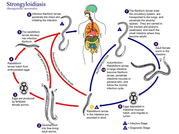
- 26. Strongyloides stercoralis The worms may be present as parasitic in the host or free living in
- 27. Life cycle Adult male and female worms live in the small intestine. After fertilization, the female
- 29. Symptoms The patient complains of mucoid diarrhea. Larvae in the lungs may cause pneumonia. Disseminated strongyloidiasis.
- 30. INTESTINAL NEMATODES WITHOUT TISSUE STAGE
- 31. Enterobius vermicularis (pin worm or thread worm) Enterobius vermicularis is a small white worm with thread-like
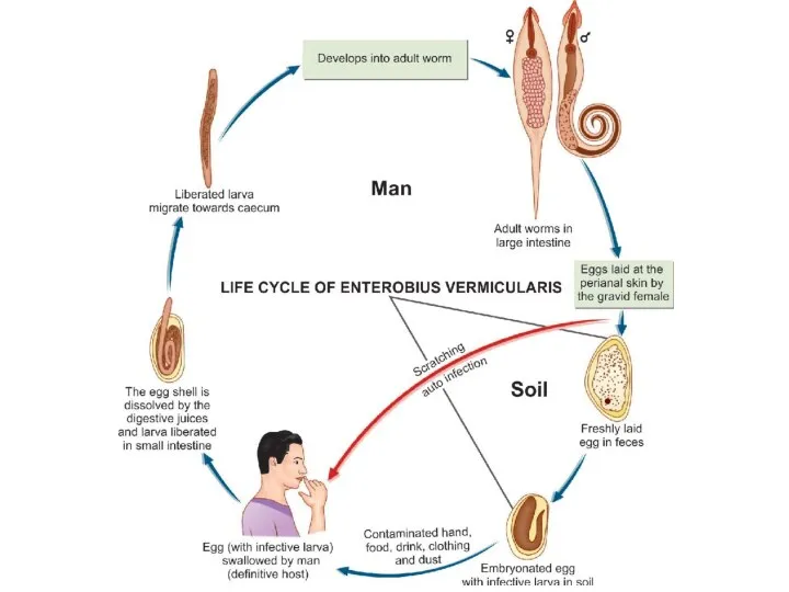
- 33. Life cycle Natural host: Man Infective form: Embryonated eggs, containing larvae with contaminated raw vegetables. Mode
- 35. Symptoms Enterobiasis occurs mostly in children. It is more common in females than in males. About
- 36. Diagnosis ♦ Detection of eggs by NIH swab and cellophane scotch tape method. Detection of eggs
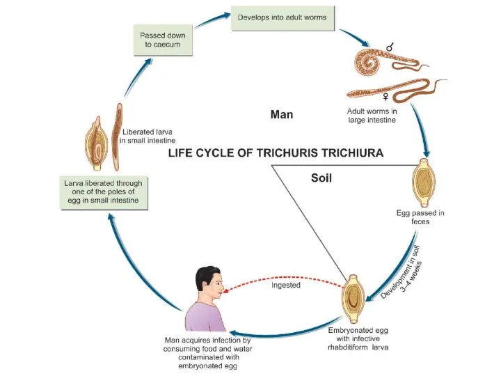
- 37. Trichuris trichiura (whip worm) The worm is divided into a thin whip-like anterior part measuring 3/5
- 39. Symptoms The patient complains of dysentery (blood and mucus in stool together with tenesmus). Rectal prolapse
- 40. Task 1. Intestinal nematodes
- 41. TISSUE NEMATODES. FILARIAL WORMS
- 42. Filarial worms This group includes the filarial worms, the guinea worm (Dranculuculus medinensis) and Trichinella spiralis.
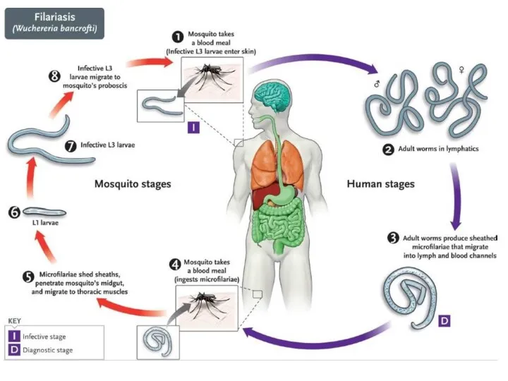
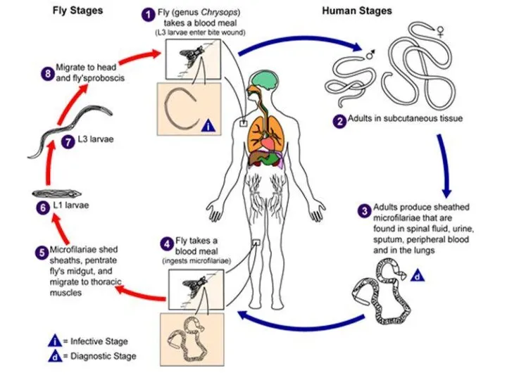
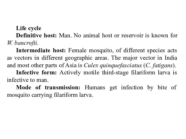
- 43. Wuchereria bancrofti This is a parasite of lymph nodes and lymphatic vessels- causing lymphatic filariasis. This
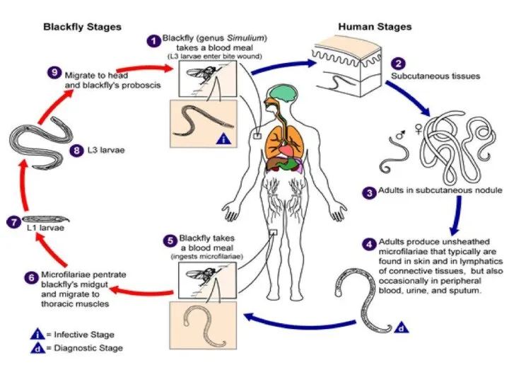
- 44. Mode of transmission and pathogenesis The filariform larvae are introduced through the skin by the bite
- 46. Symptoms ♦ The adult worm obstructs the flow of lymph in the lymph nodes and the
- 47. Treatment Diethyl carbamazine (DEC): 2 mg/kg 3x daily for 2 weeks. Endemic non-filarial elephantiasis (Podoconiosis) Non-filarial
- 48. Onchocerca volvulus Infection by this filarial worm is common in Ethiopia. Endemic foci are found in
- 50. Infective stage and mode of infection: microfilaria. Symptoms The disease, onchocerciasis or river blindness includes: •
- 51. Loa loa The eye worm, Loa loa, causes Loiasis. The insect vectors include mango flies of
- 53. Symptoms The microfilaria have a sheath. Their diurnal periodicity corresponds to the feeding pattern of the
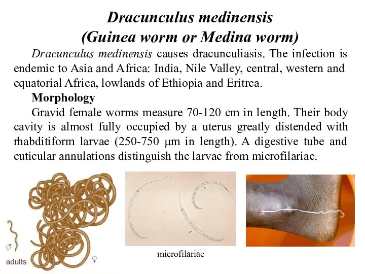
- 54. Dracunculus medinensis (Guinea worm or Medina worm) Dracunculus medinensis causes dracunculiasis. The infection is endemic to
- 55. Life cycle Definitive host: Man. No animal host or reservoir is known for W. bancrofti. Intermediate
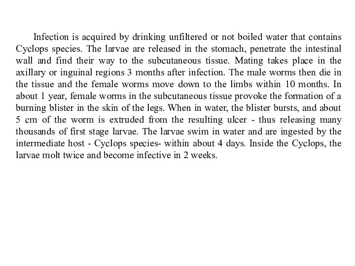
- 56. Infection is acquired by drinking unfiltered or not boiled water that contains Cyclops species. The larvae
- 58. Symptoms The female parasites in the subcutaneous tissue release toxic byproducts of histamine-like nature, which cause
- 59. Treatment Surgical excision when the worm is in the leg Niridazole (Ambilhar) or DEC Prophylaxis Eradication
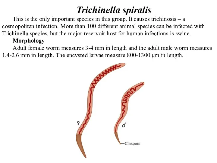
- 60. Trichinella spiralis This is the only important species in this group. It causes trichinosis – a
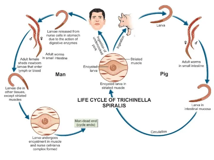
- 61. Life cycle After ingesting infected meat, the capsule of the encysted larvae is digested by gastric
- 63. Symptoms There are two clinical phases. 1. The intestinal phase: lasting 1-7 days - asymptomatic; sometimes
- 65. Скачать презентацию
All the important human parasites of the Phylum Nemathelminthes (Aschelminthes) belong
All the important human parasites of the Phylum Nemathelminthes (Aschelminthes) belong
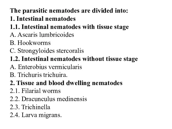
The parasitic nematodes are divided into:
1. Intestinal nematodes
1.1. Intestinal nematodes with
The parasitic nematodes are divided into:
1. Intestinal nematodes
1.1. Intestinal nematodes with
INTESTINAL NEMATODES WITH TISSUE STAGE
INTESTINAL NEMATODES WITH TISSUE STAGE
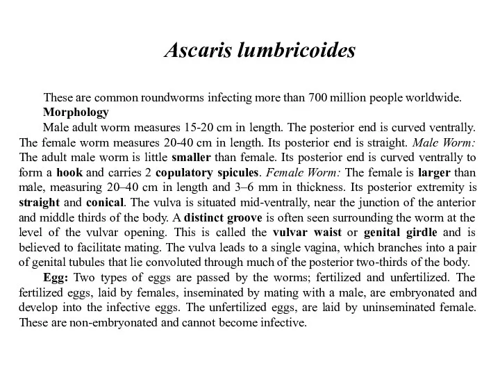
Ascaris lumbricoides
These are common roundworms infecting more than 700 million people
Ascaris lumbricoides
These are common roundworms infecting more than 700 million people
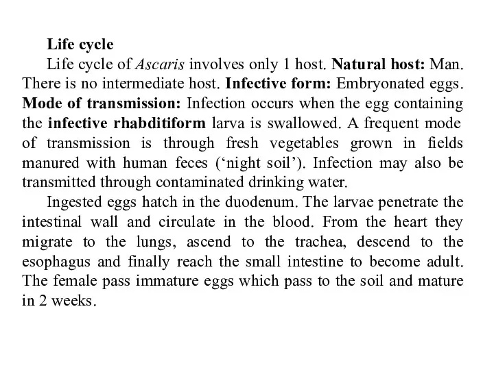
Life cycle
Life cycle of Ascaris involves only 1 host. Natural host:
Life cycle
Life cycle of Ascaris involves only 1 host. Natural host:
Symptoms
Fever, urticaria, angioneurotic edema, wheezing, and conjunctivitis.
Acute biliary obstruction or
Symptoms
Fever, urticaria, angioneurotic edema, wheezing, and conjunctivitis.
Acute biliary obstruction or

Diagnosis
Diagnosis
Treatment
pyrantel pamoate 11 mg/kg once; maximum 1 g,
albendazole 400 mg
Treatment
pyrantel pamoate 11 mg/kg once; maximum 1 g,
albendazole 400 mg
HOOK WORMS
HOOK WORMS
HOOK WORMS
There are two species of hookworm:
1. Ancylostoma duodenale
2. Necator americanus
The
HOOK WORMS
There are two species of hookworm:
1. Ancylostoma duodenale
2. Necator americanus
The

Ancylostoma duodenale
Grayish-white in color. The body is slightly ventrally curved. The
Ancylostoma duodenale
Grayish-white in color. The body is slightly ventrally curved. The


Necator americanus
This species, so called American hookworm, is found in predominantly
Necator americanus
This species, so called American hookworm, is found in predominantly
Life cycle
Adult male and female worms live in the small intestine.
Life cycle
Adult male and female worms live in the small intestine.
Symptoms
Adult worms in the intestine feed on blood causing iron deficiency
Symptoms
Adult worms in the intestine feed on blood causing iron deficiency
LARVA MIGRANS
LARVA MIGRANS

There are three types of larva migrans:
a. Cutaneous larva migrans (Creeping
There are three types of larva migrans:
a. Cutaneous larva migrans (Creeping
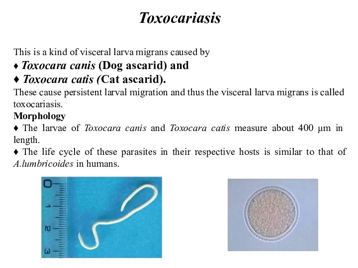
Toxocariasis
This is a kind of visceral larva migrans caused by
♦ Toxocara
Toxocariasis
This is a kind of visceral larva migrans caused by
♦ Toxocara

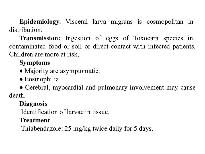
Epidemiology. Visceral larva migrans is cosmopolitan in distribution.
Transmission: Ingestion of eggs
Epidemiology. Visceral larva migrans is cosmopolitan in distribution.
Transmission: Ingestion of eggs
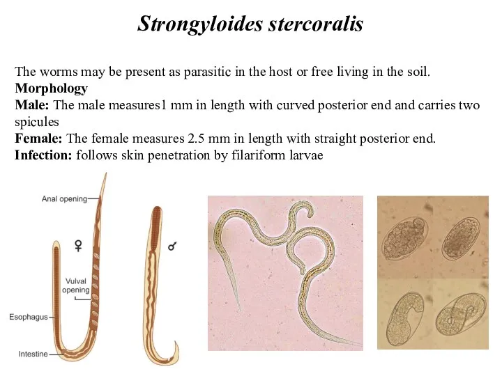
Strongyloides stercoralis
The worms may be present as parasitic in the host
Strongyloides stercoralis
The worms may be present as parasitic in the host
Life cycle
Adult male and female worms live in the small intestine.
Life cycle
Adult male and female worms live in the small intestine.
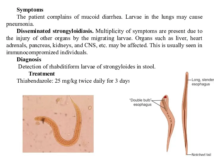
Symptoms
The patient complains of mucoid diarrhea. Larvae in the lungs may
Symptoms
The patient complains of mucoid diarrhea. Larvae in the lungs may
INTESTINAL NEMATODES WITHOUT TISSUE STAGE
INTESTINAL NEMATODES WITHOUT TISSUE STAGE
Enterobius vermicularis
(pin worm or thread worm)
Enterobius vermicularis is a small
Enterobius vermicularis
(pin worm or thread worm)
Enterobius vermicularis is a small

Life cycle
Natural host: Man
Infective form: Embryonated eggs, containing larvae with contaminated
Life cycle
Natural host: Man
Infective form: Embryonated eggs, containing larvae with contaminated
Symptoms
Enterobiasis occurs mostly in children. It is more common in females
Symptoms
Enterobiasis occurs mostly in children. It is more common in females
Diagnosis
♦ Detection of eggs by NIH swab and cellophane scotch tape
Diagnosis
♦ Detection of eggs by NIH swab and cellophane scotch tape
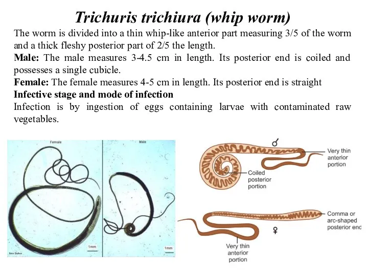
Trichuris trichiura (whip worm)
The worm is divided into a thin whip-like
Trichuris trichiura (whip worm)
The worm is divided into a thin whip-like
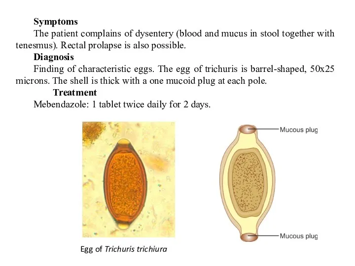
Symptoms
The patient complains of dysentery (blood and mucus in stool together
Symptoms
The patient complains of dysentery (blood and mucus in stool together
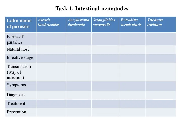
Task 1. Intestinal nematodes
Task 1. Intestinal nematodes

TISSUE NEMATODES.
FILARIAL WORMS
TISSUE NEMATODES.
FILARIAL WORMS

Filarial worms
This group includes the filarial worms, the guinea worm
Filarial worms
This group includes the filarial worms, the guinea worm
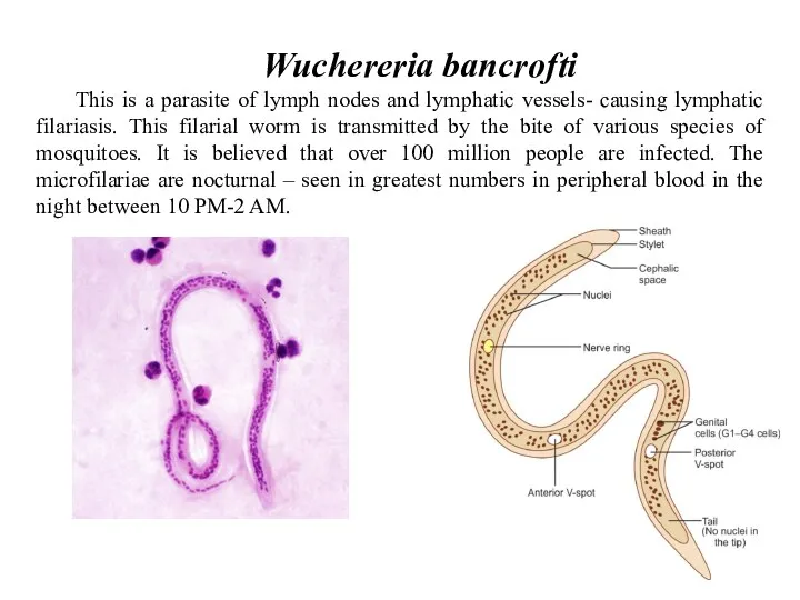
Wuchereria bancrofti
This is a parasite of lymph nodes and lymphatic vessels-
Wuchereria bancrofti
This is a parasite of lymph nodes and lymphatic vessels-
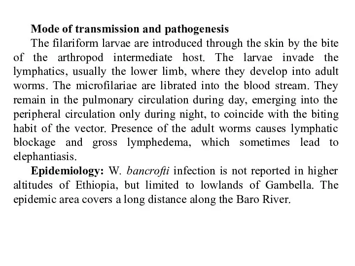
Mode of transmission and pathogenesis
The filariform larvae are introduced through the
Mode of transmission and pathogenesis
The filariform larvae are introduced through the

Symptoms
♦ The adult worm obstructs the flow of lymph in the
Symptoms
♦ The adult worm obstructs the flow of lymph in the
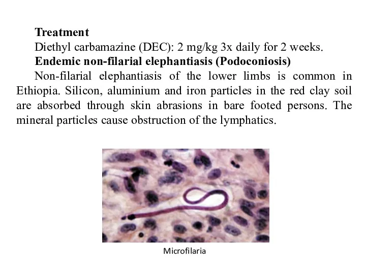
Treatment
Diethyl carbamazine (DEC): 2 mg/kg 3x daily for 2 weeks.
Endemic
Treatment
Diethyl carbamazine (DEC): 2 mg/kg 3x daily for 2 weeks.
Endemic
Onchocerca volvulus
Infection by this filarial worm is common in Ethiopia. Endemic
Onchocerca volvulus
Infection by this filarial worm is common in Ethiopia. Endemic
Infective stage and mode of infection: microfilaria.
Symptoms
The disease, onchocerciasis or river
Infective stage and mode of infection: microfilaria.
Symptoms
The disease, onchocerciasis or river
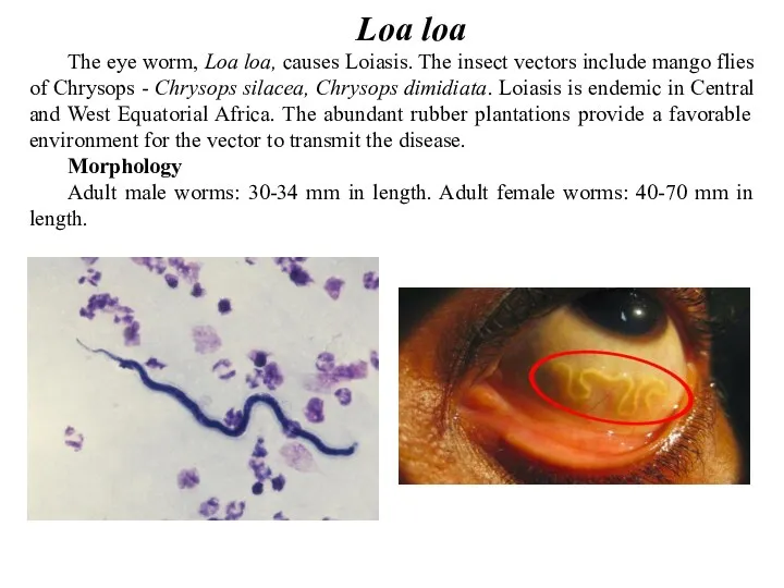
Loa loa
The eye worm, Loa loa, causes Loiasis. The insect vectors
Loa loa
The eye worm, Loa loa, causes Loiasis. The insect vectors

Symptoms
The microfilaria have a sheath. Their diurnal periodicity corresponds to the
Symptoms
The microfilaria have a sheath. Their diurnal periodicity corresponds to the
Dracunculus medinensis
(Guinea worm or Medina worm)
Dracunculus medinensis causes dracunculiasis. The
Dracunculus medinensis
(Guinea worm or Medina worm)
Dracunculus medinensis causes dracunculiasis. The
Life cycle
Definitive host: Man. No animal host or reservoir is known
Life cycle
Definitive host: Man. No animal host or reservoir is known
Infection is acquired by drinking unfiltered or not boiled water that
Infection is acquired by drinking unfiltered or not boiled water that

Symptoms
The female parasites in the subcutaneous tissue release toxic byproducts of
Symptoms
The female parasites in the subcutaneous tissue release toxic byproducts of
Treatment
Surgical excision when the worm is in the leg
Niridazole (Ambilhar) or
Treatment
Surgical excision when the worm is in the leg
Niridazole (Ambilhar) or
Trichinella spiralis
This is the only important species in this group. It
Trichinella spiralis
This is the only important species in this group. It

Life cycle
After ingesting infected meat, the capsule of the encysted larvae
Life cycle
After ingesting infected meat, the capsule of the encysted larvae

Symptoms
There are two clinical phases.
1. The intestinal phase: lasting 1-7 days
Symptoms
There are two clinical phases.
1. The intestinal phase: lasting 1-7 days
 АФО иммунной системы у детей. Методы оценки иммунной системы. Аллергодиагностика у детей. Бронхиальная астма
 Респираторный дистресс-синдром
 Железодефицитная анемия и беременность
 ИБС. Стенокардия
 Инфильтративный туберкулез легких
 Жедел бүйрек жетіспеушілігінің клиникалық көріністері
 Парентеральное питание
 Универсальный алгоритм оказания первой помощи
 Гострі пневмонії у дітей. Етіологія, патогенез. Класифікація. Клінічні форми. Лікування гострих пневмоній у дітей
 Заболевания губ у детей
 Репродуктивная система мужчины и женщины в зрелом возрасте
 Зубо-щелепні деформації
 Неотложные соматические состояния у тяжелых нетранспортабельных больных
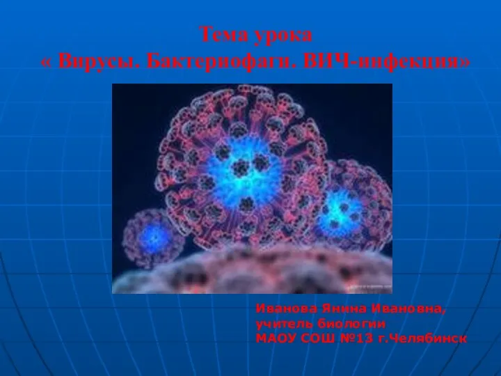 Вирусы. Бактериофаги. ВИЧ-инфекция
 Классификация дизонтогенеза. Поврежденное развитие
 Косметичні засоби
 Инструментальные методы исследования в гинекологии
 Гельминтоздар
 Когнитивно-поведенческое направление психотерапии
 Беременность и язвенная болезнь
 Первичная и вторичная профилактика депрессий у лиц пожилого возраста на базе геронтологического центра
 Лимфоидная система, как основа приобретенного антигенспецифического иммунитета
 Аутоиммунные заболевания
 Регенерация. Виды репаративной регенерации
 Национальный календарь прививок
 Нейроинфекции. Виды нейроинфекций
 Минимальды өзгеріс ауруларының емі
 Colonic Polyps