- Radiologic diagnostics of chest cavity

Содержание
- 2. Methods of respiratory organs examination: Radiography Radioscopy Bronchography Angiopulmonography Ultrasound diagnostics CT scan Magnetic resonance imaging
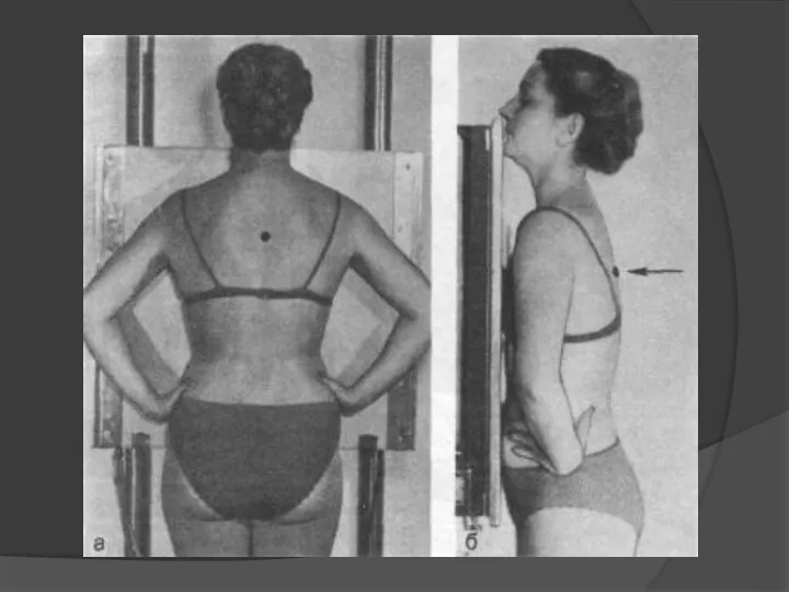
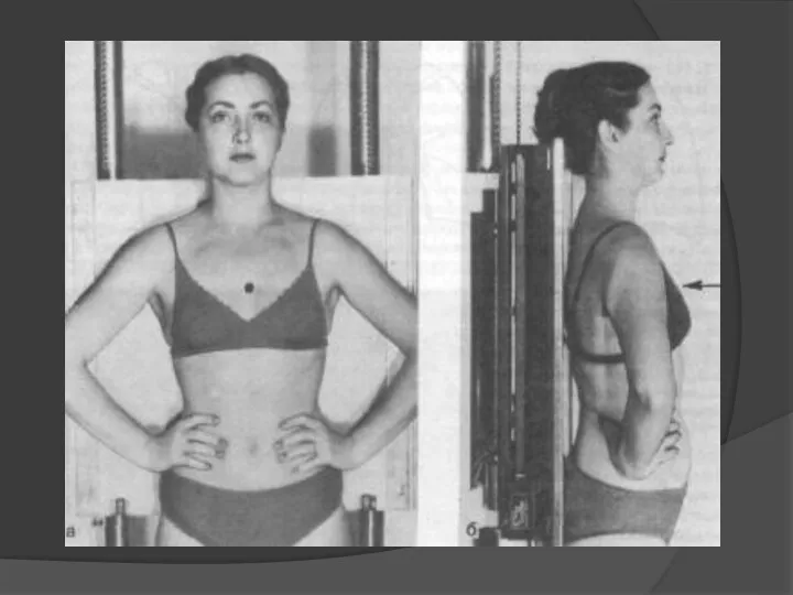
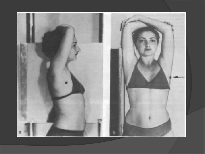
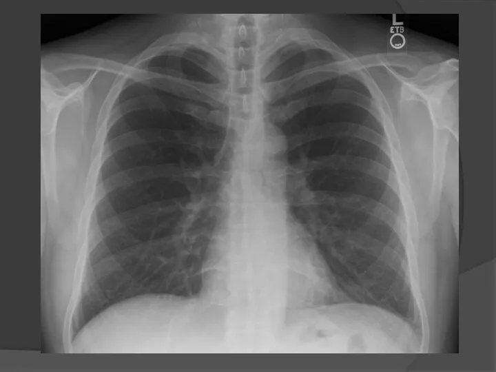
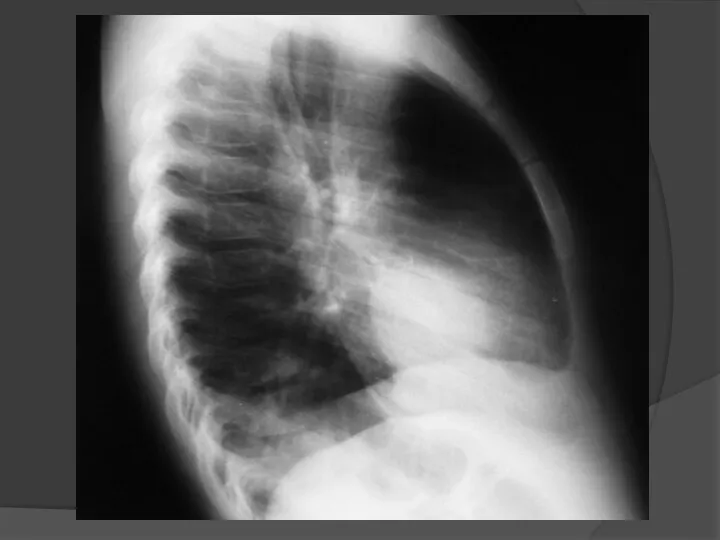
- 3. Direct projection Lateral projection Radiography of chest organs
- 4. Correct position of patient
- 5. 1 — anterior end of rib; 2 — trachea and major bronchi; 3 —ribs; 4 —right
- 6. Pulmonary fields
- 7. Roots of the lungs II-IV intercostal space II-IV ribs the left root above the right is
- 8. costodiaphragmatic sinuses Pulmonary pattern Cardiac diaphragmatic sinuses
- 9. SHADOW OF THE HEART Arc of the Heart 1- ascending aorta 2 -Arc of the right
- 10. Cardiothoracic index = transverse size of heart transverse size of thorax transverse size of heart transverse
- 11. Atriovasal angle ascending aorta Right atrium Atriovasal angle
- 12. 1 2 3 4 5 8 Segments of the lungs in a straight projection 1-2 3
- 13. 1 2 3 6 10 4 5 Lung segments in lateral projection 9 7 8 8
- 14. Segments of the lungs in a straight projection (front, back)
- 15. Median tomography 1-trachea 2-right main bronchus 3-left main bronchus 4-right upper lobe bronchus 5-right mid-lobe bronchus
- 16. Linear tomography ТРАХЕЯ правый левый
- 17. Lymph nodes of the mediastinum paratracheal lymph nodes Tracheobronchial lymph nodes Bifurcation lymph nodes Bronchopulmonary lymph
- 18. Art. pulm. sin. Arcus aortae V. anonyma sin. V. subclavia sin. Art. subclavia sin. V. jugularis
- 19. - Radiography - Fluorography - Radioscopy - Linear tomography X-ray examination methods
- 20. Radioscopy X-ray examination method in which an X-ray image of an object is obtained on the
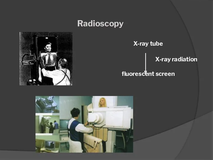
- 21. Radioscopy X-ray tube fluorescent screen X-ray radiation
- 22. Indications: - polypositional study - real-time evaluation of the function - conducting the catheterization, angioplasty under
- 23. Methods of radioscopy a - orthoscopy, b - trochoscopy, v-lateroscopy
- 25. Radiography An X-ray examination method in which a fixed X-ray image of an object is obtained
- 26. Advantages of radiography: - better detectability of small parts - less radiation load - the possibility
- 32. Fluorography The method of X-ray examination consists in photographing the image from a fluorescent screen, screen
- 33. Fluorography X-ray tube fluorescent screen camera 10 см X-ray radiation
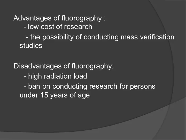
- 34. Advantages of fluorography : - low cost of research - the possibility of conducting mass verification
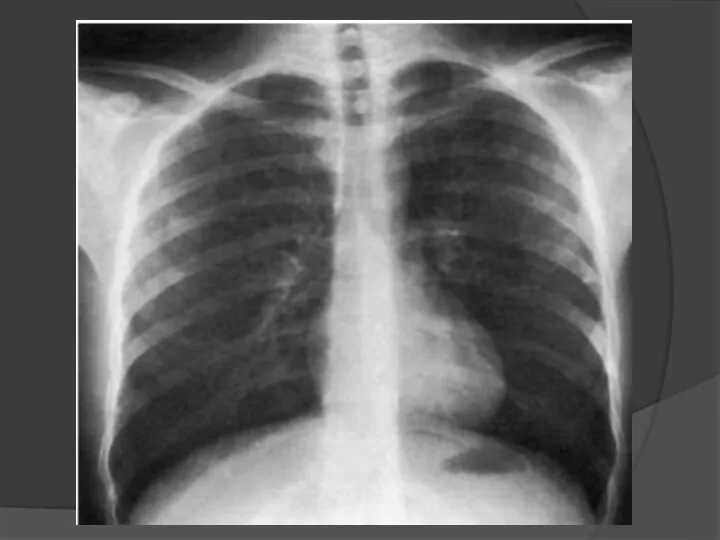
- 35. Main applications - Chest examination for early detection of tuberculosis
- 37. Linear tomography The method of X-ray examination is to obtain an image of an object at
- 40. Main applications Investigation of pulmonary parenchyma, trachea and major bronchi, intrathoracic lymph nodes, paranasal sinuses, larynx,
- 41. Tomography of chest organs
- 42. Evaluation of the quality of the radiograph Completeness of volume : the whole chest from the
- 43. Прямая проекция Критерии правильно выполненной рентгенограммы Видны легочные поля на всем протяжении и диафрагмальные синусы Изображение
- 44. Прямая проекция Ширина задних отрезков ребер значительно меньше передних Контуры задних отрезков ребер более четкие, чем
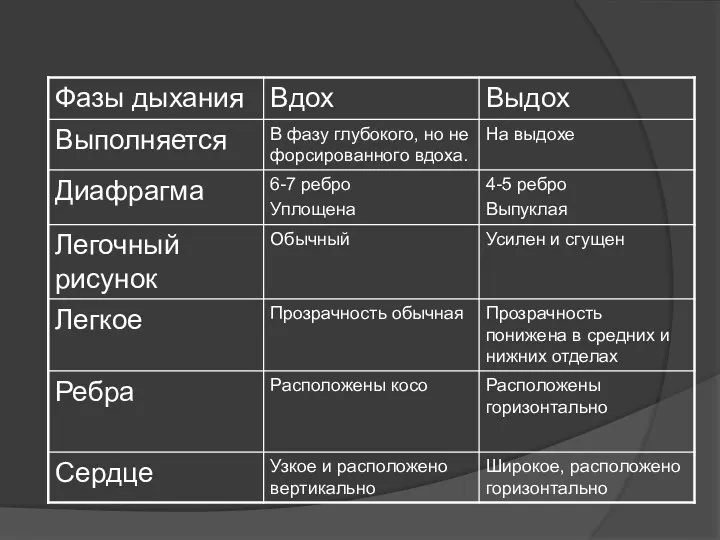
- 45. Исследование на вдохе и выдохе Рентгенография легких производится в фазу глубокого, но не форсированного вдоха. Диафрагма
- 47. Inspiration and expiration examination 6 4
- 48. Боковая проекция Критерии правильно выполненной рентгенограммы Видны легочные поля на всем протяжении (верхушки и реберно-диафрагмальные синусы).
- 50. Скачать презентацию
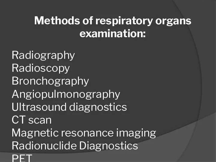
Methods of respiratory organs examination:
Radiography
Radioscopy
Bronchography
Angiopulmonography
Ultrasound diagnostics
CT scan
Magnetic resonance imaging
Radionuclide Diagnostics
PET
Methods of respiratory organs examination:
Radiography
Radioscopy
Bronchography
Angiopulmonography
Ultrasound diagnostics
CT scan
Magnetic resonance imaging
Radionuclide Diagnostics
PET
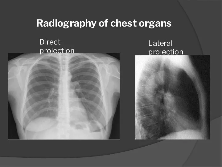
Direct projection
Lateral projection
Radiography of chest organs
Direct projection
Lateral projection
Radiography of chest organs
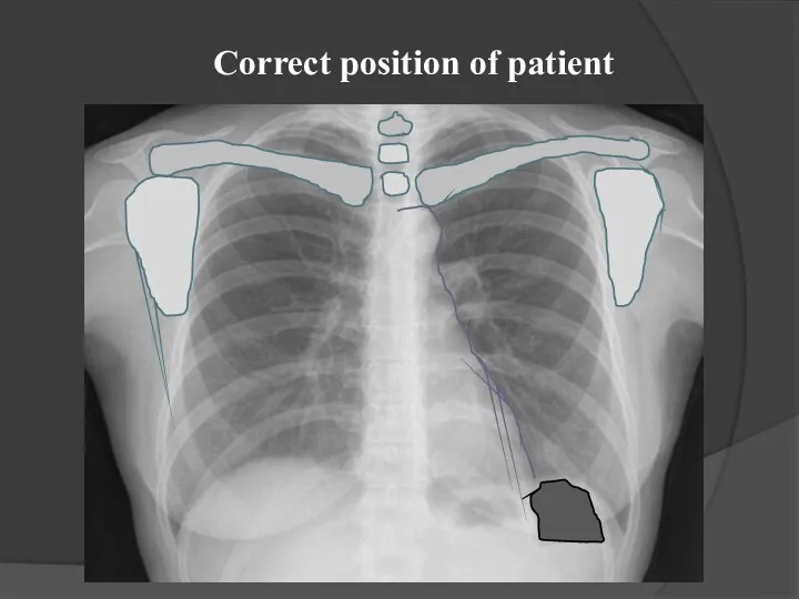
Correct position of patient
Correct position of patient
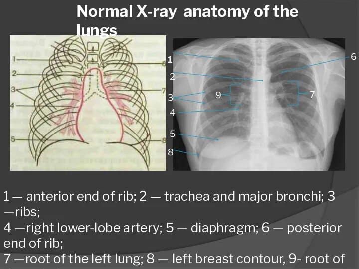
1 — anterior end of rib; 2 — trachea and major
1 — anterior end of rib; 2 — trachea and major
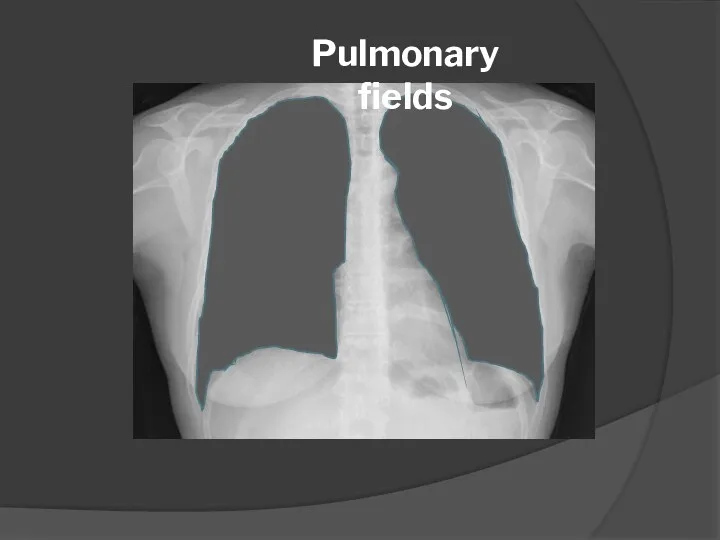
Pulmonary fields
Pulmonary fields
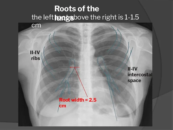
Roots of the lungs
II-IV intercostal space
II-IV
ribs
the left root above the
Roots of the lungs
II-IV intercostal space
II-IV
ribs
the left root above the
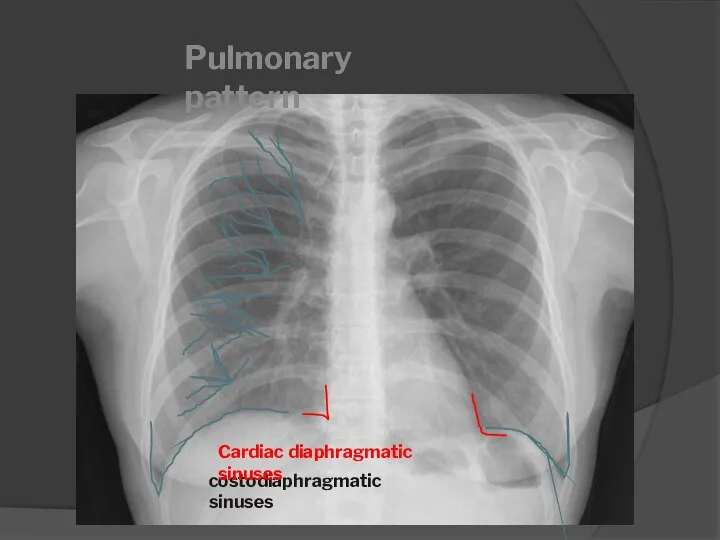
costodiaphragmatic sinuses
Pulmonary pattern
Cardiac diaphragmatic sinuses
costodiaphragmatic sinuses
Pulmonary pattern
Cardiac diaphragmatic sinuses
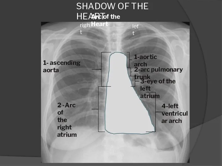
SHADOW OF THE HEART
Arc of the Heart
1- ascending aorta
2 -Arc of
SHADOW OF THE HEART
Arc of the Heart
1- ascending aorta
2 -Arc of
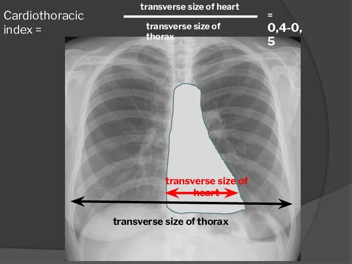
Cardiothoracic index =
transverse size of heart
transverse size of thorax
transverse size of
Cardiothoracic index =
transverse size of heart
transverse size of thorax
transverse size of
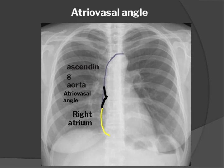
Atriovasal angle
ascending
aorta
Right atrium
Atriovasal angle
Atriovasal angle
ascending
aorta
Right atrium
Atriovasal angle
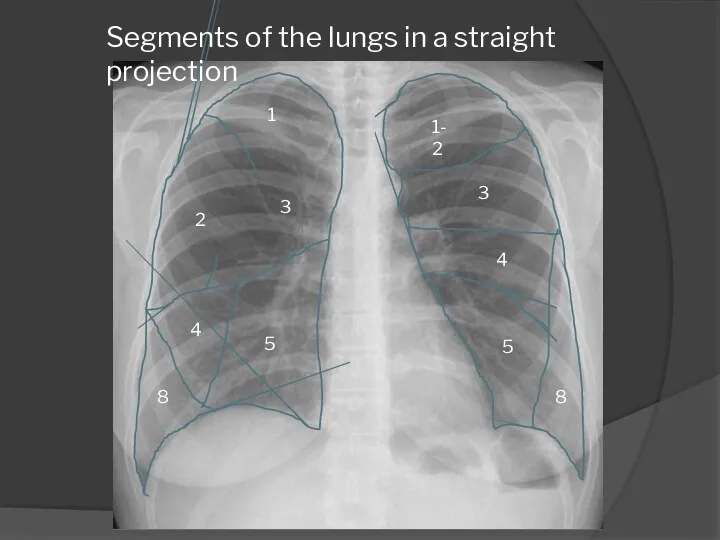
1
2
3
4
5
8
Segments of the lungs in a straight projection
1-2
3
4
5
8
1
2
3
4
5
8
Segments of the lungs in a straight projection
1-2
3
4
5
8
1
2
3
6
10
4
5
Lung segments in lateral projection
9
7
8
8
1
2
3
6
10
4
5
Lung segments in lateral projection
9
7
8
8
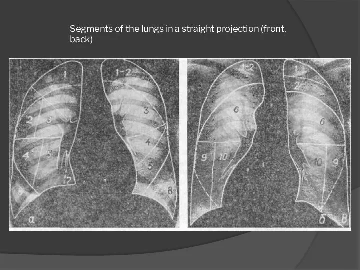
Segments of the lungs in a straight projection (front, back)
Segments of the lungs in a straight projection (front, back)
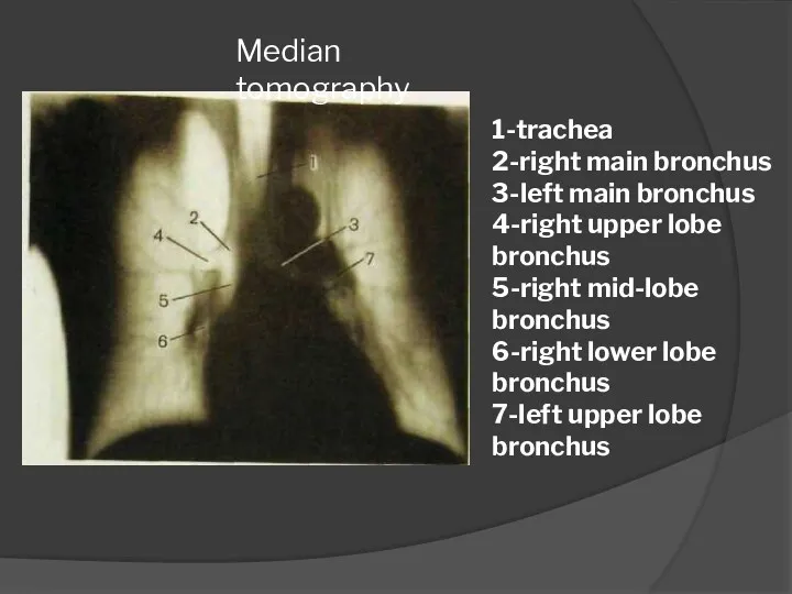
Median tomography
1-trachea
2-right main bronchus
3-left main bronchus
4-right upper lobe bronchus
5-right mid-lobe bronchus
6-right
Median tomography
1-trachea
2-right main bronchus
3-left main bronchus
4-right upper lobe bronchus
5-right mid-lobe bronchus
6-right
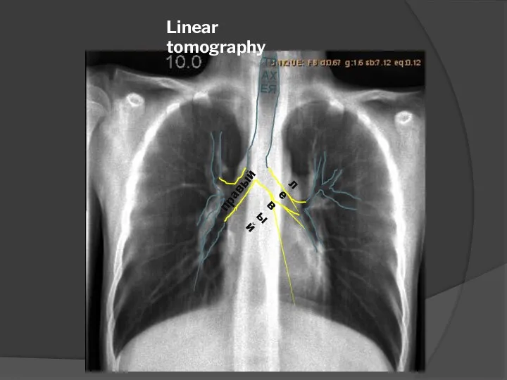
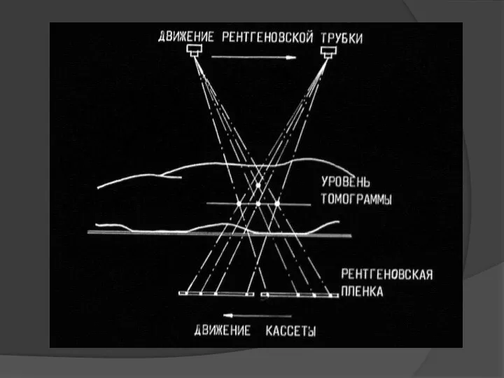
Linear tomography
ТРАХЕЯ
правый
левый
Linear tomography
ТРАХЕЯ
правый
левый
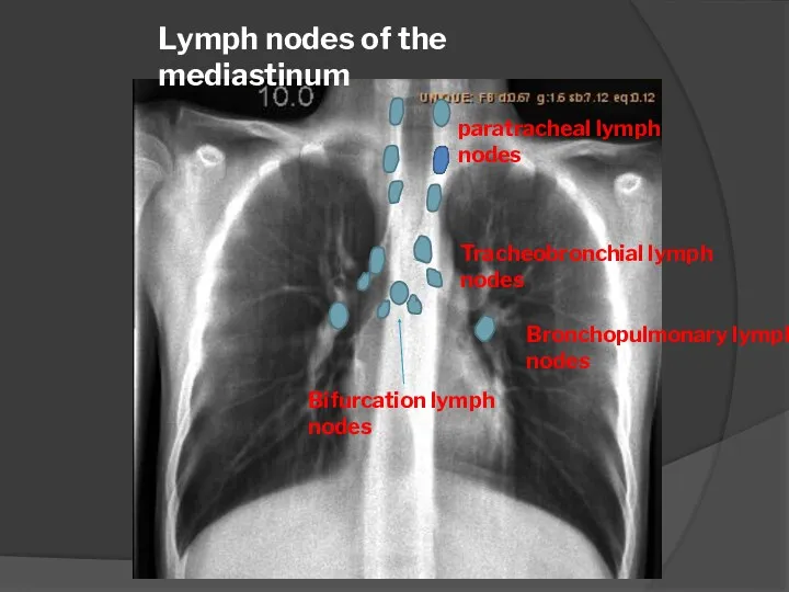
Lymph nodes of the mediastinum
paratracheal lymph nodes
Tracheobronchial lymph nodes
Bifurcation lymph nodes
Lymph nodes of the mediastinum
paratracheal lymph nodes
Tracheobronchial lymph nodes
Bifurcation lymph nodes
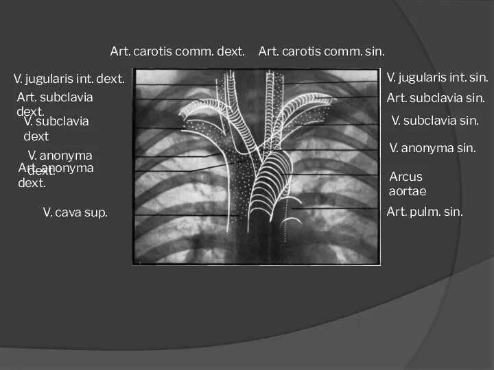
Art. pulm. sin.
Arcus aortae
V. anonyma sin.
V. subclavia sin.
Art. subclavia sin.
V. jugularis
Art. pulm. sin.
Arcus aortae
V. anonyma sin.
V. subclavia sin.
Art. subclavia sin.
V. jugularis
- Radiography - Fluorography
- Radioscopy
- Linear tomography
X-ray examination
- Radiography - Fluorography
- Radioscopy
- Linear tomography
X-ray examination
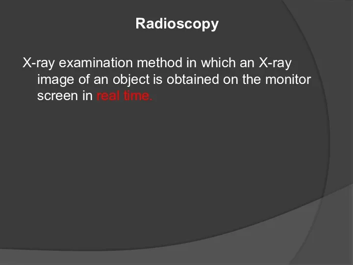
Radioscopy
X-ray examination method in which an X-ray image of an object
Radioscopy
X-ray examination method in which an X-ray image of an object
Radioscopy
X-ray tube
fluorescent screen
X-ray radiation
Radioscopy
X-ray tube
fluorescent screen
X-ray radiation
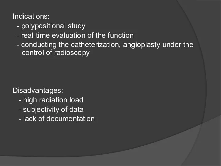
Indications:
- polypositional study
- real-time evaluation of the function
-
Indications:
- polypositional study
- real-time evaluation of the function
-
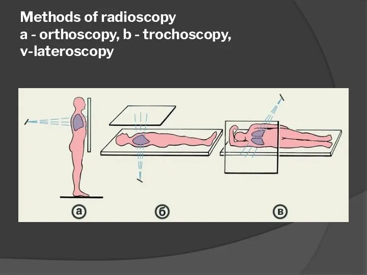
Methods of radioscopy
a - orthoscopy, b - trochoscopy, v-lateroscopy
Methods of radioscopy
a - orthoscopy, b - trochoscopy, v-lateroscopy
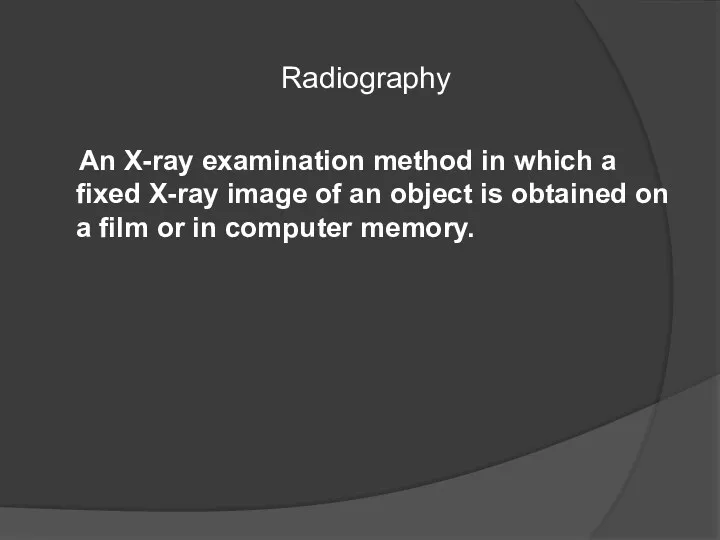
Radiography
An X-ray examination method in which a fixed X-ray
Radiography
An X-ray examination method in which a fixed X-ray
Advantages of radiography:
- better detectability of small parts
- less
Advantages of radiography:
- better detectability of small parts
- less
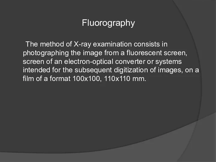
Fluorography
The method of X-ray examination consists in photographing the image from
Fluorography
The method of X-ray examination consists in photographing the image from
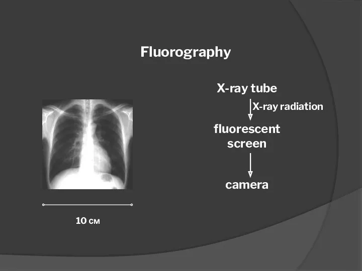
Fluorography
X-ray tube
fluorescent screen
camera
10 см
X-ray radiation
Fluorography
X-ray tube
fluorescent screen
camera
10 см
X-ray radiation
Advantages of fluorography :
- low cost of research
-
Advantages of fluorography :
- low cost of research
-
Main applications
- Chest examination for early detection of tuberculosis
Main applications
- Chest examination for early detection of tuberculosis
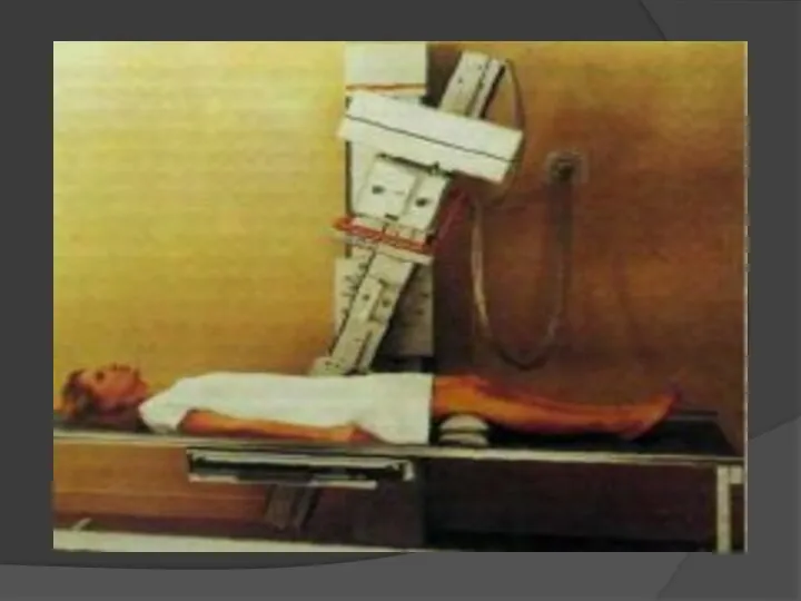
Linear tomography
The method of X-ray examination is to obtain an image
Linear tomography
The method of X-ray examination is to obtain an image
Main applications
Investigation of pulmonary parenchyma, trachea and major bronchi, intrathoracic lymph
Main applications
Investigation of pulmonary parenchyma, trachea and major bronchi, intrathoracic lymph
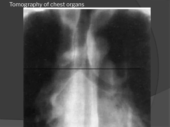
Tomography of chest organs
Tomography of chest organs
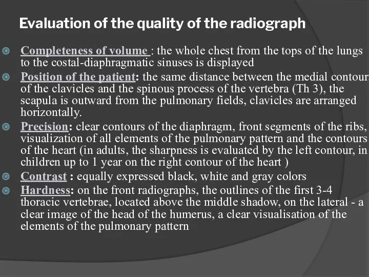
Evaluation of the quality of the radiograph
Completeness of volume : the
Evaluation of the quality of the radiograph
Completeness of volume : the
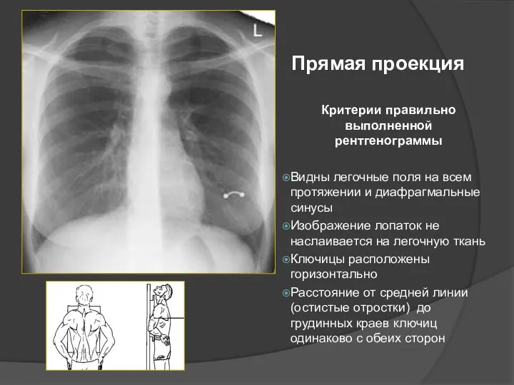
Прямая проекция
Критерии правильно выполненной рентгенограммы
Видны легочные поля на всем протяжении и
Прямая проекция
Критерии правильно выполненной рентгенограммы
Видны легочные поля на всем протяжении и
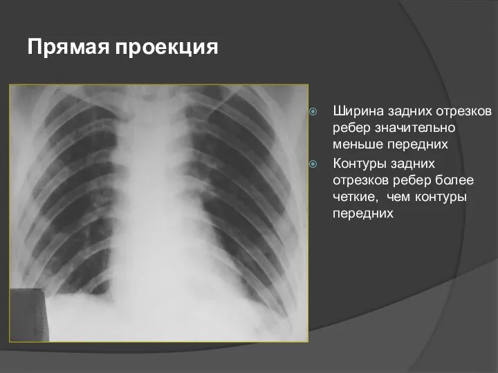
Прямая проекция
Ширина задних отрезков ребер значительно меньше передних
Контуры задних отрезков ребер
Прямая проекция
Ширина задних отрезков ребер значительно меньше передних
Контуры задних отрезков ребер
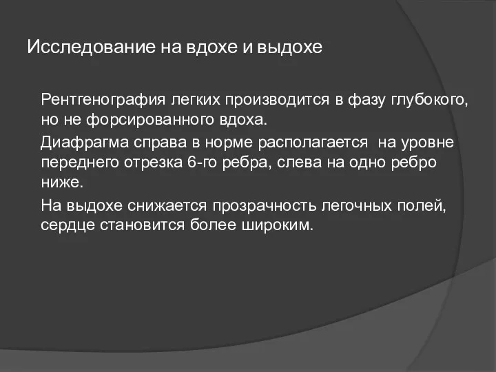
Исследование на вдохе и выдохе
Рентгенография легких производится в фазу глубокого, но
Исследование на вдохе и выдохе
Рентгенография легких производится в фазу глубокого, но
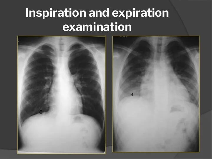
Inspiration and expiration examination
6
4
Inspiration and expiration examination
6
4
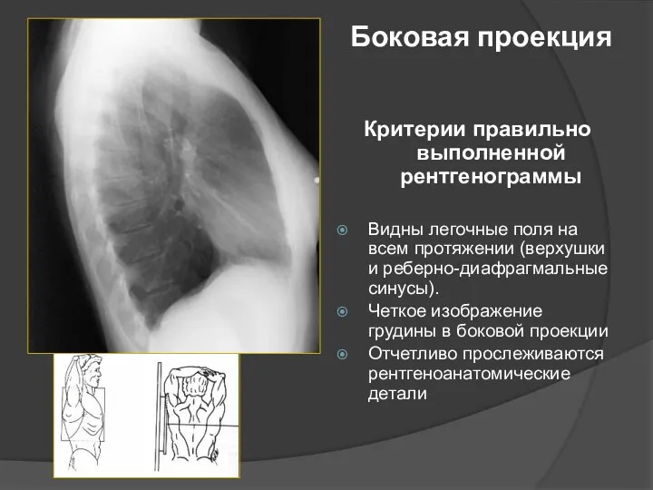
Боковая проекция
Критерии правильно выполненной рентгенограммы
Видны легочные поля на всем протяжении (верхушки
Боковая проекция
Критерии правильно выполненной рентгенограммы
Видны легочные поля на всем протяжении (верхушки
 План курса Общая нейрофизиология
 Анемии. Эритроцитопоэз
 Симптоматические артериальные гипертензии
 Cardiogenic shock
 Лекарственные растения обладающие мочегонными свойствами
 Паталогия печени
 Патогенез речевых нарушений
 Фибрилляция предсердий
 Транквилизаторы
 Зубные пасты. Жевательные резинки. Жевательные таблетки. Ополаскиватель для полости рта
 Ожоги органа зрения
 Факторы свертывающей и противосвертывающей системы крови. Геморрагические диатезы и синдромы. Тромбофилии
 Зоонозы. Туляремия
 Систематизация и диагностика васкулитов
 Травматический шок
 Иммунопрофилактика. Национальный календарь
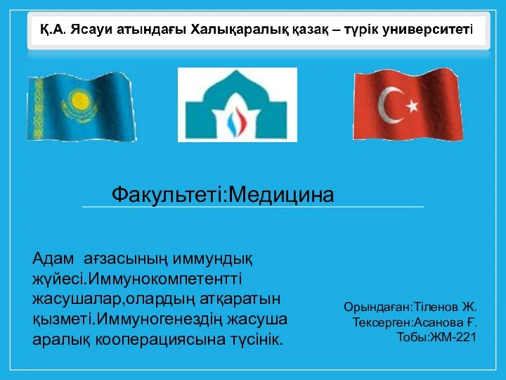 Адам ағзасының иммундық жүйесі
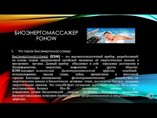 Биоэнергомассажер Fohow
 Развитие медицины в России XIX век
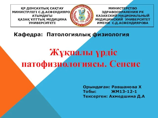 Жұқпалы үрдіс патофизиологиясы. Сепсис және сепсистік сілейменің патогенезі
 Респираторлық дистресс синдромының клинико-лабораторлық зерттеу көріністері
 Питание как стиль жизни. Основные правила и принципы
 Физическое развитие. Основные показатели физического развития в различных возрастно-половых группах, их особенности и тенденции
 Гельминтозы. Диагностика. Лечение
 Синдром Дауна
 Сүт бездерінің дисормональды аурулары туралы не білеміз?
 Жүктілік кезіндегі гипертензия
 Возможности медикаментозного обезболивания во время хирургических вмешательств