- Signaling by the neuroendocrine system

Содержание
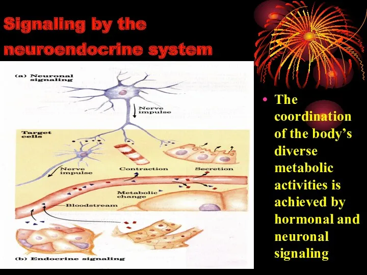
- 2. Signaling by the neuroendocrine system The coordination of the body’s diverse metabolic activities is achieved by
- 3. Hormone: A chemical substance synthesized in small amounts by an endocrine tissue and carried in the
- 4. Chemical signalling - endocrine, paracrine, autocrine and intracrine mechanisms intracrine paracrine autocrine neuroendocrine endocrine
- 5. Function of hormones HOMEOSTASIS Reproduction Growth and development Maintenance of internal environment Production, utilization and storage
- 6. At a minimum, a hormonally regulated system must include a secretary gland, a receptor for the
- 7. Hormones function in a complex hierarchy. Hormone act in hierarchy of function. The hypothala-mus functions at
- 8. CLASSIFICATION OF HORMONES
- 9. CLASSIFICATION OF HORMONES ACCORDING TO ANATOMICAL SOURCE OF THE HORMONE:
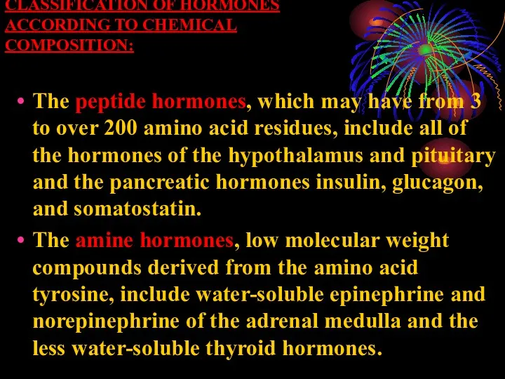
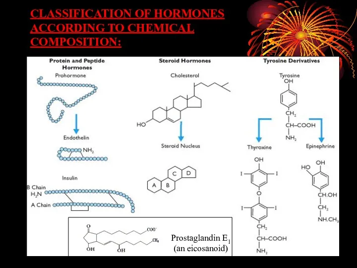
- 10. CLASSIFICATION OF HORMONES ACCORDING TO CHEMICAL COMPOSITION: The peptide hormones, which may have from 3 to
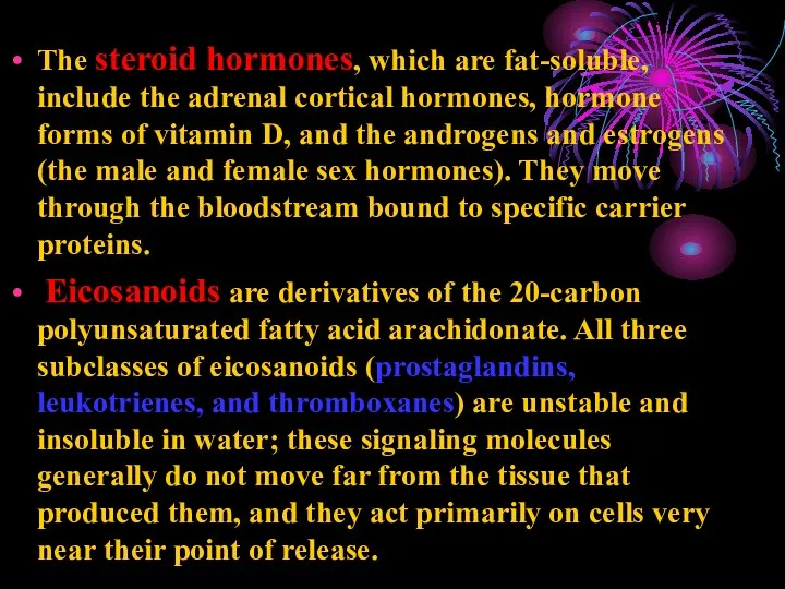
- 11. The steroid hormones, which are fat-soluble, include the adrenal cortical hormones, hormone forms of vitamin D,
- 12. CLASSIFICATION OF HORMONES ACCORDING TO CHEMICAL COMPOSITION: Prostaglandin E1 (an eicosanoid)
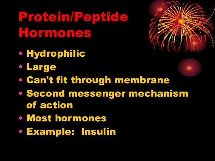
- 13. Protein/Peptide Hormones Hydrophilic Large Can't fit through membrane Second messenger mechanism of action Most hormones Example:
- 14. Steroid Hormones Small Hydrophobic/Lipophilic Travel in blood w/carrier Cytoplasmic or nuclear receptors change protein synthesis Example:
- 15. PEPTIDE HORMONES
- 16. STIMULUS Hypothalamus Releasing Hormone (Release-Inhibiting Hormone) Pituitary Stimulating Hormone Gland Hormone Target Why is the Hypothalamus
- 17. Hypothalamic hormones: Liberins or releasing hormones: 1. Corticotropin releasing factor 2. Thyrotropin releasing hormone 3. Gonadotropin
- 18. Hypothalamic hormones. Hypothalamic releasing and inhibiting hormones are carried directly to the anterior pituitary gland via
- 19. Pituitary hormones.
- 20. THE PITUITARY GLAND The pituitary gland is composed of distinctive parts: The anterior pituitary The intermediate
- 21. Anterior Pituitary Hormones and Their Hormones 1. Growth Hormone 2.Thyroid Stimulating Hormone 3.Adrenocorticotropic Hormone 4.Prolactin 5.Gonadotropins:
- 22. TARGET ORGAN OF PITUITARY HORMONE. TARGET ORGAN OF PITUITARY HORMONE.
- 23. 1. Growth Hormone (GH)
- 24. Growth Hormone (GH) Human growth hormone (somatotropin) is a protein of 191 amino acids. The GH-secreting
- 25. Growth Hormone (GH) GH promotes body growth by: binding to receptors on the surface of liver
- 26. Growth Hormone (GH) It promotes protein building in all cells (increase the transport of amino acids
- 27. A lack of GH causes dwarfism. A deficiency state can result not only from a deficiency
- 28. An excess results in gigantism or acromegaly. Gigantism, excess height and weight. Gigantism with normal body
- 29. An excess results in gigantism or acromegaly. Acromegaly , also called acromegalia. A long-term problem in
- 30. 2. Thyroid-Stimulating Hormone (Thyrotropin )
- 31. Thyroid-Stimulating Hormone (Thyrotropin ) Thyroid-stimulating hormone, also known as thyrotropin, is secreted from cells in the
- 32. Thyroid Stimulating Hormone (TSH) TSH is a glycoprotein consisting of: a beta chain of 112 amino
- 33. Regulation of secretion of thyroid-releasing hormone.
- 34. Some people develop antibodies against their own TSH receptors. When these bind the receptors, they "fool"
- 35. 3. Adrenocorticotropic Hormone (ACTH)
- 36. Adrenocorticotropic Hormone (ACTH) Adrenocorticotropic hormone is single-chain polypeptide consisting of 39 amino acids, as its name
- 37. Adrenocorticotropic hormone
- 38. Adrenocorticotropic hormone
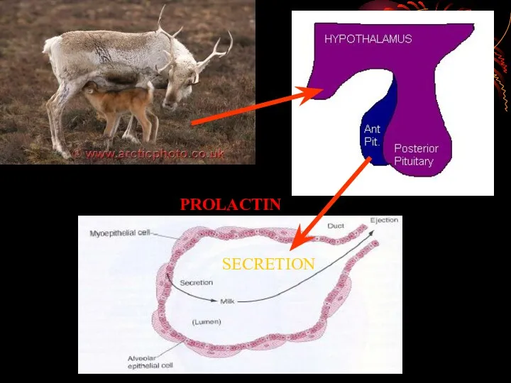
- 39. 4. PROLACTIN
- 40. Prolactin Prolactin – protein of 197 amino acids. Prolactin, acting with other hormones, starts the growth
- 41. Prolactin has two major roles in milk production: Prolactin induces lobuloalveolar growth of the mammary gland.
- 42. PROLACTIN SECRETION
- 43. Disease States Excessive secretion of prolactin - hyperprolactinemia - is a relative common disorder in humans.
- 44. 5. Gonadotropins: Luteinizing and Follicle Stimulating Hormones
- 45. Gonadotropins: Luteinizing and Follicle Stimulating Hormones Luteinizing hormone (LH) and follicle-stimulating hormone (FSH) are called gonadotropins
- 46. Gonadotropins. As describef for thyroid-simulating hormone, LH and FSH are large glycoproteins composed of alpha and
- 47. Physiologic Effects of Luteinizing Hormone In both sexes, LH stimulates secretion of sex steroids from the
- 48. Luteinizing Hormone In men In the testes, LH binds to receptors on Leydig cells, stimulating synthesis
- 49. Follicle-Stimulating Hormone As its name implies, FSH stimulates the maturation of ovarian follicles. Administration of FSH
- 50. Control of Gonadotropin Secretion The principle regulator of LH and FSH secretion is gonadotropin-releasing hormone or
- 51. Disease States Diminished secretion of LH or FSH can result in failure of gonadal function (hypogonadism).
- 52. Intermediate lobe: MSH (melanocyte stimulating hormone) Melanocyte-stimulating hormone (MSH): Known to control melanin pigmentation in the
- 53. Posterior Pituitary Hormones:
- 54. Antidiuretic Hormone (Vasopressin) Antidiuretic hormone, also known as vasopressin, is a nine amino acid peptide secreted
- 55. Antidiuretic Hormone (Vasopressin) Within hypothalamic neurons, the hormone is packaged in secretory vesicles with a carrier
- 56. Physiologic Effects of Antidiuretic Hormone A hormone that increases blood pressure with increases reabsorption of water
- 57. Physiologic Effects of Antidiuretic Hormone ADH is released when the blood volume falls, when a large
- 58. Parallel between antidiuretic hormone secretion and thirst.
- 59. Disease States Diabetes insipidus is result of a lack of ADH The most common presenting signs
- 60. Posterior Pituitary Hormones Oxytocin
- 61. Oxytocin Oxytocin in a nine amino acid peptide that is synthesized in hypothalamic neurons and transported
- 62. Physiologic Effects of Oxytocin Oxytocin stimulates contraction of myoepithelial cells, causing milk to be ejected into
- 63. OXYTOCIN
- 64. Hormones of the pancreas: insulin, glucagons, and somatostatin
- 65. The pancreas has two major biochemical functions: 1. Exocrine cells produce digestive enzymes for secretion into
- 66. Insulin Insulin is a small protein with two polypeptide chains, A and B, joined by two
- 67. Insulin decreases blood level of glucose: 1) by increase of glucose uptake into cells triggering movement
- 68. Effect of insulin on blood glucose: uptake of glucose by cells and storage as triacyglycerols and
- 69. Insulin deficiency Insulin deficiency Increased glucose utilizations Increased glycogenolysis Increased gluconeogenesis hyperglycemia Osmotic diuresis Dehydration Increased
- 70. Glucagon is a single polypeptide chain of 29 amino acid residues, and like insulin is derived
- 71. Glucagon increases blood level of glucose: 1) by stimulation of the glycogen breakdown in liver; 2)
- 72. Somatostatin is polypeptide hormone, inhibits the secretion of insulin and glucagons by the pancreas. Somatostatin is
- 73. 5’-AMP 5’-AMP cAMP Activation of cAMP-depend protein kinase 4 Inactive enzyme Active enzyme P P Active
- 74. The phosphatidylinositol pathway
- 75. The insulin receptor is a tyrosine-specific protein kinase
- 77. Скачать презентацию
Signaling by the neuroendocrine system
The coordination of the body’s diverse
Signaling by the neuroendocrine system
The coordination of the body’s diverse
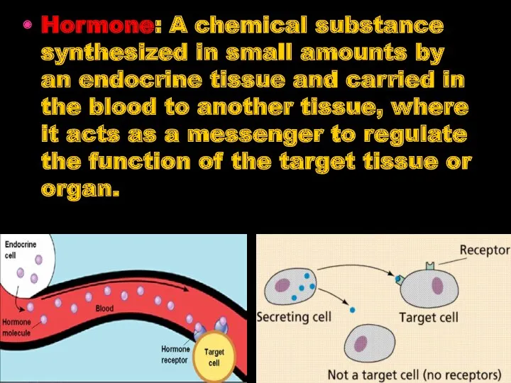
Hormone: A chemical substance synthesized in small amounts by an endocrine
Hormone: A chemical substance synthesized in small amounts by an endocrine
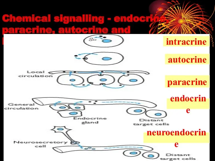
Chemical signalling - endocrine, paracrine, autocrine and
intracrine mechanisms
intracrine
paracrine
autocrine
neuroendocrine
endocrine
Chemical signalling - endocrine, paracrine, autocrine and
intracrine mechanisms
intracrine
paracrine
autocrine
neuroendocrine
endocrine
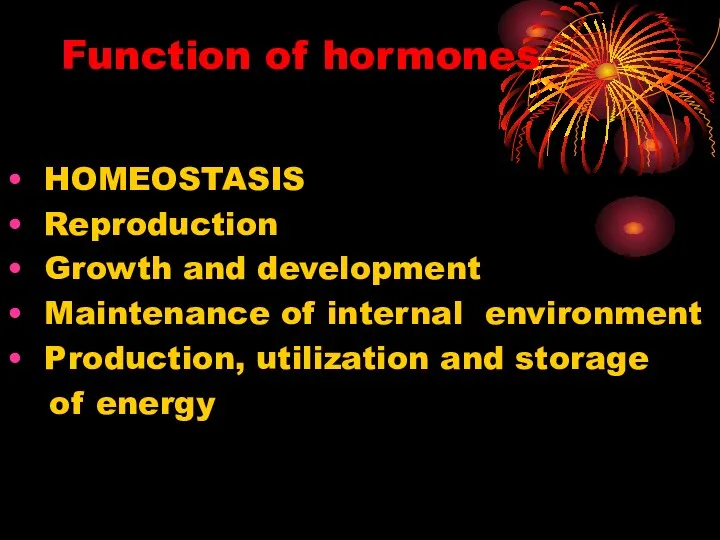
Function of hormones
HOMEOSTASIS
Reproduction
Growth and development
Maintenance of internal
Function of hormones
HOMEOSTASIS
Reproduction
Growth and development
Maintenance of internal
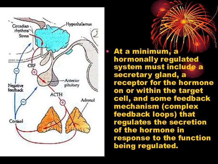
At a minimum, a hormonally regulated system must include a secretary
At a minimum, a hormonally regulated system must include a secretary
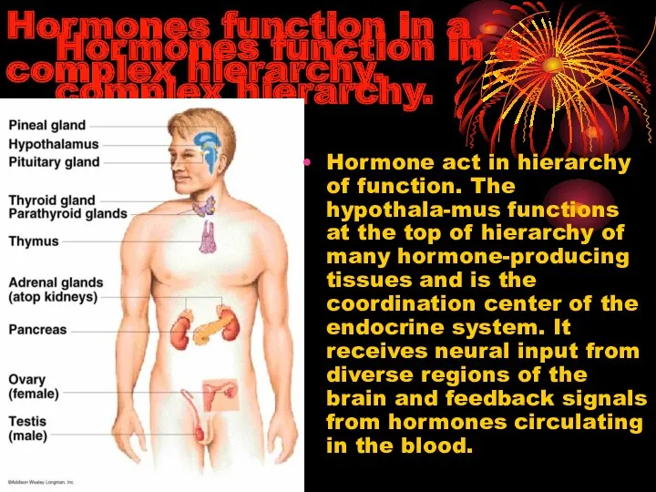
Hormones function in a complex hierarchy.
Hormone act in hierarchy of function.
Hormones function in a complex hierarchy.
Hormone act in hierarchy of function.
CLASSIFICATION OF HORMONES
CLASSIFICATION OF HORMONES
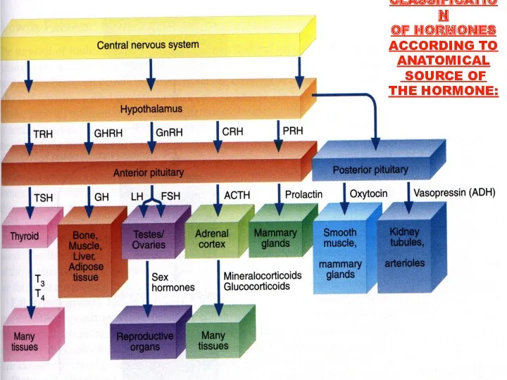
CLASSIFICATION
OF HORMONES
ACCORDING TO
ANATOMICAL
SOURCE OF
THE HORMONE:
CLASSIFICATION
OF HORMONES
ACCORDING TO
ANATOMICAL
SOURCE OF
THE HORMONE:
CLASSIFICATION OF HORMONES
ACCORDING TO CHEMICAL
COMPOSITION:
The peptide hormones, which may have
CLASSIFICATION OF HORMONES
ACCORDING TO CHEMICAL
COMPOSITION:
The peptide hormones, which may have
The steroid hormones, which are fat-soluble, include the adrenal cortical hormones,
The steroid hormones, which are fat-soluble, include the adrenal cortical hormones,
CLASSIFICATION OF HORMONES
ACCORDING TO CHEMICAL
COMPOSITION:
Prostaglandin E1
(an eicosanoid)
CLASSIFICATION OF HORMONES
ACCORDING TO CHEMICAL
COMPOSITION:
Prostaglandin E1
(an eicosanoid)
Protein/Peptide Hormones
Hydrophilic
Large
Can't fit through membrane
Second messenger mechanism of action
Most hormones
Example: Insulin
Protein/Peptide Hormones
Hydrophilic
Large
Can't fit through membrane
Second messenger mechanism of action
Most hormones
Example: Insulin
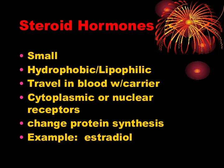
Steroid Hormones
Small
Hydrophobic/Lipophilic
Travel in blood w/carrier
Cytoplasmic or nuclear receptors
change protein synthesis
Example: estradiol
Steroid Hormones
Small
Hydrophobic/Lipophilic
Travel in blood w/carrier
Cytoplasmic or nuclear receptors
change protein synthesis
Example: estradiol
PEPTIDE HORMONES
PEPTIDE HORMONES
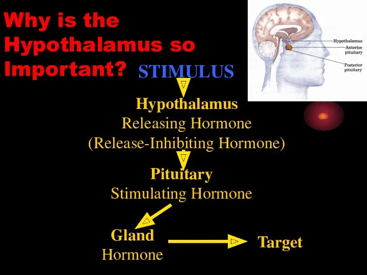
STIMULUS
Hypothalamus
Releasing Hormone
(Release-Inhibiting Hormone)
Pituitary
Stimulating Hormone
Gland
Hormone
Target
Why is the Hypothalamus so Important?
STIMULUS
Hypothalamus
Releasing Hormone
(Release-Inhibiting Hormone)
Pituitary
Stimulating Hormone
Gland
Hormone
Target
Why is the Hypothalamus so Important?
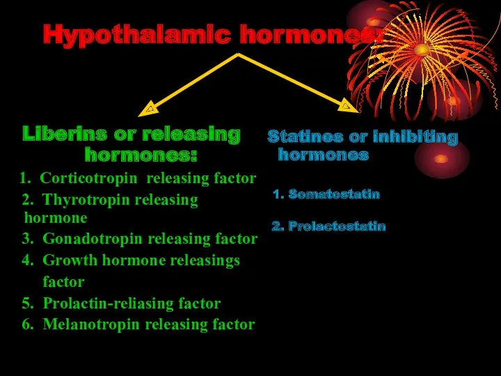
Hypothalamic hormones:
Liberins or releasing hormones:
1. Corticotropin releasing factor
2. Thyrotropin releasing hormone
Hypothalamic hormones:
Liberins or releasing hormones:
1. Corticotropin releasing factor
2. Thyrotropin releasing hormone
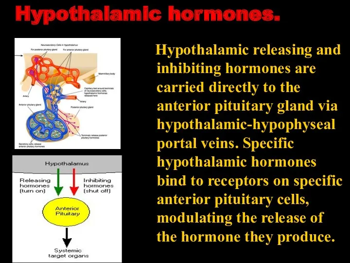
Hypothalamic hormones.
Hypothalamic releasing and inhibiting hormones are carried directly to
Hypothalamic hormones.
Hypothalamic releasing and inhibiting hormones are carried directly to
Pituitary hormones.
Pituitary hormones.
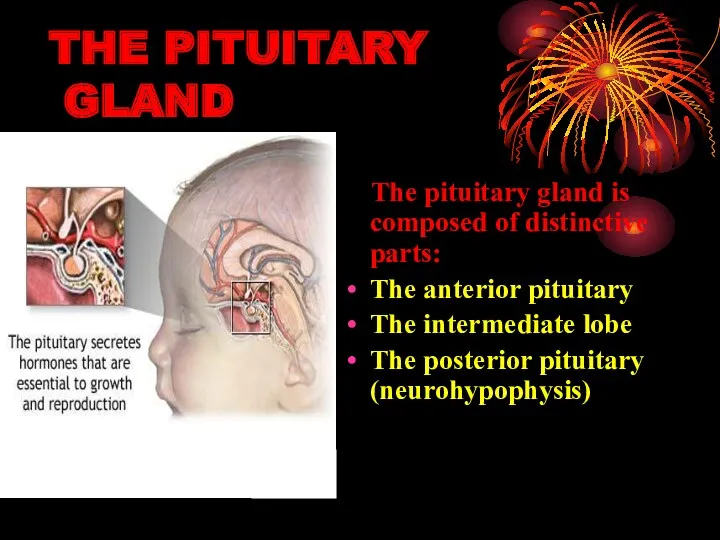
THE PITUITARY
GLAND
The pituitary gland is composed of distinctive
THE PITUITARY
GLAND
The pituitary gland is composed of distinctive
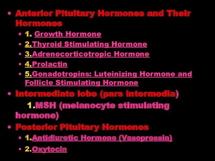
Anterior Pituitary Hormones and Their Hormones
1. Growth Hormone
2.Thyroid Stimulating
Anterior Pituitary Hormones and Their Hormones
1. Growth Hormone
2.Thyroid Stimulating
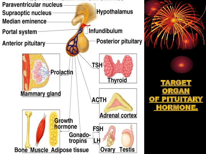
TARGET
ORGAN
OF PITUITARY
HORMONE.
TARGET
ORGAN
OF PITUITARY
HORMONE.
TARGET
ORGAN
OF PITUITARY
HORMONE.
TARGET
ORGAN
OF PITUITARY
HORMONE.
1.
Growth Hormone (GH)
1.
Growth Hormone (GH)
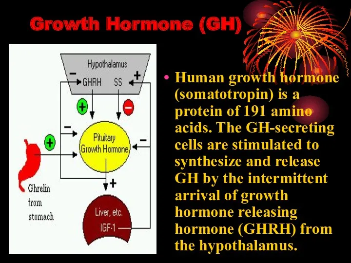
Growth Hormone (GH)
Human growth hormone (somatotropin) is a protein of 191
Growth Hormone (GH)
Human growth hormone (somatotropin) is a protein of 191
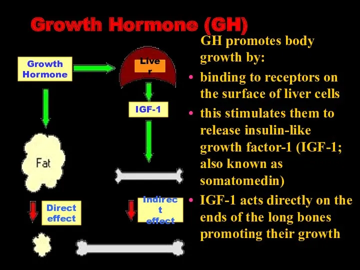
Growth Hormone (GH)
GH promotes body growth by:
binding to receptors
Growth Hormone (GH)
GH promotes body growth by:
binding to receptors
Growth Hormone (GH)
It promotes protein building in all cells (increase the
Growth Hormone (GH)
It promotes protein building in all cells (increase the
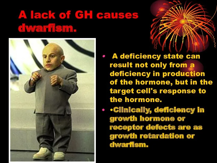
A lack of GH causes dwarfism.
A deficiency state can
A lack of GH causes dwarfism.
A deficiency state can
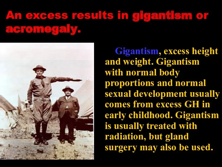
An excess results in gigantism or acromegaly.
Gigantism, excess height
An excess results in gigantism or acromegaly.
Gigantism, excess height
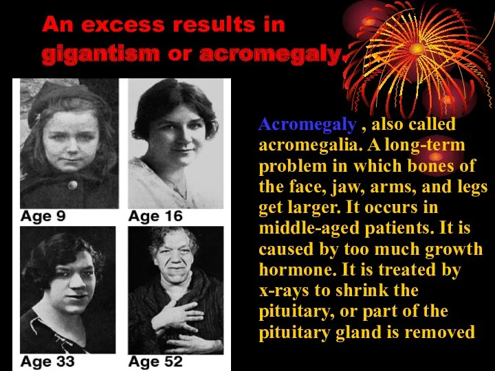
An excess results in
gigantism or acromegaly.
Acromegaly , also
An excess results in
gigantism or acromegaly.
Acromegaly , also
2.
Thyroid-Stimulating Hormone (Thyrotropin )
2.
Thyroid-Stimulating Hormone (Thyrotropin )
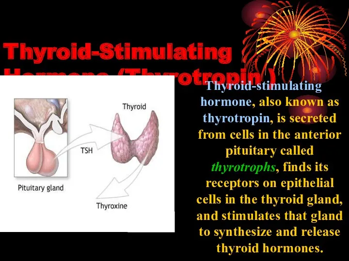
Thyroid-Stimulating Hormone (Thyrotropin )
Thyroid-stimulating hormone, also known as thyrotropin, is secreted
Thyroid-Stimulating Hormone (Thyrotropin )
Thyroid-stimulating hormone, also known as thyrotropin, is secreted
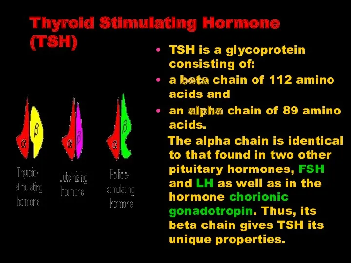
Thyroid Stimulating Hormone (TSH)
TSH is a glycoprotein consisting of:
a beta
Thyroid Stimulating Hormone (TSH)
TSH is a glycoprotein consisting of:
a beta
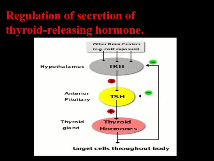
Regulation of secretion of thyroid-releasing hormone.
Regulation of secretion of thyroid-releasing hormone.
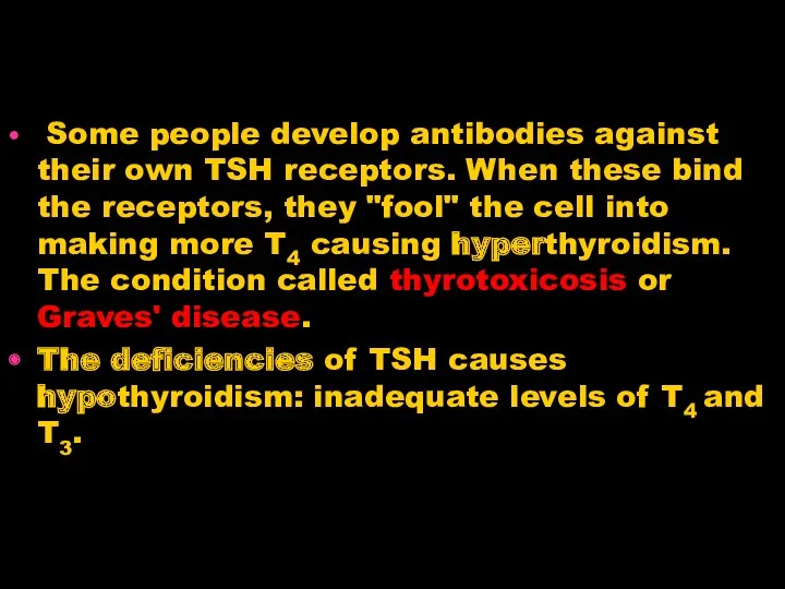
Some people develop antibodies against their own TSH receptors. When
Some people develop antibodies against their own TSH receptors. When
3.
Adrenocorticotropic Hormone
(ACTH)
3.
Adrenocorticotropic Hormone
(ACTH)
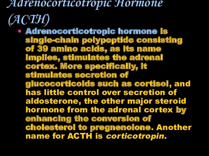
Adrenocorticotropic Hormone (ACTH)
Adrenocorticotropic hormone is single-chain polypeptide consisting of 39 amino
Adrenocorticotropic Hormone (ACTH)
Adrenocorticotropic hormone is single-chain polypeptide consisting of 39 amino
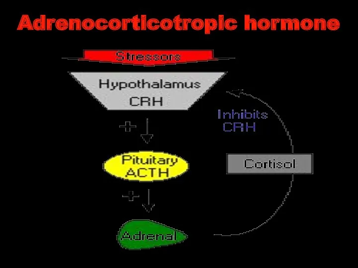
Adrenocorticotropic hormone
Adrenocorticotropic hormone
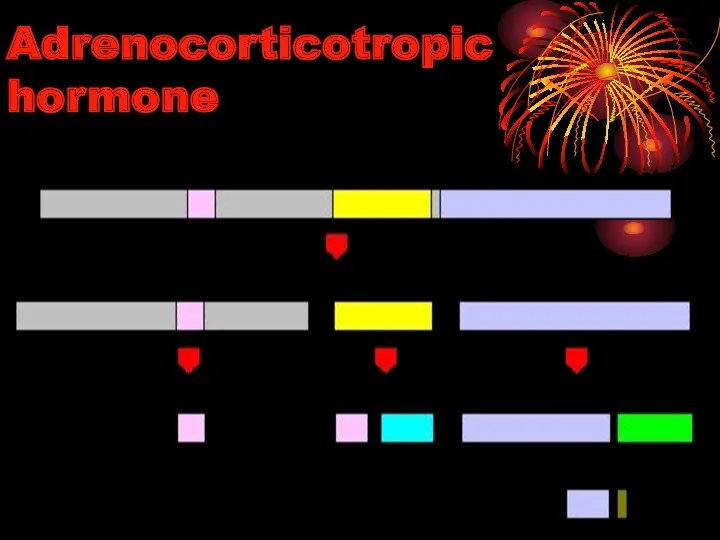
Adrenocorticotropic hormone
Adrenocorticotropic hormone
4.
PROLACTIN
4.
PROLACTIN
Prolactin
Prolactin – protein of 197 amino acids.
Prolactin, acting with other
Prolactin
Prolactin – protein of 197 amino acids.
Prolactin, acting with other
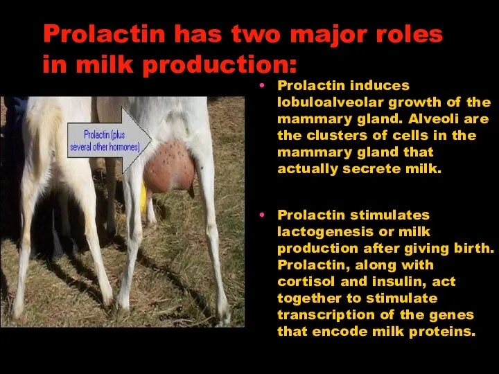
Prolactin has two major roles in milk production:
Prolactin induces lobuloalveolar
Prolactin has two major roles in milk production:
Prolactin induces lobuloalveolar
PROLACTIN
SECRETION
PROLACTIN
SECRETION
Disease States
Excessive secretion of prolactin - hyperprolactinemia - is a relative
Disease States
Excessive secretion of prolactin - hyperprolactinemia - is a relative
5.
Gonadotropins: Luteinizing and Follicle Stimulating Hormones
5.
Gonadotropins: Luteinizing and Follicle Stimulating Hormones
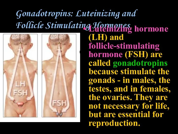
Gonadotropins: Luteinizing and Follicle Stimulating Hormones
Luteinizing hormone (LH) and follicle-stimulating hormone
Gonadotropins: Luteinizing and Follicle Stimulating Hormones
Luteinizing hormone (LH) and follicle-stimulating hormone
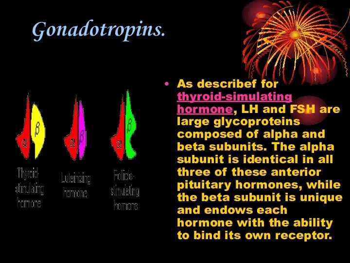
Gonadotropins.
As describef for thyroid-simulating hormone, LH and FSH are large glycoproteins
Gonadotropins.
As describef for thyroid-simulating hormone, LH and FSH are large glycoproteins
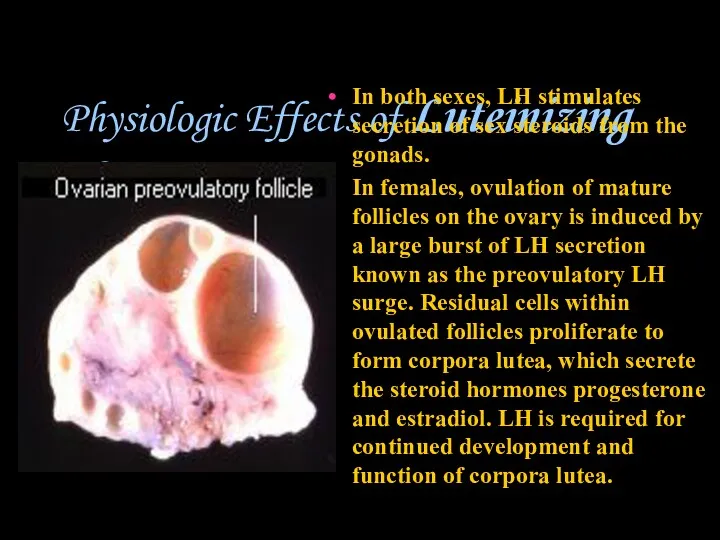
Physiologic Effects of Luteinizing Hormone
In both sexes, LH stimulates secretion
Physiologic Effects of Luteinizing Hormone
In both sexes, LH stimulates secretion
Luteinizing Hormone
In men In the testes, LH binds to receptors
Luteinizing Hormone
In men In the testes, LH binds to receptors
Follicle-Stimulating Hormone
As its name implies, FSH stimulates the maturation of ovarian
Follicle-Stimulating Hormone
As its name implies, FSH stimulates the maturation of ovarian
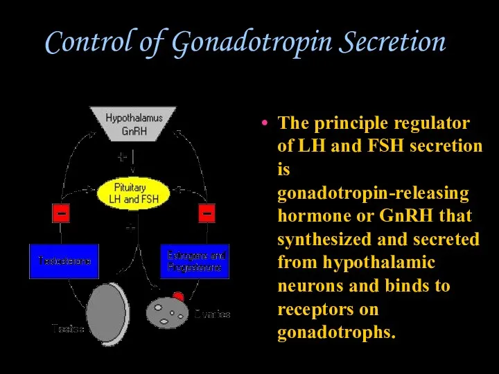
Control of Gonadotropin Secretion
The principle regulator of LH and FSH
Control of Gonadotropin Secretion
The principle regulator of LH and FSH
Disease States
Diminished secretion of LH or FSH can result in failure
Disease States
Diminished secretion of LH or FSH can result in failure
Intermediate lobe: MSH (melanocyte stimulating hormone)
Melanocyte-stimulating hormone (MSH): Known to control
Intermediate lobe: MSH (melanocyte stimulating hormone)
Melanocyte-stimulating hormone (MSH): Known to control
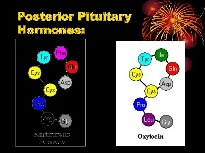
Posterior Pituitary Hormones:
Posterior Pituitary Hormones:
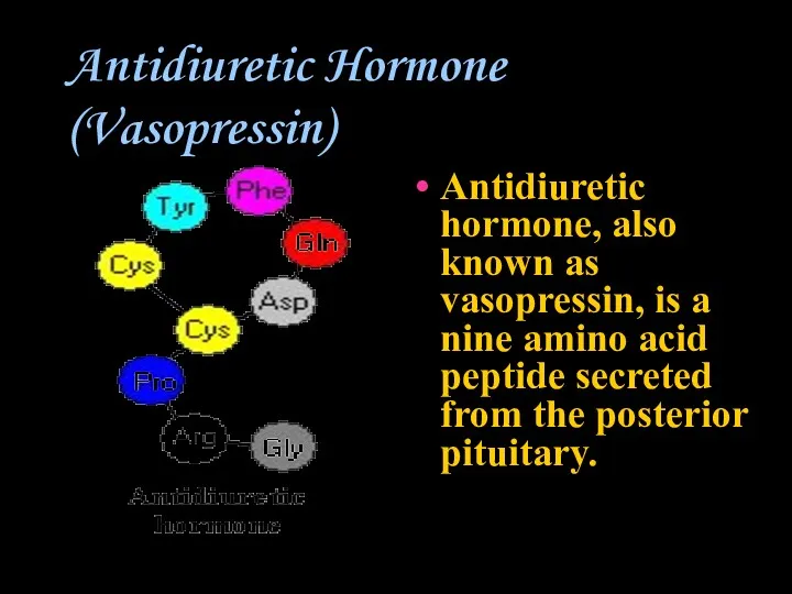
Antidiuretic Hormone (Vasopressin)
Antidiuretic hormone, also known as vasopressin, is a nine
Antidiuretic Hormone (Vasopressin)
Antidiuretic hormone, also known as vasopressin, is a nine
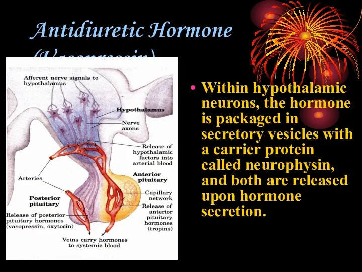
Antidiuretic Hormone (Vasopressin)
Within hypothalamic neurons, the hormone is packaged in secretory
Antidiuretic Hormone (Vasopressin)
Within hypothalamic neurons, the hormone is packaged in secretory
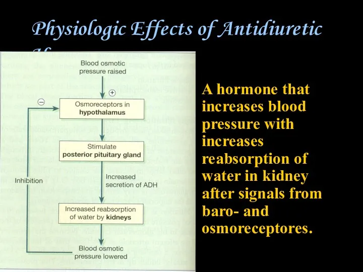
Physiologic Effects of Antidiuretic Hormone
A hormone that increases blood pressure with
Physiologic Effects of Antidiuretic Hormone
A hormone that increases blood pressure with
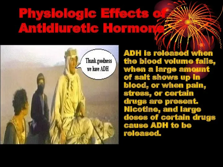
Physiologic Effects of Antidiuretic Hormone
ADH is released when the blood volume
Physiologic Effects of Antidiuretic Hormone
ADH is released when the blood volume
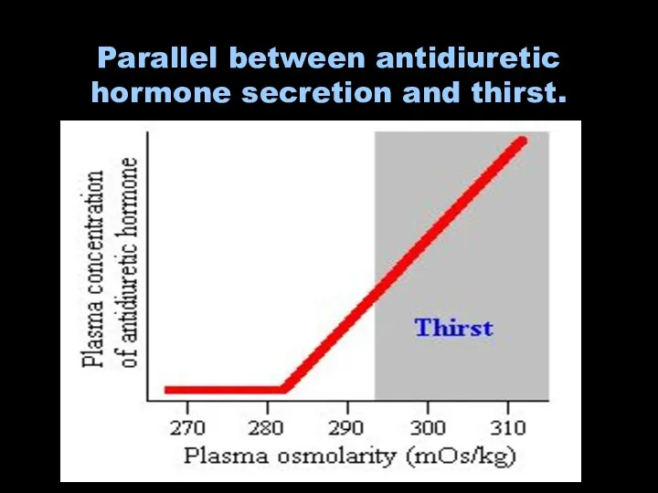
Parallel between antidiuretic hormone secretion and thirst.
Parallel between antidiuretic hormone secretion and thirst.
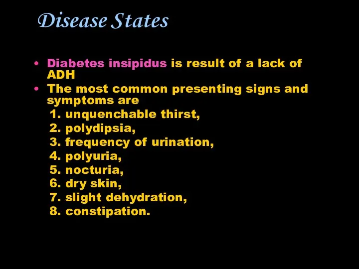
Disease States
Diabetes insipidus is result of a lack of ADH
The most
Disease States
Diabetes insipidus is result of a lack of ADH
The most
Posterior Pituitary Hormones
Oxytocin
Posterior Pituitary Hormones
Oxytocin
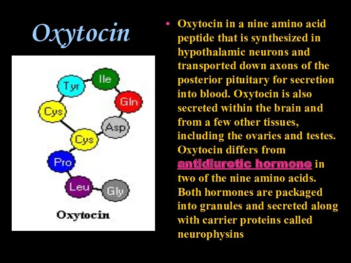
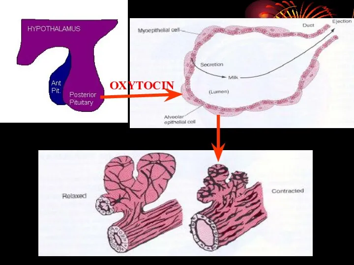
Oxytocin
Oxytocin in a nine amino acid peptide that is synthesized in
Oxytocin
Oxytocin in a nine amino acid peptide that is synthesized in
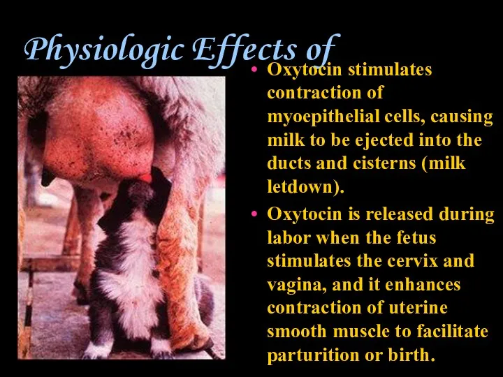
Physiologic Effects of Oxytocin
Oxytocin stimulates contraction of myoepithelial cells, causing milk
Physiologic Effects of Oxytocin
Oxytocin stimulates contraction of myoepithelial cells, causing milk
OXYTOCIN
OXYTOCIN
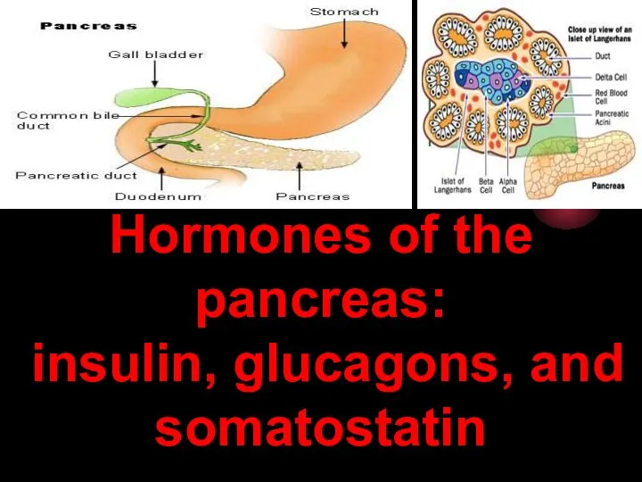
Hormones of the pancreas:
insulin, glucagons, and somatostatin
Hormones of the pancreas:
insulin, glucagons, and somatostatin

The pancreas has two major biochemical functions:
1. Exocrine cells produce digestive
The pancreas has two major biochemical functions:
1. Exocrine cells produce digestive
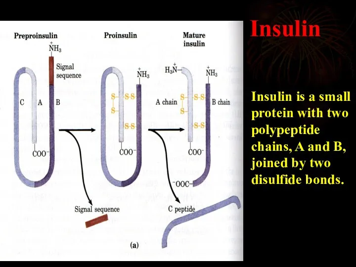
Insulin
Insulin is a small protein with two polypeptide chains, A and
Insulin
Insulin is a small protein with two polypeptide chains, A and
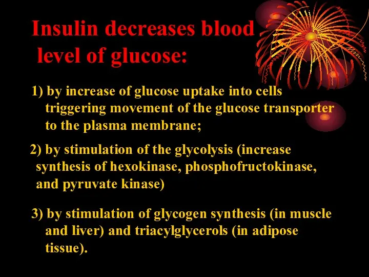
Insulin decreases blood
level of glucose:
1) by increase of glucose uptake
Insulin decreases blood
level of glucose:
1) by increase of glucose uptake
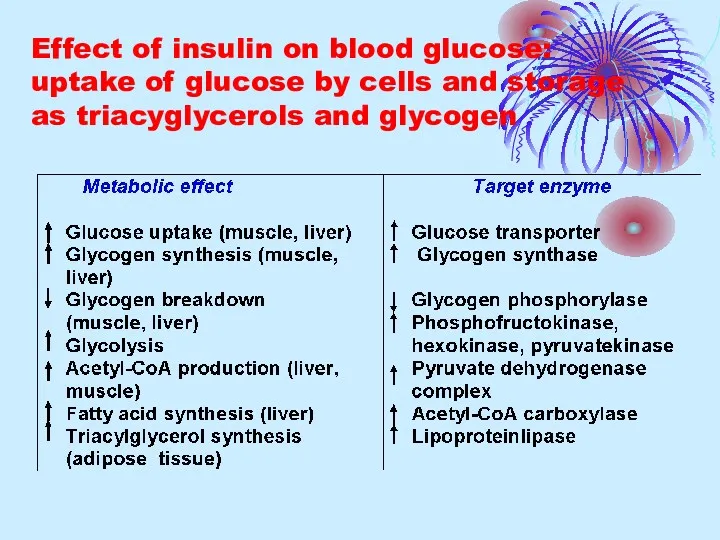
Effect of insulin on blood glucose: uptake of glucose by cells
Effect of insulin on blood glucose: uptake of glucose by cells
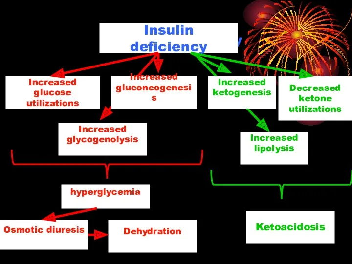
Insulin deficiency
Insulin deficiency
Increased
glucose utilizations
Increased
glycogenolysis
Increased
gluconeogenesis
hyperglycemia
Osmotic diuresis
Dehydration
Increased
ketogenesis
Decreased
ketone
utilizations
Increased
lipolysis
Ketoacidosis
Insulin deficiency
Insulin deficiency
Increased
glucose utilizations
Increased
glycogenolysis
Increased
gluconeogenesis
hyperglycemia
Osmotic diuresis
Dehydration
Increased
ketogenesis
Decreased
ketone
utilizations
Increased
lipolysis
Ketoacidosis
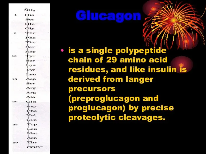
Glucagon
is a single polypeptide chain of 29 amino acid residues, and
Glucagon
is a single polypeptide chain of 29 amino acid residues, and
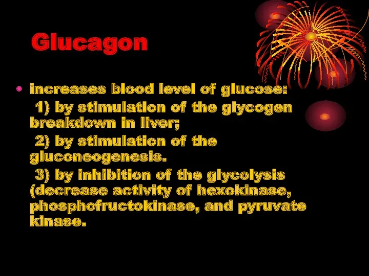
Glucagon
increases blood level of glucose:
1) by stimulation of the glycogen
Glucagon
increases blood level of glucose:
1) by stimulation of the glycogen
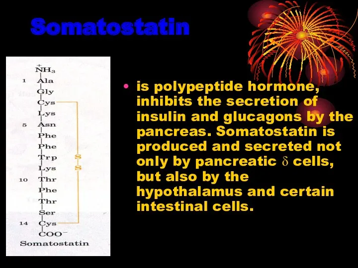
Somatostatin
is polypeptide hormone, inhibits the secretion of insulin and glucagons by
Somatostatin
is polypeptide hormone, inhibits the secretion of insulin and glucagons by
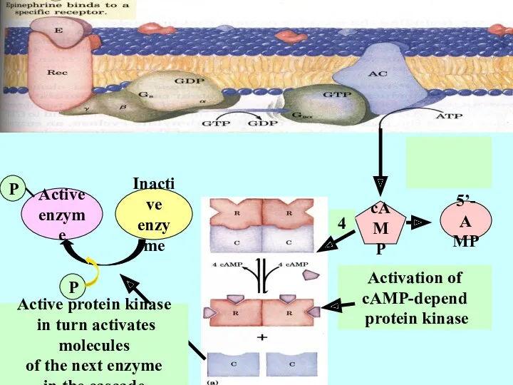
5’-AMP
5’-AMP
cAMP
Activation of
cAMP-depend
protein kinase
4
Inactive
enzyme
Active
enzyme
P
P
Active protein kinase
in turn activates molecules
5’-AMP
5’-AMP
cAMP
Activation of
cAMP-depend
protein kinase
4
Inactive
enzyme
Active
enzyme
P
P
Active protein kinase
in turn activates molecules
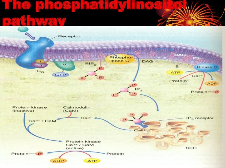
The phosphatidylinositol pathway
The phosphatidylinositol pathway
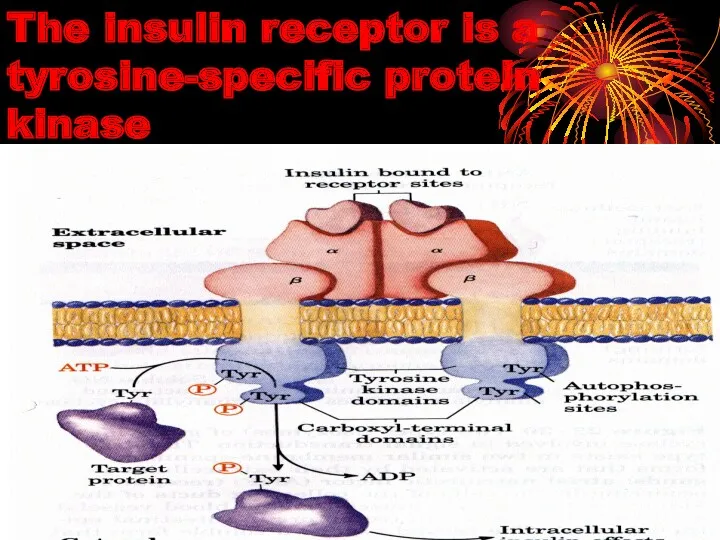
The insulin receptor is a tyrosine-specific protein kinase
The insulin receptor is a tyrosine-specific protein kinase
 Посттрансплантационные осложнения. Что такое РТПХ? Иммуносупрессивная терапия
 Правила личной гигиены и здоровья
 Роль зовнішніх факторів у патології. Патогенна дія фізичних факторів на організм. (Лекція 3)
 Оказание первой помощи. Современные требования. Часть 1
 Кардиомиопатии (КМП)
 Глубокое резцовое перекрытие и дистальное смещение нижней челюсти
 Акушериядағы қан кетулер. Босанғаннан кейінгі ҚК
 Развитие зубов. Закладка зачатков зубов
 Местное лечение заболеваний пародонта
 Зубы. Твердые и мягкие ткани зуба. Поддерживающий аппарат зуба
 Жынысты көбею. Мейоз, оның биологиялық маңызы
 Повреждения острыми предметами
 Синтетические противомикробные средства
 Алгоритм ранней диагностики злокачественных новообразований на уровне ПМСП
 Новые горизонты в лечении ИБС
 Хроническая обструктивная болезнь легких
 12 жұп бас ми нервтері
 Синдром Марфана
 Ампутации и экзартикуляции конечностей
 Психологія діагностичного процесу
 The hormonal regulation of the body
 Оба қоздырғышы
 Бригаданың медициналық қызметі
 Александр Николаевич Кудрин
 The physiology of childbirth
 Приоритеты в медикаментах
 Доказательная профилактика. Основные виды, проблемы внедрения и анализа результатов скрининговых программ
 Семейная наследственная гиперхолестеринемия